Free with trial Vector round icons of human organs Flat design. Structure long bone vectors Vector set of flat icons with human organs. Vector round icons of human organs Flat design
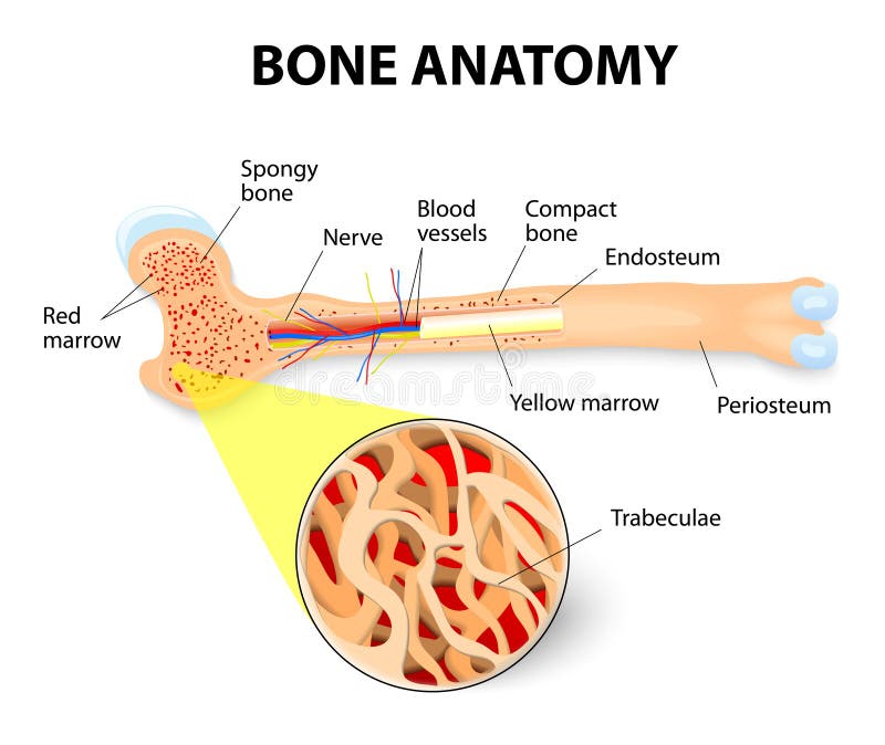
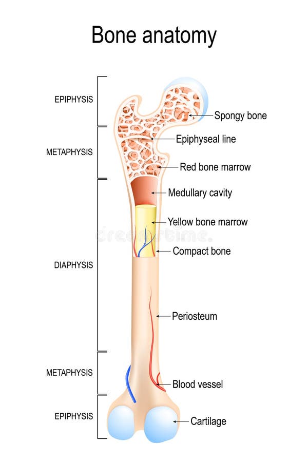
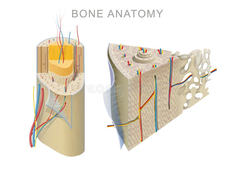
Free with trial Anatomy of the Long Bone. Periosteum, endosteum, bone marrow and trabeculae. Structure long bone vectors Bone anatomy. Anatomy of the Long Bone. Periosteum, endosteum, bone marrow and trabeculae.
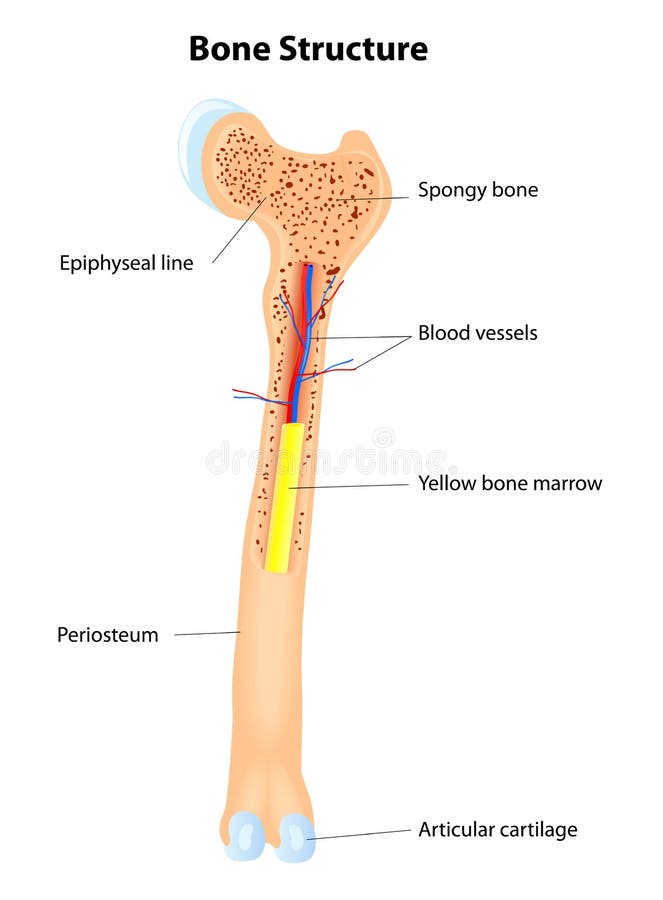
Free with trial Anatomy of a long bone, eps8, gradient and mesh printing compatible. Structure long bone vectors Bone structure. Anatomy of a long bone, eps8, gradient and mesh printing compatible
Free with trial Vector illustration of long bone basic structure. Structure long bone vectors Bone structure. Vector illustration of long bone basic structure.
Free with trial Long Bone Anatomy. Vector scheme. illustration of human bone anatomy. Structure long bone vectors Bone structure details. Long Bone Anatomy. Vector scheme. illustration of human bone anatomy
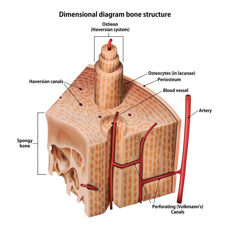
Free with trial Three-dimensional diagram bone structure on whate. Structure long bone illustrations Three-dimensional diagram bone structure
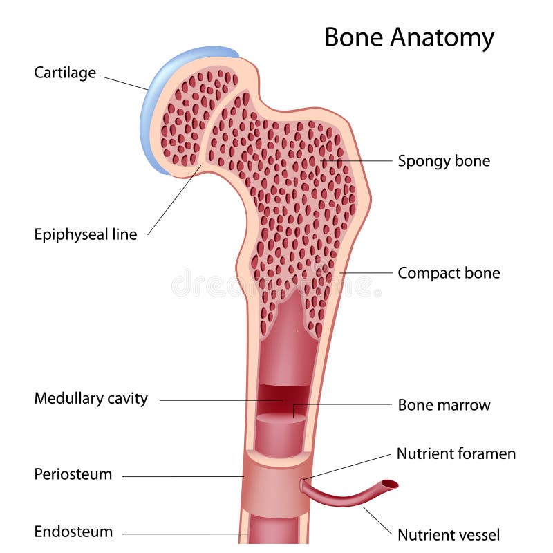
Free with trial Bone anatomy. Structure of a Long Bone. illustration for medical, educational and science use. Structure long bone illustrations Structure of a Long Bone.
Free with trial Bone is not a uniformly solid material, but is mostly a matrix. The primary tissue of bone, osseous tissue, is relatively hard and lightweight. Structure long bone illustrations Bone structure. Bone is not a uniformly solid material, but is mostly a matrix. The primary tissue of bone, osseous tissue, is relatively hard and lightweight.
Free with trial Long Bone Anatomy. Vector scheme. Structure long bone vectors Bone Structure. Vector scheme. Long Bone Anatomy. Vector scheme
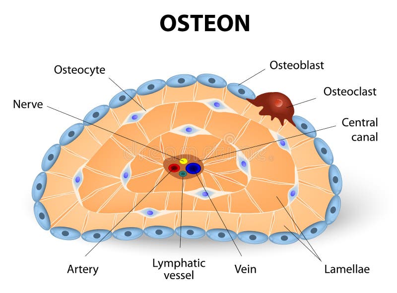
Free with trial Osteon development and structure. Osteoblast, osteocyte, and osteoclast. Structure long bone vectors Osteon development and structure
Free with trial Bones are rigid organs that constitute part of the endoskeleton of vertebrates. They support and protect the various organs of the body, produce red and white blood cells and store minerals. Structure long bone illustrations Structure of bone. Bones are rigid organs that constitute part of the endoskeleton of vertebrates. They support and protect the various organs of the body, produce red and white blood cells and store minerals.
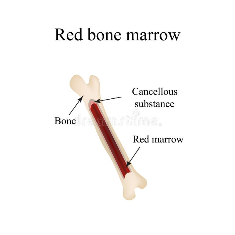
Free with trial Bone marrow is the spongy tissue inside some of the bones in the body, including the hip and thigh bones. Structure long bone illustrations Bone marrow
Free with trial Bone Marrow Yellow, Red and blood cells erythrocyte, lymphocyte, monocyte, esinophil, basophil, neurophil. Vector diagram for your design, educational, biological, science and medical use. Structure long bone vectors Bone Marrow Yellow, Red and blood cells
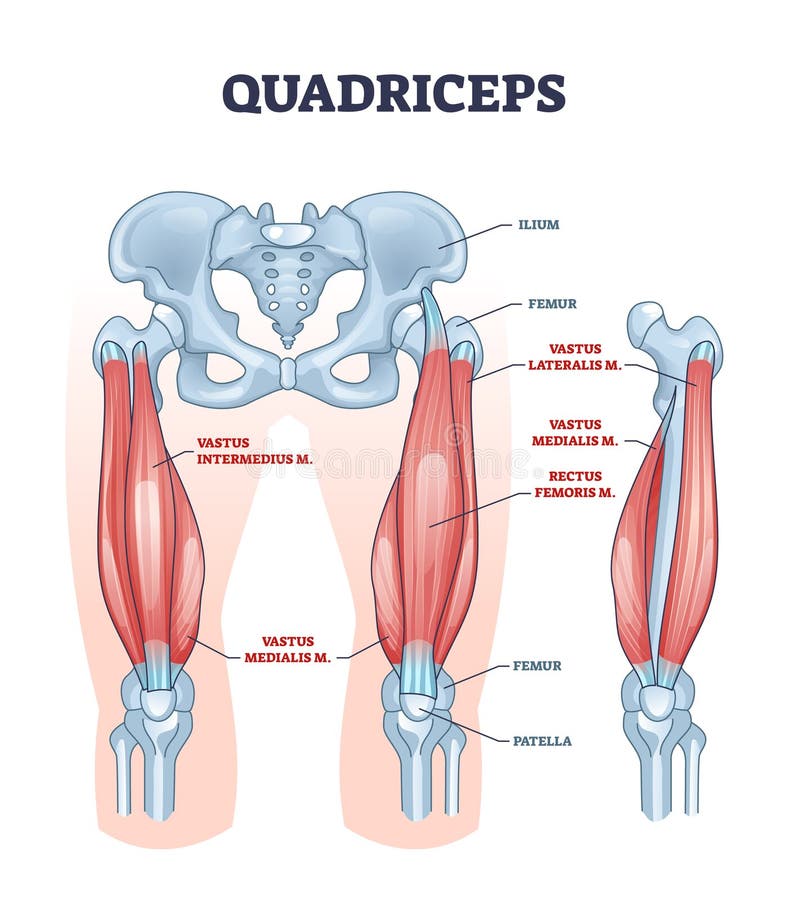
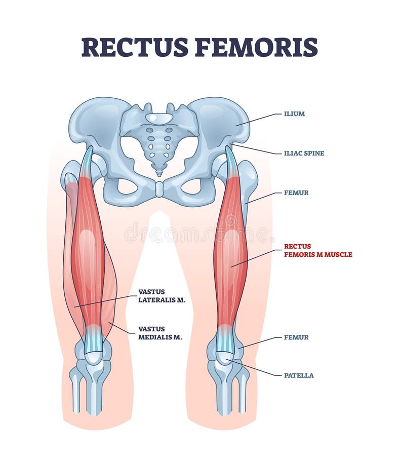
Free with trial Quadriceps muscle and quads leg muscular or bone anatomy outline diagram. Labeled educational medical scheme with vastus intermedius, medialis, lateralis or rectus femoris location vector illustration. Structure long bone vectors Quadriceps muscle or quads leg muscular anatomical structure outline diagram. Quadriceps muscle and quads leg muscular or bone anatomy outline diagram. Labeled educational medical scheme with vastus intermedius, medialis, lateralis or rectus femoris location vector illustration
Free with trial The human rib cage is made up of 12 paired rib bones; each are symmetrically paired on a right and left side. Of all 24 ribs, the first seven pairs are often labeled as `true. ` These bones are connected to the costal cartilage, while the five other `false` sets are not. The ribcage also encloses the thoracic cavity and helps protect the heart and lungs from damage. There are 24 ribs in the human body, divided into two sets of 12 curved, flat bones. Each one is attached by cartilage at the back to the thoracic vertebrae. MEN and women have 12 pairs of ribs a few individuals have 13 or 11 pairs. The idea that men have fewer ribs than women is widespread but wrong, perhaps deriving from the biblical story of Eve being made from one of Adam`s ribs. Both men and women have 24 ribs, twelve on each side. Floating rib: One of the last two ribs. A rib is said to be `floating` if it does not attach to the sternum the breast bone or to another rib. There are usually 12 pairs of ribs in all. Each pair of ribs is attached to the building blocks of the spine the vertebrae in the back. The ribs partially enclose and protect the chest cavity, where many vital organs including the heart and the lungs are located. The rib cage is collectively made up of long, curved individual bones with joint-connections to the spinal vertebrae. Structure long bone illustrations Ribs with Ligments anterior view. The human rib cage is made up of 12 paired rib bones; each are symmetrically paired on a right and left side. Of all 24 ribs, the first seven pairs are often labeled as `true.` These bones are connected to the costal cartilage, while the five other `false` sets are not. The ribcage also encloses the thoracic cavity and helps protect the heart and lungs from damage. There are 24 ribs in the human body, divided into two sets of 12 curved, flat bones. Each one is attached by cartilage at the back to the thoracic vertebrae. MEN and women have 12 pairs of ribs a few individuals have 13 or 11 pairs. The idea that men have fewer ribs than women is widespread but wrong, perhaps deriving from the biblical story of Eve being made from one of Adam`s ribs. Both men and women have 24 ribs, twelve on each side. Floating rib: One of the last two ribs. A rib is said to be `floating` if it does not attach to the sternum the breast bone or to another rib. There are usually 12 pairs of ribs in all. Each pair of ribs is attached to the building blocks of the spine the vertebrae in the back. The ribs partially enclose and protect the chest cavity, where many vital organs including the heart and the lungs are located. The rib cage is collectively made up of long, curved individual bones with joint-connections to the spinal vertebrae.
Free with trial Types of bones. Long Humerus. Shirt Heel. Flat Scapula. Mixed Vertebra. Structure long bone illustrations Types of bones
Free with trial Vector illustration of icons of internal human organs and systems. Flat design. Structure long bone vectors Vector flat icons with human organs and systems. Vector illustration of icons of internal human organs and systems. Flat design
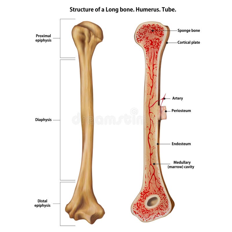
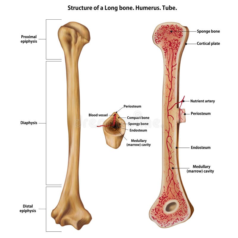
Free with trial Structure of a Long bone. Humerus. Tube. Structure long bone illustrations Structure of a Long bone.
Free with trial Structure of a long bone and cells of bone tissue(useful for education in schools and clinics ) - vector illustration. Structure long bone vectors Structure of a long bone
Free with trial Biceps muscle with anatomical skeletal medical arm structure outline diagram. Labeled educational explanation with inner muscular and bone description vector illustration. Hand physiology scheme. Structure long bone vectors Biceps muscle with anatomical skeletal medical arm structure outline diagram
Free with trial Coracobrachialis muscle with anatomical and medical structure outline diagram. Labeled educational scheme with inner skeletal and muscular system in human body vector illustration. Bone physiology. Structure long bone vectors Coracobrachialis muscle with anatomical and medical structure outline diagram
Free with trial Humerus bone labeled vector illustration diagram. Long bone type in the upper arm. Skeleton anatomy scheme with greater tubercle, deltoid tuberosity, medial epicondyle, trochlea and other parts. Structure long bone vectors Humerus bone labeled vector illustration diagram
Free with trial Soleus muscle with anatomical leg bones skeletal structure outline diagram. Labeled educational scheme with posterior view of right human leg vector illustration. Human foot detailed description. Structure long bone vectors Soleus muscle with anatomical leg bones skeletal structure outline diagram
Free with trial Bone marrow is the spongy tissue inside some of the bones in the body, including the hip and thigh bones. Structure long bone illustrations Bone marrow Aerial View. Bone marrow is the spongy tissue inside some of the bones in the body, including the hip and thigh bones.
Free with trial Bone marrow is the spongy tissue inside some of the bones in the body, including the hip and thigh bones. Structure long bone illustrations Bone marrow
Free with trial Palmaris longus skeletal and muscular body structure for human arm outline diagram. Labeled educational scheme with anatomical and medical hand inner parts physical description vector illustration. Structure long bone vectors Palmaris longus skeletal and muscular structure for human arm outline diagram. Palmaris longus skeletal and muscular body structure for human arm outline diagram. Labeled educational scheme with anatomical and medical hand inner parts physical description vector illustration.
Free with trial Peroneus brevis leg muscle with longus and tertius muscular part location outline diagram. Labeled educational foot skeletal structure from later view vector illustration. Tibia and metatarsal bone. Structure long bone vectors Peroneus brevis leg muscle with longus and tertius location outline diagram. Peroneus brevis leg muscle with longus and tertius muscular part location outline diagram. Labeled educational foot skeletal structure from later view vector illustration. Tibia and metatarsal bone.
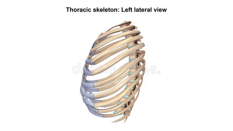
Free with trial Thoracic Skeleton The human rib cage is made up of 12 paired rib bones; each are symmetrically paired on a right and left side. Of all 24 ribs, the first seven pairs are often labeled as `true. ` These bones are connected to the costal cartilage, while the five other `false` sets are not. The ribcage also encloses the thoracic cavity and helps protect the heart and lungs from damage. There are 24 ribs in the human body, divided into two sets of 12 curved, flat bones. Each one is attached by cartilage at the back to the thoracic vertebrae. MEN and women have 12 pairs of ribs a few individuals have 13 or 11 pairs. The idea that men have fewer ribs than women is widespread but wrong, perhaps deriving from the biblical story of Eve being made from one of Adam`s ribs. Both men and women have 24 ribs, twelve on each side. Floating rib: One of the last two ribs. A rib is said to be `floating` if it does not attach to the sternum the breast bone or to another rib. There are usually 12 pairs of ribs in all. Each pair of ribs is attached to the building blocks of the spine the vertebrae in the back. The ribs partially enclose and protect the chest cavity, where many vital organs including the heart and the lungs are located. The rib cage is collectively made up of long, curved individual bones with joint-connections to the spinal vertebrae. Structure long bone illustrations Thoracic Skeleton Lateral view. Thoracic Skeleton The human rib cage is made up of 12 paired rib bones; each are symmetrically paired on a right and left side. Of all 24 ribs, the first seven pairs are often labeled as `true.` These bones are connected to the costal cartilage, while the five other `false` sets are not. The ribcage also encloses the thoracic cavity and helps protect the heart and lungs from damage. There are 24 ribs in the human body, divided into two sets of 12 curved, flat bones. Each one is attached by cartilage at the back to the thoracic vertebrae. MEN and women have 12 pairs of ribs a few individuals have 13 or 11 pairs. The idea that men have fewer ribs than women is widespread but wrong, perhaps deriving from the biblical story of Eve being made from one of Adam`s ribs. Both men and women have 24 ribs, twelve on each side. Floating rib: One of the last two ribs. A rib is said to be `floating` if it does not attach to the sternum the breast bone or to another rib. There are usually 12 pairs of ribs in all. Each pair of ribs is attached to the building blocks of the spine the vertebrae in the back. The ribs partially enclose and protect the chest cavity, where many vital organs including the heart and the lungs are located. The rib cage is collectively made up of long, curved individual bones with joint-connections to the spinal vertebrae.
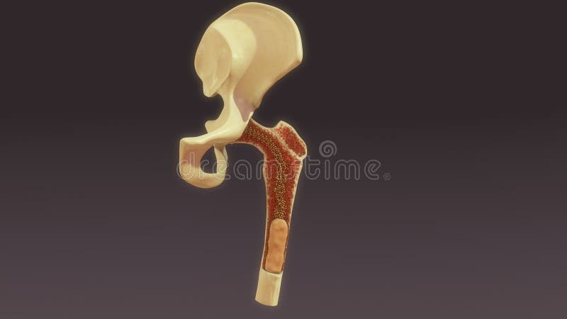
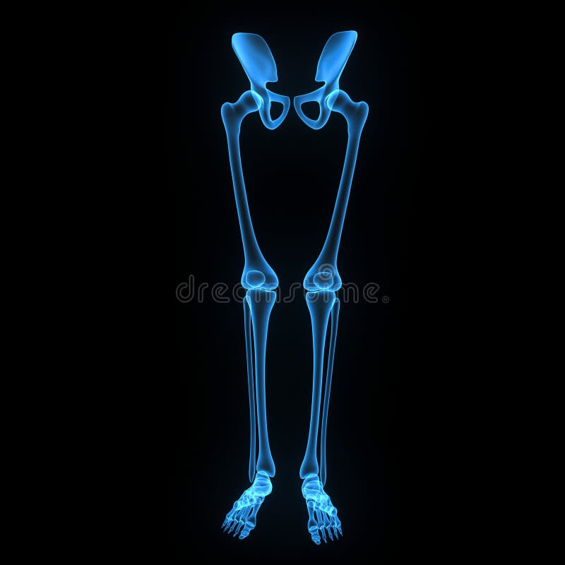
Free with trial The hip joint is one of the most important joints in the human body. It allows us to walk, run, and jump. It bears our body’s weight and the force of the strong muscles of the hip and leg. Yet the hip joint is also one of our most flexible joints and allows a greater range of motion than all other joints in the body except for the shoulder. The hip joint is a ball-and-socket synovial joint formed between the os coxa (hip bone) and the femur. A round, cup-shaped structure on the os coax, known as the acetabulum, forms the socket for the hip joint. The rounded head of the femur forms the ball of the joint. The tibia, sometimes known as the shin bone, is the larger and stronger of the two lower leg bones. It forms the knee joint with the femur and the ankle joint with the fibula and tarsus. The fibula is the long, thin and lateral bone of the lower leg. It runs parallel to the tibia, or shin bone, and plays a significant role in stabilizing the ankle and supporting the muscles of the lower leg. The bones of the ankle and foot form the most distal region of the lower limb in the appendicular skeleton. These bones are responsible for the propulsion, balance, and support of the body’s weight through many diverse activities, such as standing, walking, running, and jumping. Structure long bone illustrations Skeleton: Hip, Femur, Tibia, Fibula, Ankle and Foot bones. The hip joint is one of the most important joints in the human body. It allows us to walk, run, and jump. It bears our body’s weight and the force of the strong muscles of the hip and leg. Yet the hip joint is also one of our most flexible joints and allows a greater range of motion than all other joints in the body except for the shoulder. The hip joint is a ball-and-socket synovial joint formed between the os coxa (hip bone) and the femur. A round, cup-shaped structure on the os coax, known as the acetabulum, forms the socket for the hip joint. The rounded head of the femur forms the ball of the joint. The tibia, sometimes known as the shin bone, is the larger and stronger of the two lower leg bones. It forms the knee joint with the femur and the ankle joint with the fibula and tarsus. The fibula is the long, thin and lateral bone of the lower leg. It runs parallel to the tibia, or shin bone, and plays a significant role in stabilizing the ankle and supporting the muscles of the lower leg. The bones of the ankle and foot form the most distal region of the lower limb in the appendicular skeleton. These bones are responsible for the propulsion, balance, and support of the body’s weight through many diverse activities, such as standing, walking, running, and jumping.
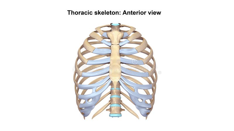
Free with trial Thoracic Skeleton The human rib cage is made up of 12 paired rib bones; each are symmetrically paired on a right and left side. Of all 24 ribs, the first seven pairs are often labeled as `true. ` These bones are connected to the costal cartilage, while the five other `false` sets are not. The ribcage also encloses the thoracic cavity and helps protect the heart and lungs from damage. There are 24 ribs in the human body, divided into two sets of 12 curved, flat bones. Each one is attached by cartilage at the back to the thoracic vertebrae. MEN and women have 12 pairs of ribs a few individuals have 13 or 11 pairs. The idea that men have fewer ribs than women is widespread but wrong, perhaps deriving from the biblical story of Eve being made from one of Adam`s ribs. Both men and women have 24 ribs, twelve on each side. Floating rib: One of the last two ribs. A rib is said to be `floating` if it does not attach to the sternum the breast bone or to another rib. There are usually 12 pairs of ribs in all. Each pair of ribs is attached to the building blocks of the spine the vertebrae in the back. The ribs partially enclose and protect the chest cavity, where many vital organs including the heart and the lungs are located. The rib cage is collectively made up of long, curved individual bones with joint-connections to the spinal vertebrae. Structure long bone illustrations Thoracic Skeleton Anterior view. Thoracic Skeleton The human rib cage is made up of 12 paired rib bones; each are symmetrically paired on a right and left side. Of all 24 ribs, the first seven pairs are often labeled as `true.` These bones are connected to the costal cartilage, while the five other `false` sets are not. The ribcage also encloses the thoracic cavity and helps protect the heart and lungs from damage. There are 24 ribs in the human body, divided into two sets of 12 curved, flat bones. Each one is attached by cartilage at the back to the thoracic vertebrae. MEN and women have 12 pairs of ribs a few individuals have 13 or 11 pairs. The idea that men have fewer ribs than women is widespread but wrong, perhaps deriving from the biblical story of Eve being made from one of Adam`s ribs. Both men and women have 24 ribs, twelve on each side. Floating rib: One of the last two ribs. A rib is said to be `floating` if it does not attach to the sternum the breast bone or to another rib. There are usually 12 pairs of ribs in all. Each pair of ribs is attached to the building blocks of the spine the vertebrae in the back. The ribs partially enclose and protect the chest cavity, where many vital organs including the heart and the lungs are located. The rib cage is collectively made up of long, curved individual bones with joint-connections to the spinal vertebrae.
Free with trial Brachioradialis muscle medical location with anatomical bones outline diagram. Labeled educational scheme with hands inner structure and muscular description vector illustration. Body and arm parts. Structure long bone vectors Brachioradialis muscle medical location with anatomical bones outline diagram
Free with trial The spinal cord is a long, thin, tubular bundle of nervous tissue and support cells that extends from the brain (the medulla oblongata specifically). Structure long bone illustrations Spinal cord
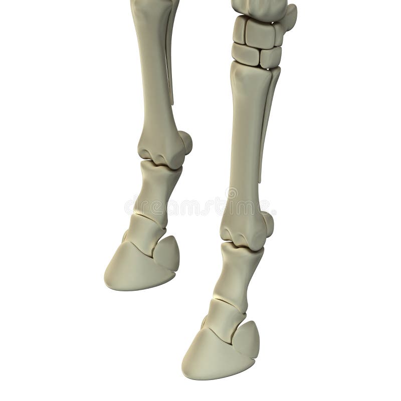
Free with trial Horse Front Leg Bones - Horse Equus Anatomy - isolated on white. Structure long bone illustrations Horse Front Leg Bones - Horse Equus Anatomy - isolated on white
Free with trial The spinal cord is a long, thin, tubular bundle of nervous tissue and support cells that extends from the brain (the medulla oblongata specifically). Structure long bone illustrations Spinal cord
Free with trial Rectus femoris muscle as one of quadriceps muscular group outline diagram. Labeled educational scheme with skeletal upper leg anatomy vector illustration. Body vastus lateralis and medialis location. Structure long bone vectors Rectus femoris muscle as one of quadriceps muscular group outline diagram
Free with trial The spinal cord is a long, thin, tubular bundle of nervous tissue and support cells that extends from the brain (the medulla oblongata specifically). Structure long bone illustrations Spinal cord
Free with trial Rotator cuff impingement and anatomical shoulder muscle outline diagram. Labeled educational muscular and skeletal description with injury example vector illustration. Supraspinatus body part location. Structure long bone vectors Rotator cuff impingement and anatomical shoulder muscle outline diagram
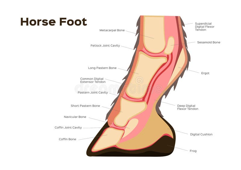
Free with trial Horse foot and leg anatomy / infographic chart vector on white. Structure long bone vectors Horse foot and leg anatomy / infographic chart vector
Free with trial Old woman with osteoporosis problem on the blue background. Structure long bone vectors Old woman with osteoporosis
Free with trial Old man with osteoporosis problem on the blue background. Structure long bone vectors Old man with osteoporosis
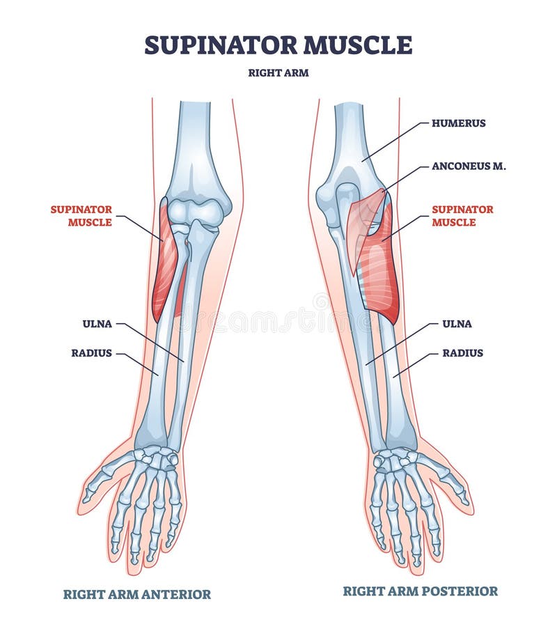
Free with trial Supinator muscle with right arm anatomical bone structure outline diagram. Labeled educational scheme with medical anterior and posterior hand view vector illustration. Humerus, anconeus and ulna xray. Structure long bone vectors Supinator muscle with right arm anatomical bone structure outline diagram
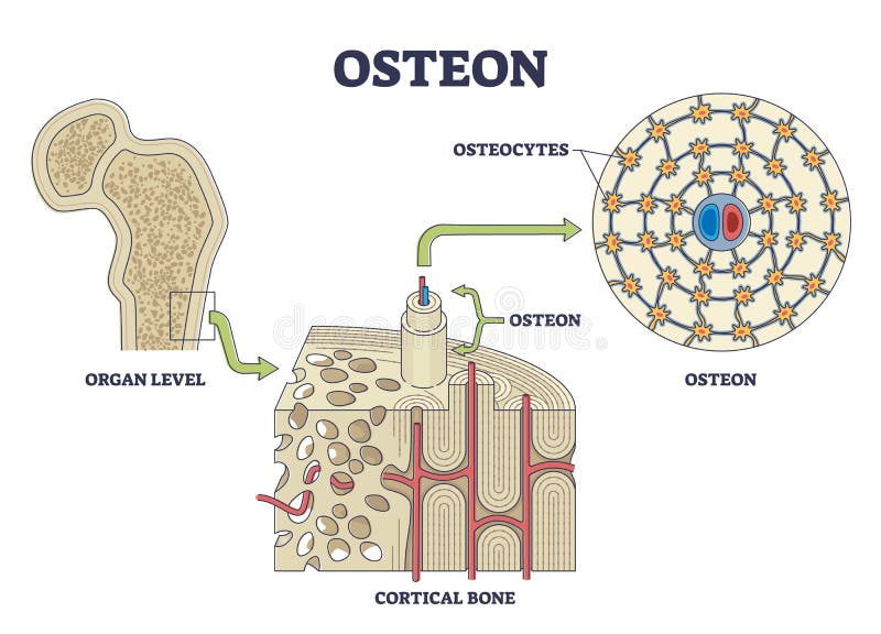
Free with trial Osteon or haversian system with compact bone structure outline diagram. Labeled educational scheme with cortical bone or organ level anatomy vector illustration. Osteocytes location from cross section. Structure long bone vectors Osteon or haversian system with compact bone structure outline diagram
Free with trial Layer of a Long bone. Humerus. Tube. Isolated on white. Structure long bone illustrations Layer of a Long bone. Humerus. Tube.
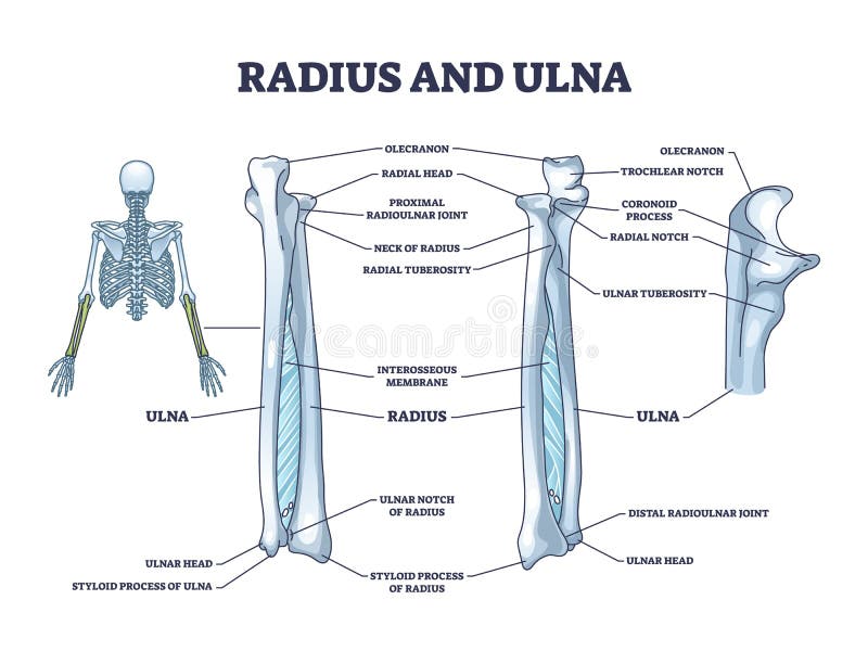
Free with trial Radius and ulna bone anatomy with arm skeletal structure outline diagram. Labeled educational scheme with upper body parts and hand long bones vector illustration. Detailed physiological description. Structure long bone vectors Radius and ulna bone anatomy with arm skeletal structure outline diagram
Free with trial Infraspinatus muscle and bone skeletal structure in human shoulder outline diagram. Labeled educational scheme with supraspinatus, teres minor and major inner physical body parts vector illustration. Structure long bone vectors Infraspinatus muscle and bone skeletal structure in shoulder outline diagram. infraspinatus muscle and bone skeletal structure in human shoulder outline diagram. Labeled educational scheme with supraspinatus, teres minor and major inner physical body parts vector illustration.
Free with trial Bone cross section and isolated anatomical detailed structure outline diagram. Labeled educational medical body description with distal and proximal epiphysis and osteon closeup vector illustration. Structure long bone vectors Bone cross section and isolated anatomical detailed structure outline diagram
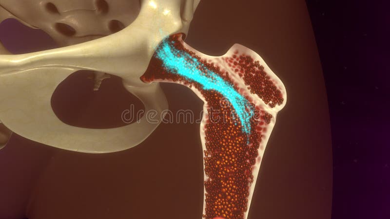
Free with trial Bone anatomy. Structure of a femur. Close-up of a cross section of Spongy Trabeculated bone tissue with Red bone marrow. vector illustration. Structure long bone vectors Bone anatomy. Structure of a femur
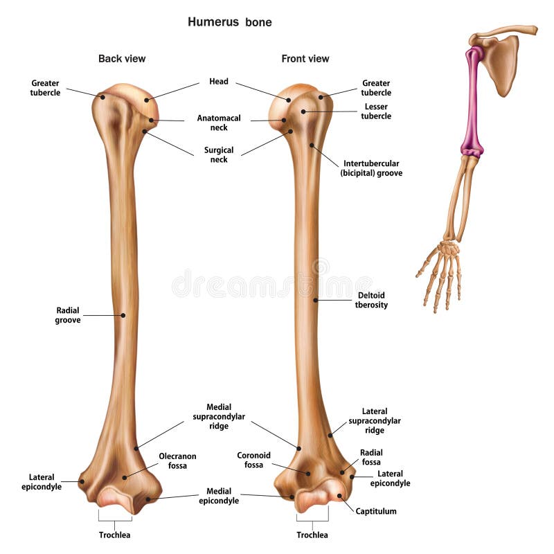
Free with trial Structure of the humerus bone with the name and description of all sites. Back and front view. Human anatomy. Structure long bone illustrations Structure of the humerus bone with the name and description of all sites.
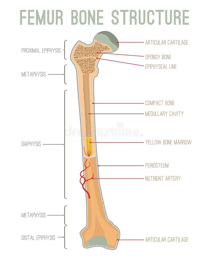
Free with trial Femur bone structure. Human health concept useful for medical, anatomy and biology educational poster design. Vector illustration with detailed information isolated on a white background. Structure long bone vectors Femur Bone Structure
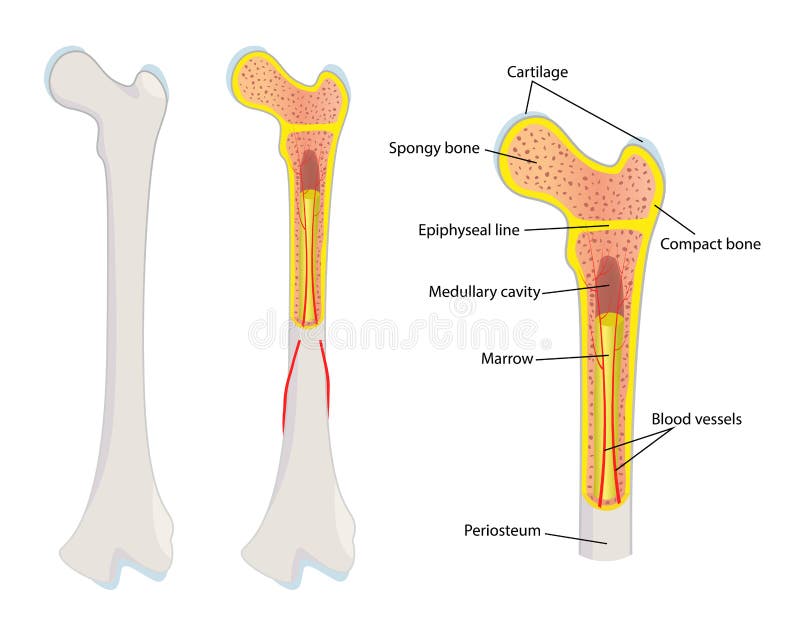
Free with trial The outer shell of the long bone is made of cortical bone also known as compact bone. This is covered by a membrane of connective tissue called the periosteum. Beneath the cortical bone layer is a layer of spongy cancellous bone. Inside this is the medullary cavity which has an inner core of bone marrow, it contains nutrients and help in formation of cells, made up of yellow marrow in the adult and red marrow in the child. Structure long bone illustrations Anatomy of a Long Bone. The outer shell of the long bone is made of cortical bone also known as compact bone. This is covered by a membrane of connective tissue called the periosteum. Beneath the cortical bone layer is a layer of spongy cancellous bone. Inside this is the medullary cavity which has an inner core of bone marrow, it contains nutrients and help in formation of cells, made up of yellow marrow in the adult and red marrow in the child.
Free with trial Bone marrow flat line icon. Vector thin pictogram of human skeleton structure, outline illustration for orthopedic clinic. Structure long bone vectors Bone marrow flat line icon. Vector thin pictogram of human skeleton structure, outline illustration for orthopedic
Free with trial Femur bone structure. Human health concept useful for medical, anatomy and biology educational poster design. Vector illustration with detailed information isolated on a black background. Structure long bone vectors Femur Bone Structure
Free with trial The structure of the bone marrow. Infographics. Vector illustration. Structure long bone vectors The structure of the bone marrow. Infographics. Vector illustration
Free with trial Human bone anatomy. Long bone structure diagram. Part of skeletal system. Medical, educational, science poster vector illustrationn. Structure long bone vectors Human bone anatomy
Free with trial Bone cross section. Anatomical detailed structure of bone tissue. Spongy and compact bone with blood vessels. Flat vector illustration. Structure long bone vectors Bone cross section. Anatomical detailed structure of bone tissue.
Free with trial Detailed anatomical view of a long bone's internal structure, highlighting compact bone, spongy bone, medullary cavity, red and yellow bone marrow, nerves, and blood vessels. Structure long bone illustrations Cross section of a long bone showing internal anatomy and marrow. Detailed anatomical view of a long bone's internal structure, highlighting compact bone, spongy bone, medullary cavity, red and yellow bone marrow, nerves, and blood vessels.
Free with trial Bone marrow set anatomical poster. Human bone structure and clinic logo. Doctors appointment, consultation and medical exam flat vector illustration. Human skeleton x ray scan medical banner. Structure long bone vectors Bone marrow set
Free with trial The spinal cord is a long, thin, tubular structure made up of nervous tissue, which extends from the medulla obligation in the brain stem to the lumbar region of the vertebral column. It encloses the central canal of the spinal cord, which contains cerebrovascular fluid. The brain and spinal cord together make up the central nervous system. In humans, the spinal cord begins at the occidental bone, passing through the foreman magnum and entering the spinal canal at the beginning of the cervical vertebrae. Structure long bone illustrations 3D illustration of Spine - Part of Human Organic. The spinal cord is a long, thin, tubular structure made up of nervous tissue, which extends from the medulla obligation in the brain stem to the lumbar region of the vertebral column. It encloses the central canal of the spinal cord, which contains cerebrovascular fluid. The brain and spinal cord together make up the central nervous system . In humans, the spinal cord begins at the occidental bone, passing through the foreman magnum and entering the spinal canal at the beginning of the cervical vertebrae.
Free with trial The spinal cord is a long, thin, tubular structure made up of nervous tissue, which extends from the medulla obligation in the brain stem to the lumbar region of the vertebral column. Structure long bone illustrations 3d render of human body spinal bone anatomy. The spinal cord is a long, thin, tubular structure made up of nervous tissue, which extends from the medulla obligation in the brain stem to the lumbar region of the vertebral column.
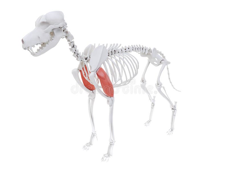
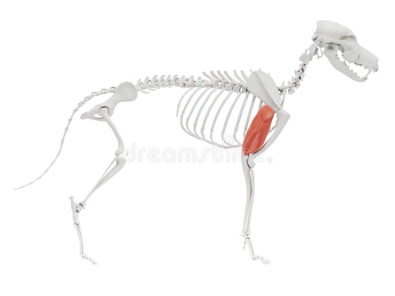
Free with trial 3d rendered illustration of the dog muscle anatomy - triceps long head. Structure long bone illustrations Triceps long head
Free with trial 3d rendered illustration of the dog muscle anatomy - triceps long head. Structure long bone illustrations Triceps long head
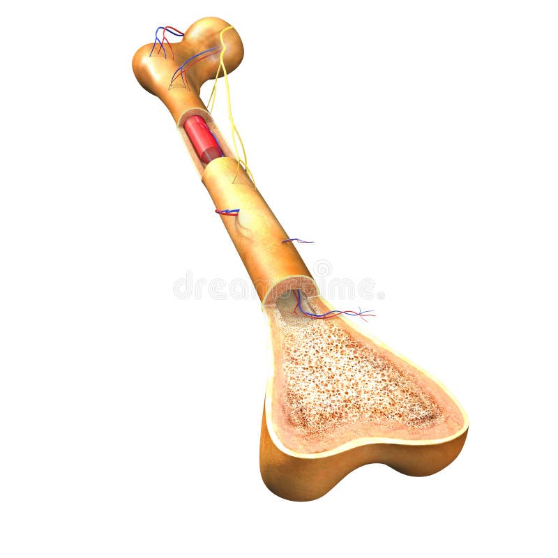
Free with trial A bone is a rigid organ that constitutes part of the vertebral skeleton. Structure long bone illustrations Bone Section. A bone is a rigid organ that constitutes part of the vertebral skeleton.
Free with trial A bone is a rigid organ that constitutes part of the vertebral skeleton. Structure long bone illustrations Bone Section. A bone is a rigid organ that constitutes part of the vertebral skeleton.
Free with trial A bone is a rigid organ that constitutes part of the vertebral skeleton. Structure long bone illustrations Bone Section. A bone is a rigid organ that constitutes part of the vertebral skeleton.
Free with trial Bone marrow is the spongy tissue inside some of the bones in the body, including the hip and thigh bones. Structure long bone illustrations Bone marrow Front View. Bone marrow is the spongy tissue inside some of the bones in the body, including the hip and thigh bones.
Free with trial Cartoon doctor with bone on the blue background. Structure long bone vectors Cartoon doctor with bone
Free with trial A bone is a rigid organ that constitutes part of the vertebral skeleton. Structure long bone illustrations Bone Section. A bone is a rigid organ that constitutes part of the vertebral skeleton.
Free with trial A bone is a rigid organ that constitutes part of the vertebral skeleton. Structure long bone illustrations Bone Section. A bone is a rigid organ that constitutes part of the vertebral skeleton.
Free with trial Bone with health concept on the blue background. Structure long bone vectors Bone with health concept
Free with trial Bone with health concept on the blue background. Structure long bone vectors Bone with health concept
Free with trial Bone with health concept on the blue background. Structure long bone vectors Bone with health concept
Free with trial Compact bone consists of closely packed osteons or haversian systems. The osteon consists of a central canal called the osteonic (haversian) canal, which is surrounded by concentric rings (lamellae) of matrix. Between the rings of matrix, the bone cells (osteocytes) are located in spaces called lacunae. Structure long bone illustrations Anatomy Of Compact Bone. Compact bone consists of closely packed osteons or haversian systems. The osteon consists of a central canal called the osteonic (haversian) canal, which is surrounded by concentric rings (lamellae) of matrix. Between the rings of matrix, the bone cells (osteocytes) are located in spaces called lacunae
Free with trial Limbo platelets in the bone marrow. Dieback of platelets in the spleen, the liver. The life of the platelet. Infographics. Vector illustration on isolated background. Structure long bone vectors Limbo platelets in the bone marrow. Dieback of platelets in the spleen, the liver. The life of the platelet. Infographics.
Free with trial A skeleton rests on a cracked surface in a deserted area, showcasing intricate bone structure and dirt. The setting suggests a long-abandoned site with visible wear. Structure long bone illustrations A skeleton rests on a cracked surface in a deserted area, showcasing intricate bone. Structure and dirt. The setting suggests a long-abandoned site with visible
Free with trial Bone marrow anatomical icon or logo. Human bone structure. Laboratory research, tests, surgery and medical exam. Human skeleton x ray scan. Medical poster for clinic. Isolated flat vector illustration. Structure long bone vectors Human internal organs. Bone marrow anatomical icon or logo. Human bone structure. Laboratory research, tests, surgery and medical exam. Human skeleton x ray scan. Medical poster for clinic. Isolated flat vector illustration
Free with trial Bone marrow anatomical icon. Human bone structure and clinic logo. Doctors appointment, consultation and medical exam flat vector illustration. Human skeleton x ray scan medical poster for hospital. Structure long bone vectors Bones template concept. Bone marrow anatomical icon. Human bone structure and clinic logo. Doctors appointment, consultation and medical exam flat vector illustration. Human skeleton x ray scan medical poster for hospital.
Free with trial Bone marrow anatomical icon. Human bone structure and clinic logo. Doctors appointment, consultation and medical exam flat vector illustration. Human skeleton x ray scan medical poster for hospital. Structure long bone vectors Bones template concept. Bone marrow anatomical icon. Human bone structure and clinic logo. Doctors appointment, consultation and medical exam flat vector illustration. Human skeleton x ray scan medical poster for hospital.
Free with trial Skeleton femur thigh bone Human front view with two arm poses with partly transparent bones position. Realistic flat natural color concept Vector illustration of anatomy isolated on white background. Structure long bone vectors Skeleton femur thigh bone Human front view with two arm poses with partly transparent bones position. Realistic flat
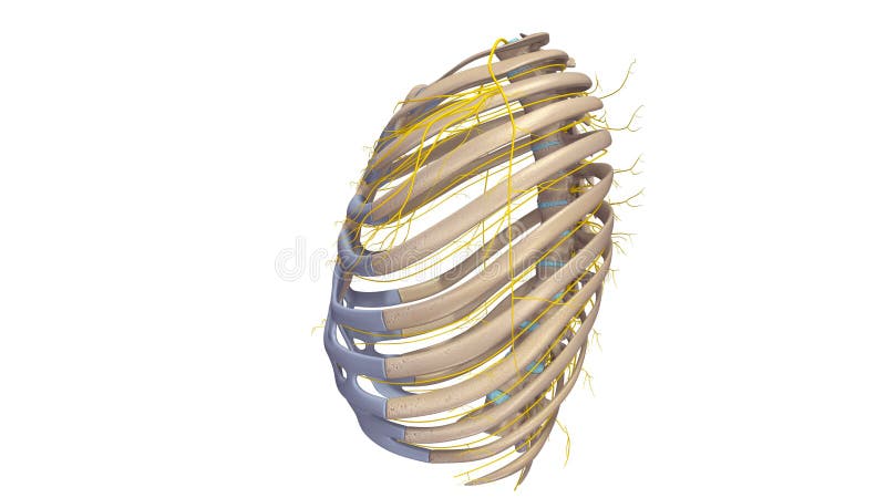
Free with trial The human rib cage is made up of 12 paired rib bones; each are symmetrically paired on a right and left side. Of all 24 ribs, the first seven pairs are often labeled as `true. ` These bones are connected to the costal cartilage, while the five other `false` sets are not. The ribcage also encloses the thoracic cavity and helps protect the heart and lungs from damage. There are 24 ribs in the human body, divided into two sets of 12 curved, flat bones. Each one is attached by cartilage at the back to the thoracic vertebrae. MEN and women have 12 pairs of ribs a few individuals have 13 or 11 pairs. The idea that men have fewer ribs than women is widespread but wrong, perhaps deriving from the biblical story of Eve being made from one of Adam`s ribs. Both men and women have 24 ribs, twelve on each side. Floating rib: One of the last two ribs. A rib is said to be `floating` if it does not attach to the sternum the breast bone or to another rib. There are usually 12 pairs of ribs in all. Each pair of ribs is attached to the building blocks of the spine the vertebrae in the back. The ribs partially enclose and protect the chest cavity, where many vital organs including the heart and the lungs are located. The rib cage is collectively made up of long, curved individual bones with joint-connections to the spinal vertebrae. Structure long bone illustrations Ribs with Nerves lateral view. The human rib cage is made up of 12 paired rib bones; each are symmetrically paired on a right and left side. Of all 24 ribs, the first seven pairs are often labeled as `true.` These bones are connected to the costal cartilage, while the five other `false` sets are not. The ribcage also encloses the thoracic cavity and helps protect the heart and lungs from damage. There are 24 ribs in the human body, divided into two sets of 12 curved, flat bones. Each one is attached by cartilage at the back to the thoracic vertebrae. MEN and women have 12 pairs of ribs a few individuals have 13 or 11 pairs. The idea that men have fewer ribs than women is widespread but wrong, perhaps deriving from the biblical story of Eve being made from one of Adam`s ribs. Both men and women have 24 ribs, twelve on each side. Floating rib: One of the last two ribs. A rib is said to be `floating` if it does not attach to the sternum the breast bone or to another rib. There are usually 12 pairs of ribs in all. Each pair of ribs is attached to the building blocks of the spine the vertebrae in the back. The ribs partially enclose and protect the chest cavity, where many vital organs including the heart and the lungs are located. The rib cage is collectively made up of long, curved individual bones with joint-connections to the spinal vertebrae.