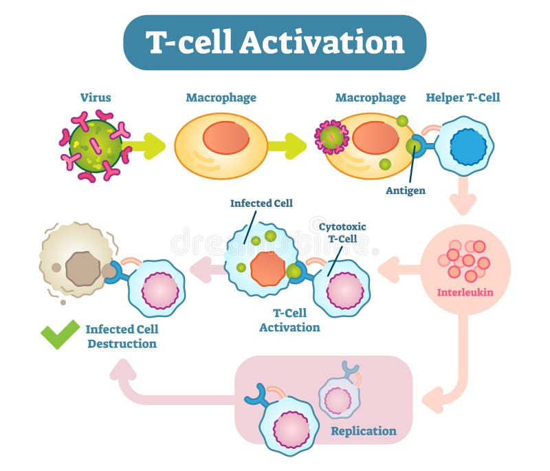
Free with trial A T cell, or T lymphocyte, is a type of lymphocyte a subtype of white blood cell that plays a central role in cell-mediated immunity. T cell activation vectors T-Cell activation diagram, vector scheme illustration. A T cell, or T lymphocyte, is a type of lymphocyte a subtype of white blood cell that plays a central role in cell-mediated immunity.
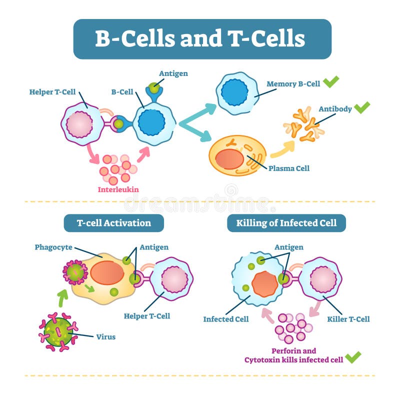
Free with trial B-cells and T-cells schematic diagram, vector illustration, immune system cell functions. T cell activation vectors B-cells and T-cells schematic diagram, vector illustration. B-cells and T-cells schematic diagram, vector illustration, immune system cell functions.
Free with trial Adaptive immune system from Antigen presentation to activation of other immune cells. specific immune. T-helper and T-killer cells. Memory and Effector cells. Viruse, Lymphocyte, antibody and antigen. Vector diagram for educational, biological, and science use. T cell activation vectors Adaptive immune system from Antigen presentation to activation o
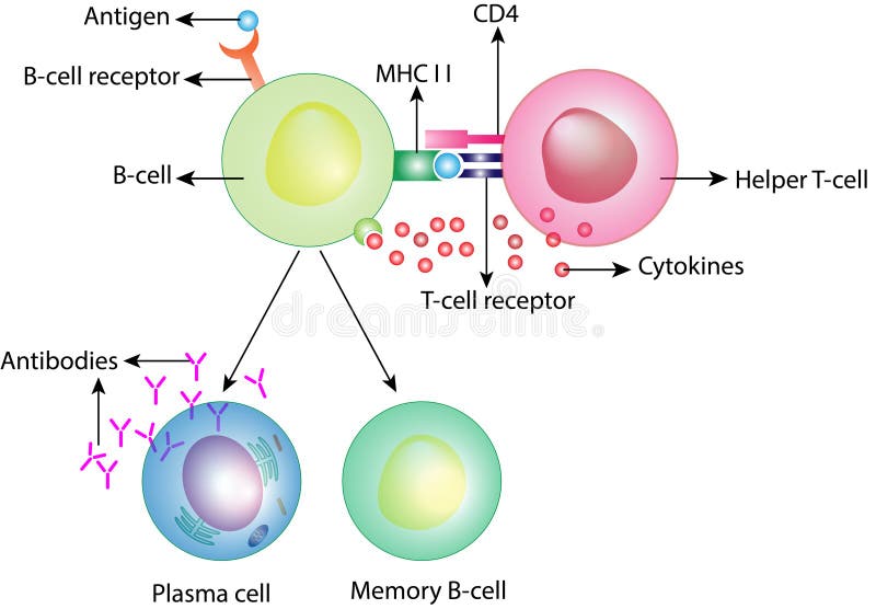
Free with trial B-cell and T helper cells. Basic B-cells function: bind an antigen, receive help from a T helper cell, and differentiate into a plasma cell that secretes large amounts of antibodies. Human immune system. T cell activation vectors B-cell and T helper cells function. B-cell and T helper cells. Basic B-cells function: bind an antigen, receive help from a T helper cell, and differentiate into a plasma cell that secretes large amounts of antibodies. Human immune system
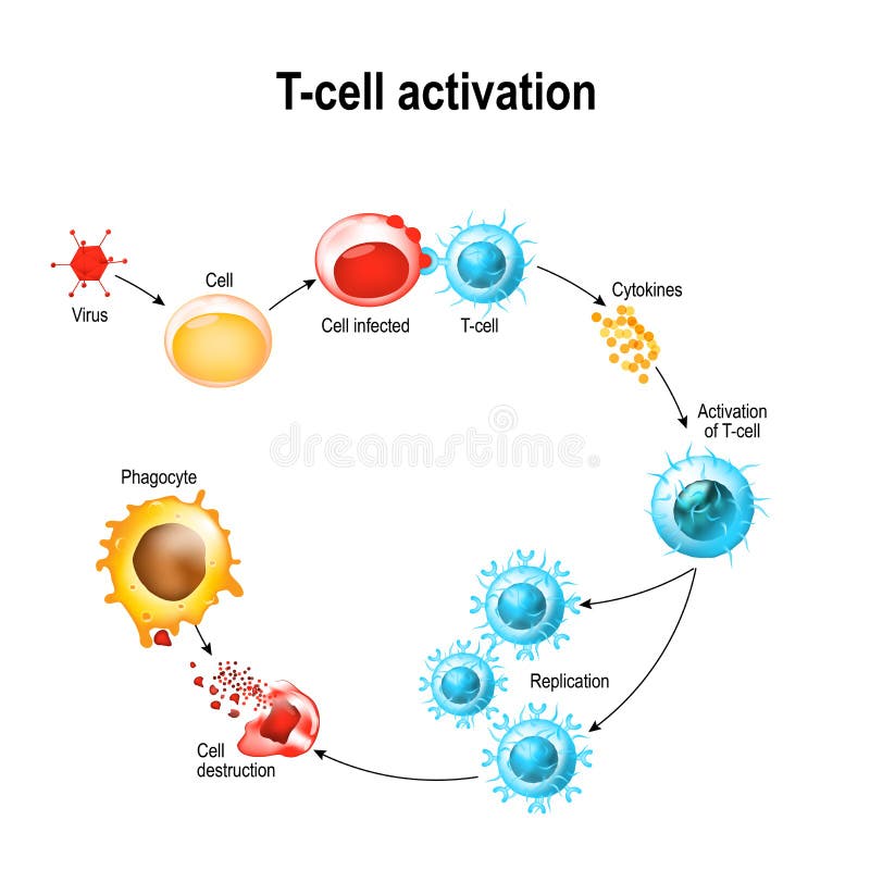
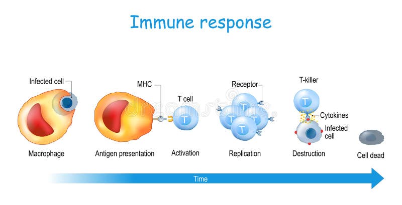
Free with trial T-cell. Activation and lysis of the leukocytes. T-cell encounters its cognate antigen on the surface of an infected cell. T cells direct and regulate immune responses and attack infected or cancerous cells. T cell activation vectors T-cell. Activation and lysis of the leukocytes.
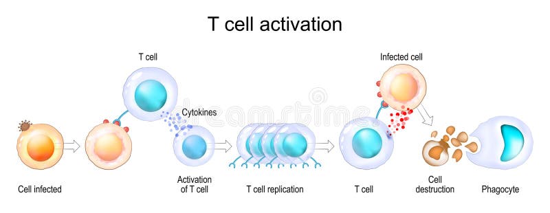
Free with trial Activation of T-cell leukocytes. T-cell encounters its cognate antigen on the surface of an infected cell. T cells direct and regulate immune responses and attack infected or cancerous cells. T cell activation vectors Activation of T-cell leukocytes
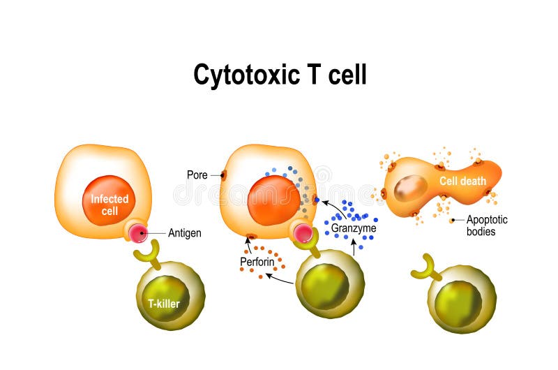
Free with trial Cytotoxic T cell. T-cell regulate immune responses, release the perforin and granzymes, and attack infected or cancerous cells. Through the action of perforin, granzymes enter the cytoplasm of the target cell, and lead to apoptosis cell death. T cell activation vectors Cytotoxic T cell
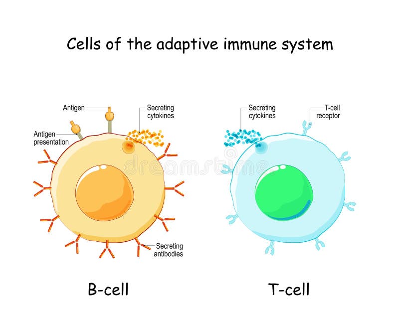
Free with trial Cells of Adaptive immune system immune response. B lymphocyte and T-cell. Types, and function of lymphocytes. Infographics. Vector illustration on white background. T cell activation vectors B-cell and T-cell. Adaptive immune system. Cells of Adaptive immune system immune response. B lymphocyte and T-cell. Types, and function of lymphocytes. Infographics. Vector illustration on white background
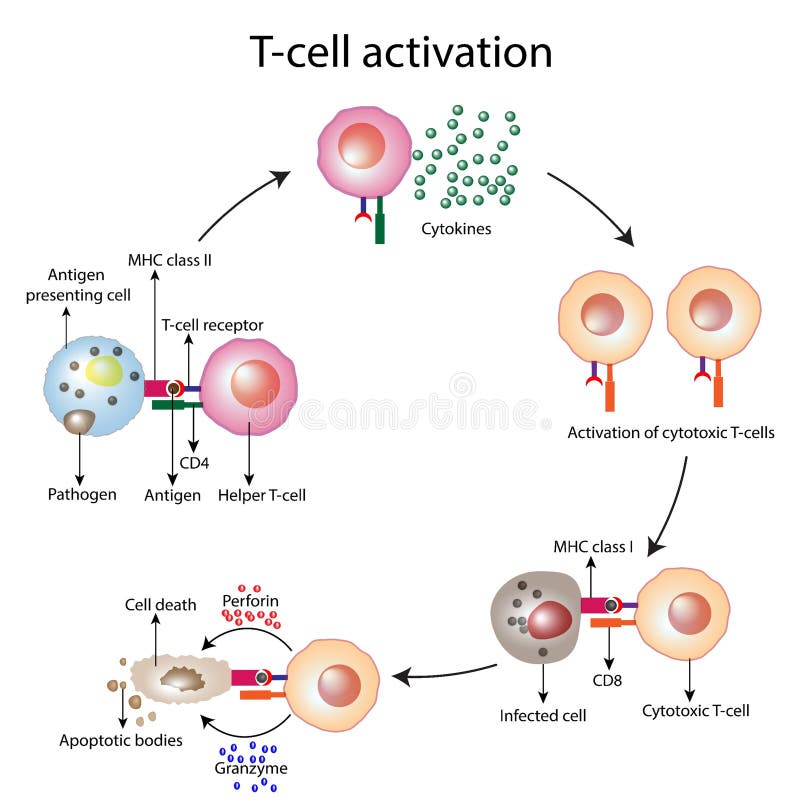
Free with trial Dendritic cells present antigens green to lymphocytes through their membran bound MHC-molecules violet. CD4 molecules light blue bind to other portions of the MHC, strengthening the interaction. After binding to the MHC-antigen complex, The T-cell receptor blue sends a signal cascade through an attached G-protein into the T-lymphocyte cell, that activates an immune response. T cell activation illustrations Activation of the immune response: antigen presenting cell activates T-lymphocytes (smaller c. Dendritic cells present antigens green to lymphocytes through their membran bound MHC-molecules violet. CD4 molecules light blue bind to other portions of the MHC, strengthening the interaction. After binding to the MHC-antigen complex, The T-cell receptor blue sends a signal cascade through an attached G-protein into the T-lymphocyte cell, that activates an immune response.
Free with trial T cell Activation. T lymphocyte, is a white blood cell. cell-mediated immunity. T cell activation vectors T cell Activation
Free with trial Activation of leukocytes. T-cell encounters its cognate antigen on the surface of an infected cell. T-cells direct and regulate immune responses and attack infected or cancerous cells. Cell-mediated immunity. The Adaptive and Innate immune system. vector poster. T cell activation vectors Activation of T cell leukocytes. Immune response. Activation of leukocytes. T-cell encounters its cognate antigen on the surface of an infected cell. T-cells direct and regulate immune responses and attack infected or cancerous cells. Cell-mediated immunity. The Adaptive and Innate immune system. vector poster
Free with trial Activation of B-cell leukocytes. B lymphocyte differentiation. Plasma cell and memory B cell. B cell and T cell interaction. adaptive immune system. Activated B cell producing antibodies. T cell activation vectors Activation of B-cell. adaptive immune system. plasma cell and memory B cell. Activation of B-cell leukocytes. B lymphocyte differentiation. Plasma cell and memory B cell. B cell and T cell interaction. adaptive immune system. Activated B cell producing antibodies
Free with trial An activated T helper cell segregates the cytokines IL-4, IL-5, IL-6, IL-9, IL-10 and IL-13, leading to B cell antibody class switching, activation of cytotoxic T cells, and maximazing the bactericidal activity of phagocytes such as macrophages. Source: PDB entries 2B8U, 3VA2, 1ALU, 2H24, 3BPO. T cell activation illustrations After activation by an antigen presenting cell, a T helper cell segregates several cytokines. An activated T helper cell segregates the cytokines IL-4, IL-5, IL-6, IL-9, IL-10 and IL-13, leading to B cell antibody class switching, activation of cytotoxic T cells, and maximazing the bactericidal activity of phagocytes such as macrophages. Source: PDB entries 2B8U, 3VA2, 1ALU, 2H24, 3BPO
Free with trial Lymphocytes. B cell for humoral immunity. T-cell for adaptive immune response. Vector illustration. Poster. T cell activation vectors Lymphocytes. B cell and T-cell. Lymphocytes. B cell for humoral immunity. T-cell for adaptive immune response. Vector illustration. Poster
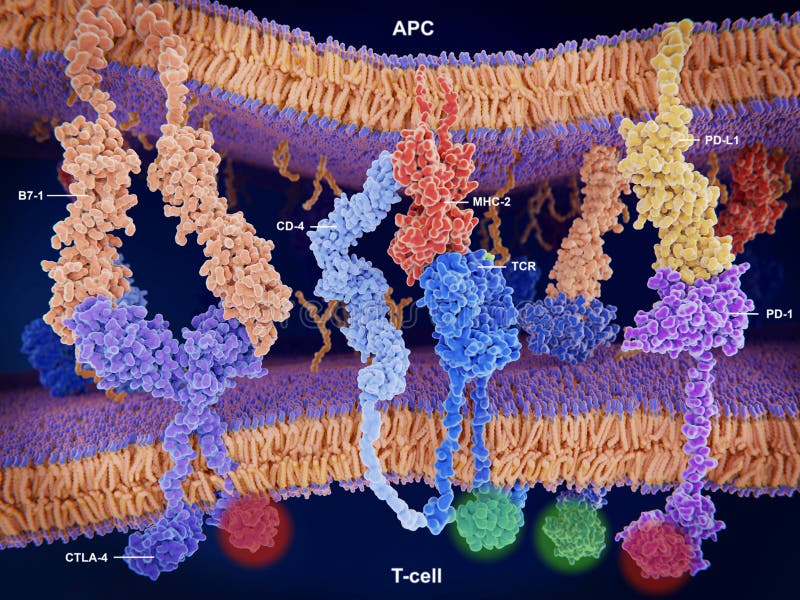
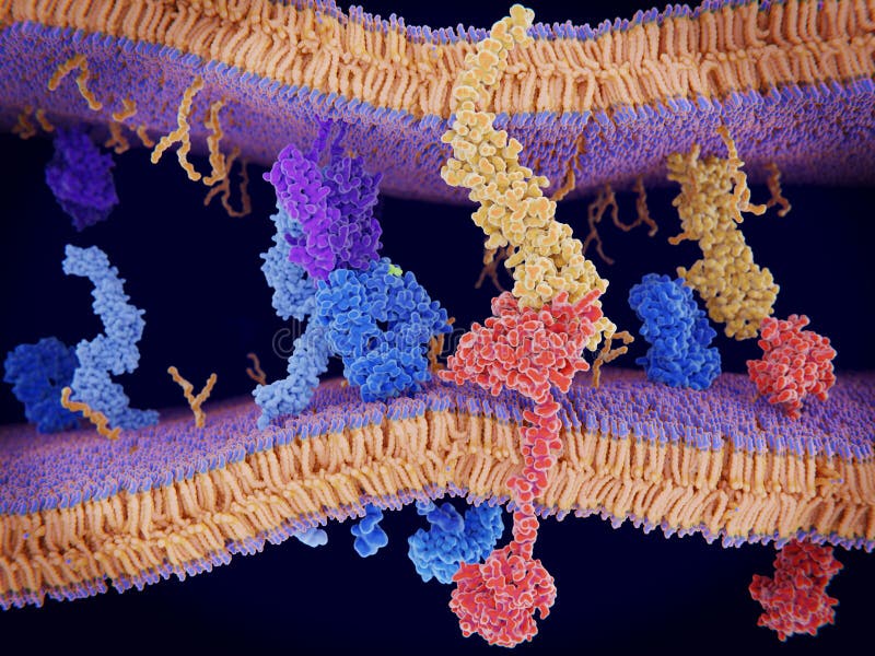
Free with trial Immunologically active proteins on a T-cell. TCR blue, CD-4 light blue, CD-28 dark blue, PD-1 magenta, CTLA-4 violet, Ca-channel dark violet. The T-cell receptor, CD-4 and CD-28 activate T-cells, while PD-1 and CTLA-4 inhibit the activation of T-cells. T cell activation illustrations T-cell receptor, CD-4, CD-28, PD-1 and CTLA-4 and a calcium chan. Immunologically active proteins on a T-cell. TCR blue, CD-4 light blue, CD-28 dark blue, PD-1 magenta, CTLA-4 violet, Ca-channel dark violet. The T-cell receptor, CD-4 and CD-28 activate T-cells, while PD-1 and CTLA-4 inhibit the activation of T-cells.
Free with trial Interactions of MHC-II with the T-cell receptor and CD4 and B7-1 with CD-28 activates T-cells while the interactions of P7-1 with CTLA-4 and PD-L1 with PD-1 deactivates T-cells. T cell activation illustrations Activation and inhibition of the immune response on T-cells. Interactions of MHC-II with the T-cell receptor and CD4 and B7-1 with CD-28 activates T-cells while the interactions of P7-1 with CTLA-4 and PD-L1 with PD-1 deactivates T-cells.
Free with trial Immune response and Antigen presentation. T-cell Activation. T lymphocyte, is a white blood cell. cell-mediated immunity. T cell activation vectors Immune response and Antigen presentation
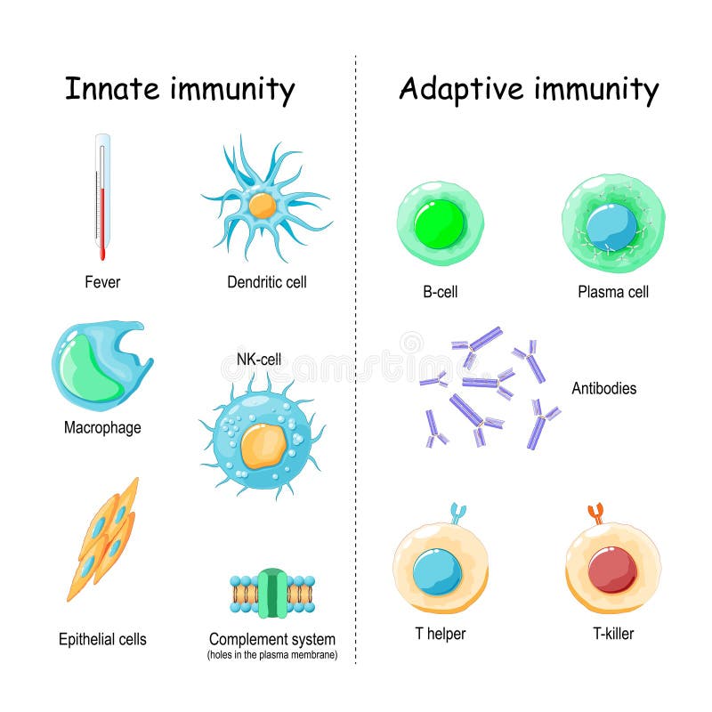
Free with trial Innate immunity from Fever and Complement system protein for holes in the plasma membrane, to Macrophage, NK and Dendritic cells. Adaptive immunity from Antibodies and Plasma cell to B-cell, T helper, T-killer. comparison and difference. T cell activation vectors Innate and Adaptive immunity. comparison and difference. Innate immunity from Fever and Complement system protein for holes in the plasma membrane, to Macrophage, NK and Dendritic cells. Adaptive immunity from Antibodies and Plasma cell to B-cell, T helper, T-killer. comparison and difference
Free with trial B cell activation pathway showing naive B cell, helper T cell, and antibodies, antigen signals drive clonal expansion to memory and plasma cells. Outline diagram. T cell activation vectors B cell activation pathway showing naive B cell, helper T ... B cell activation pathway showing naive B cell, helper T cell, and antibodies, antigen signals drive clonal expansion to memory and plasma cells. Outline diagram
Free with trial T cell activation diagram, T lymphocyte activation, helper T-cell and cytotoxic T-cell vector illustration, scheme diagram, white blood cells, immune response to infection. T-cell regulate immune responses, Cytotoxic t cell produce perforin and granzymes which enter the cytoplasm of the target infected cell, and lead to apoptosis cell death. T cell activation vectors T cell activation diagram, t lymphpcytes, helper T-cell and cytotoxic T-cell vector illustration, white blood cells. T cell activation diagram, T lymphocyte activation, helper T-cell and cytotoxic T-cell vector illustration, scheme diagram, white blood cells, immune response to infection. T-cell regulate immune responses, Cytotoxic t cell produce perforin and granzymes which enter the cytoplasm of the target infected cell, and lead to apoptosis cell death.
Free with trial T cell activation, T lymphocyte activation, helper T-cell and cytotoxic T cell vector illustration, scheme diagram, white blood cells, immune response to infection. T cell regulate immune responses, Cytotoxic t cell produce perforin and granzymes which enter the cytoplasm of the target infected cell, and lead to apoptosis cell death. T cell activation vectors T cell activation diagram, t lymphocytes, helper T-cell and cytotoxic T-cell vector illustration, white blood cells. T cell activation, T lymphocyte activation, helper T-cell and cytotoxic T cell vector illustration, scheme diagram, white blood cells, immune response to infection. T cell regulate immune responses, Cytotoxic t cell produce perforin and granzymes which enter the cytoplasm of the target infected cell, and lead to apoptosis cell death.
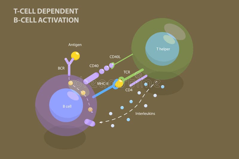
Free with trial 3D Isometric Flat Vector Illustration of T-cell Dependent B-cell Activation, Adaptive Immune System. T cell activation vectors 3D Isometric Flat Vector Illustration of T-cell Dependent B-cell Activation
Free with trial T-cell dependent b-cell activation. B lymphocyte cell and T-helper. Close-up of White blood cells, leucocytes. Immune response. Adaptive immunity. Humoral immunity. vector illustration. T cell activation vectors T-cell dependent b-cell activation
Free with trial T-Cell Activation and Cancer Cell Killing. Illustrates the process of T-cell activation to target and kill a cancer cell, including a mechanism of cancer immune evasion. T cell activation vectors T-Cell Activation and Cancer Cell Killing.
Free with trial B cell activation diagram. Process of recognizing an antigen and binds to it. Bidirectional activation signals. Allergic diseases concept. Human immune system medical flat vector illustration. T cell activation vectors B cell activation
Free with trial B cell activation diagram. Process of recognizing an antigen and binds to it. Bidirectional activation signals. Allergic diseases concept. Human immune system medical flat vector illustration. T cell activation vectors B cell activation
Free with trial CD3 protein (epsilon/delta ectodomain dimer). CD3 is present on the surface of T-lymphocytes and is required for T-cell activation. 3D rendering based on protein data bank entry 1xiw. Cartoon representation combined with semi-transparent surfaces. Dark background. T cell activation illustrations CD3 protein (epsilon/delta ectodomain dimer). CD3 is present on the surface of T-lymphocytes and is required for T-cell activation
Free with trial Cytotoxic T cell. T-cell regulate immune responses, release the perforin and granzymes, and attack infected or cancerous cells. Through the action of perforin, granzymes enter the cytoplasm of the target cell, and lead to apoptosis cell death. T cell activation vectors Cytotoxic T cell
Free with trial Activation of the immune response to antigenes through the complex between a T-cell receptor blue, an MHC IIred-antigen green and a CD4 protein light blue. The T-cell receptor activates the immune response to antigens in T-lymphocytes. T cell activation illustrations Complex within a MHC II-antigen, a T cell receptor and CD4. Activation of the immune response to antigenes through the complex between a T-cell receptor blue, an MHC IIred-antigen green and a CD4 protein light blue.The T-cell receptor activates the immune response to antigens in T-lymphocytes
Free with trial T cell receptor molecular structure design. Cell Signaling receptors. T cell activation illustrations T cell receptor molecular structure design. T cell receptor molecular structure design. Cell Signaling receptors
Free with trial Immune response. Cell-mediated immunity. activation of phagocytes, antigen-specific cytotoxic T-lymphocytes, and the release of cytokines in response to an antigen. Vector poster for educatio. T cell activation vectors Immune response. Cell-mediated immunity
Free with trial CAR-T-cell cancer therapy. Process of a T cell reprogramming. Immunotherapy of a Chimeric Antigen Receptor CAR. Cancer treatment. Genetic engineering. vector illustration. T cell activation vectors CAR-T-cell cancer therapy. Process of a T cell reprogramming
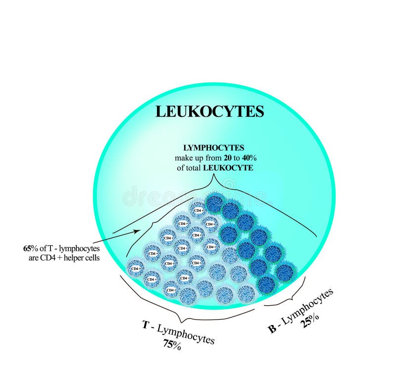
Free with trial Lymphocytes make up from 20 to 40 percent of the total number of leukocytes. T Lymphocytes and B Lymphocytes. Cell killers. Immunity Helper Cells. Infographics. Vector illustration on isolated background. T cell activation vectors Lymphocytes make up from 20 to 40 percent of the total number of leukocytes. T Lymphocytes and B Lymphocytes. Cell
Free with trial PD-1 red extends from the surface of a T-cell and interacts with the ligand protein PD-L1 yellow from a antigen presenting cell. Although the T-cell has been activated through the interaction of a T-cell receptor blue and a MHC protein violett, PD-1 regulates these activation down. It is a checkpoint to slow down T-cells. T cell activation illustrations The protein PD-1 is a checkpoint to slow down T-cells. PD-1 red extends from the surface of a T-cell and interacts with the ligand protein PD-L1 yellow from a antigen presenting cell. Although the T-cell has been activated through the interaction of a T-cell receptor blue and a MHC protein violett, PD-1 regulates these activation down. It is a checkpoint to slow down T-cells.
Free with trial Cancer therapy. CAR T immunotherapy. Artificial T-cell receptors are proteins that have been engineered for cancer therapy killing of tumor cells. genetically engineered. Vector diagram for medical and science use. T cell activation vectors Cancer therapy. CAR T immunotherapy
Free with trial PD-1 red extends from the surface of a T-cell and interacts with the ligand protein PD-L1 yellow from a antigen presenting cell. Although the T-cell has been activated through the interaction of a T-cell receptor blue and a MHC protein violett, PD-1 regulates these activation down. It is a checkpoint to slow down T-cells. T cell activation illustrations The protein PD-1 is a checkpoint to slow down T-cells. PD-1 red extends from the surface of a T-cell and interacts with the ligand protein PD-L1 yellow from a antigen presenting cell. Although the T-cell has been activated through the interaction of a T-cell receptor blue and a MHC protein violett, PD-1 regulates these activation down. It is a checkpoint to slow down T-cells.
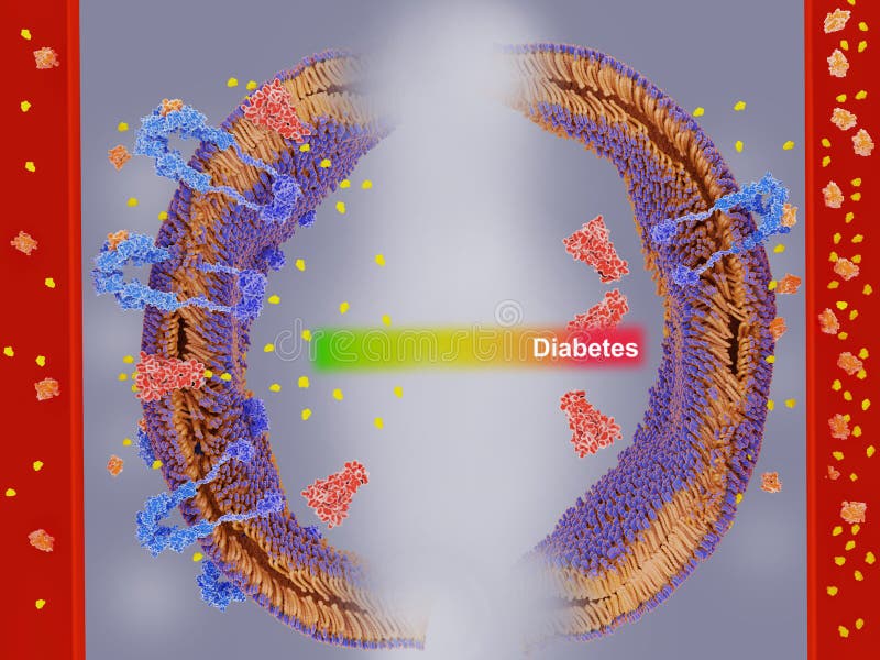
Free with trial In the pathological condition insulin resistance, the cells fail to respond properly to the hormone insulin orange. Resistant cells produce less insulin receptors blue. Their response to insulin binding doesn`t lead any more to the activation of glucose transporter proteins red, which transport glucose into the cell under normal conditions. T cell activation illustrations Insulin resistance, insulin receptors cannot respond properly to insulin binding. In the pathological condition insulin resistance, the cells fail to respond properly to the hormone insulin orange. Resistant cells produce less insulin receptors blue. Their response to insulin binding doesn`t lead any more to the activation of glucose transporter proteins red, which transport glucose into the cell under normal conditions
Free with trial Immune response and Antigen presentation. Cell-mediated immunity is an immune response that does not involve antibodies. T-cell Activation. Vector poster. T cell activation vectors Immune response. Cell-mediated immunity. Immune response and Antigen presentation. Cell-mediated immunity is an immune response that does not involve antibodies. T-cell Activation. Vector poster
Free with trial CAR T cells attacking cancer cells in a medical illustration. Concept Medical Illustration, Cancer Treatment, Immunotherapy, CAR T Cells, Cell Biology. T cell activation illustrations Concept Medical Illustration, Cancer CAR T cells attacking cancer cells in a medical illustration. CAR T cells attacking cancer cells in a medical illustration. Concept Medical Illustration, Cancer Treatment, Immunotherapy, CAR T Cells, Cell Biology
Free with trial This diagram illustrates the immune system's complex processes, including B cell activation, T cell interactions, and antibody production in response to antigens. T cell activation illustrations Diagram illustrating the process of the immune system response. This diagram illustrates the immune system's complex processes, including B cell activation, T cell interactions, and antibody production in response to antigens
Free with trial Interactions of MHC-II with the T-cell receptor and CD4 and B7-1 with CD-28 activates T-cells while the interactions of P7-1 with CTLA-4 and PD-L1 with PD-1 deactivates T-cells. T cell activation illustrations Membrane proteins involved in the activation and inhibition of. Interactions of MHC-II with the T-cell receptor and CD4 and B7-1 with CD-28 activates T-cells while the interactions of P7-1 with CTLA-4 and PD-L1 with PD-1 deactivates T-cells.
Free with trial Interleukin 5 is produced by T helper cells. It stimulates B cell growth, increases immunoglobulin secretion and mediates eosinophil activation. T cell activation illustrations Interleukin 5 molecules. Interleukin 5 is produced by T helper cells. It stimulates B cell growth, increases immunoglobulin secretion and mediates eosinophil activation
Free with trial T-cells and red blood cells on red background. vector Illustration easy editable for Your color. lymphocyte in immune response. T cell activation vectors T-cells and red blood cells on red background
Free with trial Types of t Lymphocyte isolated, 3d rendering. T cell activation illustrations Types of t Lymphocyte isolated
Free with trial Illustration of immune system T cells attacking cancer cells. T cell activation illustrations T cells attacking cancer cells
Free with trial 3D Isometric Flat Vector Conceptual Illustration of Immune Response, Cells of The Immune System. T cell activation vectors 3D Isometric Flat Vector Conceptual Illustration of Immune Response
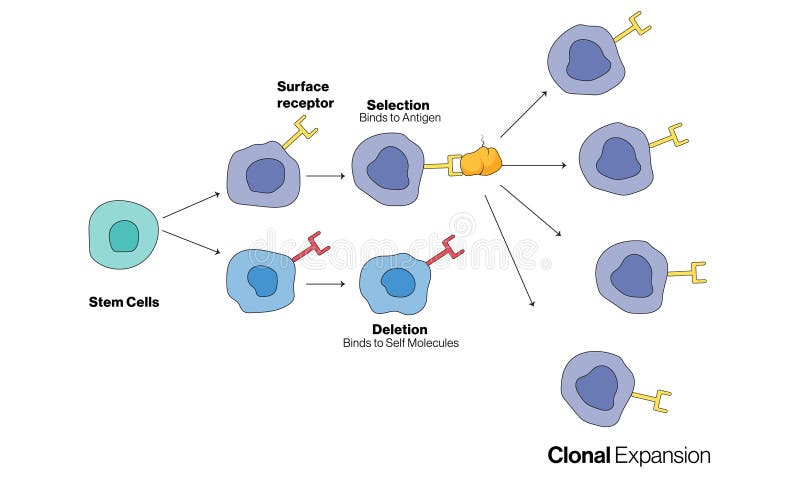
Free with trial This vector illustration depicts the clonal selection theory, a fundamental concept in immunology. It explains how specific B and T lymphocytes are selected and activated by antigens to produce a targeted immune response. The diagram shows the process of clonal expansion, where activated lymphocytes proliferate and differentiate into effector and memory cells, enhancing the body's ability to fight infections. This detailed and educational illustration is ideal for use in academic materials, scientific publications, and educational resources to visually communicate the mechanisms of adaptive immunity. Set against a clean white background, the illustration highlights key components and steps involved in the clonal selection process, making it an excellent resource for students, educators, and professionals in the field of immunology. T cell activation vectors Illustration of Clonal Selection Theory in Immunology on a White Background. This vector illustration depicts the clonal selection theory, a fundamental concept in immunology. It explains how specific B and T lymphocytes are selected and activated by antigens to produce a targeted immune response. The diagram shows the process of clonal expansion, where activated lymphocytes proliferate and differentiate into effector and memory cells, enhancing the body's ability to fight infections. This detailed and educational illustration is ideal for use in academic materials, scientific publications, and educational resources to visually communicate the mechanisms of adaptive immunity. Set against a clean white background, the illustration highlights key components and steps involved in the clonal selection process, making it an excellent resource for students, educators, and professionals in the field of immunology.
Free with trial T-cell Activation and Response. Adaptive immunity. Vector illustration. Medical poster. Schematic diagram. T cell activation vectors T-cell Activation and Response. Adaptive immunity
Free with trial T cell functions outline depicts helper CD4, cytotoxic CD8, and regulatory cells guiding activation, killing infected cells, and suppression with arrows and labels. Outline diagram. T cell activation vectors T cell functions outline depicts helper CD4, cytotoxic CD8, and ... T cell functions outline depicts helper CD4, cytotoxic CD8, and regulatory cells guiding activation, killing infected cells, and suppression with arrows and labels. Outline diagram
Free with trial B lymphocyte activation shown as stepwise immune response, B cell binds antigen, receives T helper signals, differentiates to plasma cell producing antibodies. Outline diagram. T cell activation vectors B lymphocyte activation shown as stepwise immune response, B cell ... B lymphocyte activation shown as stepwise immune response, B cell binds antigen, receives T helper signals, differentiates to plasma cell producing antibodies. Outline diagram
Free with trial T-cell activation process diagram poster healthcare, activation of leukocytes, T-cell encounters its cognate antigen on infected cell, immune response, labeled phagocyte, t-cell replication, infected. T cell activation vectors T-cell activation process diagram poster healthcare, activation of leukocytes, T-cell encounters its cognate antigen on infected
Free with trial T-cell activation process diagram poster healthcare, activation of leukocytes, T-cell encounters its cognate antigen on infected cell, immune response, labeled phagocyte, t-cell replication, infected. T cell activation vectors T-cell activation process diagram poster healthcare, activation of leukocytes, T-cell encounters its cognate antigen on infected
Free with trial Cell mediated immunity diagram process, Immune response. activation of phagocytes, labeled macrophage, pathogen, t-lymphocyte, release cytokines in response to antigen, cytotoxic. Biology education. T cell activation vectors Cell mediated immunity diagram process, Immune response. activation of phagocytes, labeled macrophage, pathogen, t-lymphocyte
Free with trial Visualize the intricate dance of the human immune system with this stunning, futuristic digital illustration. The image showcases T-cell activation and pathogen neutralization on a sleek digital interface. Bathed in a mesmerizing blue glow with neon accents, the hologram-like display offers a dynamic visualization of the immune response. This medical illustration is ideal for healthcare, science,. T cell activation illustrations A futuristic visual depiction of the human immune system displaying t cell activation and pathogen neutralization on a digital. Visualize the intricate dance of the human immune system with this stunning, futuristic digital illustration. The image showcases T-cell activation and pathogen neutralization on a sleek digital interface. Bathed in a mesmerizing blue glow with neon accents, the hologram-like display offers a dynamic visualization of the immune response. This medical illustration is ideal for healthcare, science,
Free with trial This isometric illustration visualizes the process of modern biological research, including genetic sequencing and bioinformatics analysis. It progresses through neoantigen design, vaccine production with molecular models, and finally immune cell profiling and T-cell activation visualization for drug discovery and medical advancement. T cell activation illustrations Genomic Research DNA Sequencing, Vaccine Production, Immune Cell Profiling, and Bioinformatics. This isometric illustration visualizes the process of modern. This isometric illustration visualizes the process of modern biological research, including genetic sequencing and bioinformatics analysis. It progresses through neoantigen design, vaccine production with molecular models, and finally immune cell profiling and T-cell activation visualization for drug discovery and medical advancement.
Free with trial B-cell leukocyte activation by Antigen. From antigen binding to B cell receptor, and Chemical Signal of T-cell helper to Becomes plasma cell and Antibodies Releases. White blood cell. Vector illustration. T cell activation vectors B-cell leukocyte activation by Antigen
Free with trial B cell activation process. Humoral immunity. T cell-dependent activation. Adaptive immunity. Vector illustration. Medical poster. Schematic diagram. T cell activation vectors B cell activation process. Humoral immunity
Free with trial Biological anatomy of T - Cell. T cell activation vectors T-cell. Biological anatomy of T - Cell
Free with trial 3d-rendered illustration of immune cell signaling--T-cell activation pathways. Image using saturated teal, maroon, and ivory tones. Close-up of cell membranes, receptors, and kinase cascades. T cell activation illustrations 3d-rendered illustration of immune cell signaling--T-cell activation pathways. Close-up of cell membranes, receptors. 3d-rendered illustration of immune cell signaling--T-cell activation pathways. Image using saturated teal, maroon, and ivory tones. Close-up of cell membranes, receptors, and kinase cascades.
Free with trial A large blue T cell aggressively targets and eliminates a foreign red virus after being activated by a vaccine, illustrating the immune response in action. T cell activation illustrations Successful T cell activation and viral attack showcasing immune response in human body. A large blue T cell aggressively targets and eliminates a foreign red virus after being activated by a vaccine, illustrating the immune response in action
Free with trial Visualize the intricate workings of the human immune system with this stunning conceptual representation. The digital display showcases a vibrant 3D rendering highlighting T-cell activation and pathogen neutralization processes. This medical illustration employs a futuristic aesthetic with neon blue and glowing elements, offering a clear understanding of immunity at a cellular level. The image is. T cell activation illustrations Human immune system conceptual visualization depicting t cell activation and pathogen neutralization processes in a digital. Visualize the intricate workings of the human immune system with this stunning conceptual representation. The digital display showcases a vibrant 3D rendering highlighting T-cell activation and pathogen neutralization processes. This medical illustration employs a futuristic aesthetic with neon blue and glowing elements, offering a clear understanding of immunity at a cellular level. The image is
Free with trial T lymphocytes diagram shows helper, cytotoxic, and regulatory T cells coordinating immunity, key objects, helper T cell, cytotoxic T cell, infected cell. Outline diagram. T cell activation vectors T lymphocytes diagram shows helper, cytotoxic, and regulatory T cells ... T lymphocytes diagram shows helper, cytotoxic, and regulatory T cells coordinating immunity, key objects, helper T cell, cytotoxic T cell, infected cell. Outline diagram
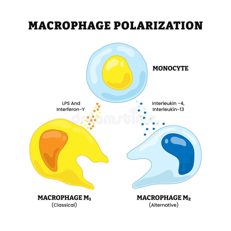
Free with trial Macrophage polarization process, labeled t cell, macrophage m2, m1, interleukin,interferon, lps Chronic inflammation. Metabolic reprogramming. Pro-inflammatory, Anti-inflammatory response. T cell activation vectors Macrophage polarization process, labeled t cell, macrophage m2, m1, interleukin,interferon, lps Chronic inflammation. Metabolic
Free with trial Macrophage polarization process, labeled t cell, macrophage m2, m1, interleukin,interferon, lps Chronic inflammation. Metabolic reprogramming. Pro-inflammatory, Anti-inflammatory response. T cell activation vectors Macrophage polarization process, labeled t cell, macrophage m2, m1, interleukin,interferon, lps Chronic inflammation. Metabolic
Free with trial A close up view of human T cell attacking cancer cells, showcasing intricate details and vibrant colors. This highlights dynamic interaction between immune cells and cancer, emphasizing importance. T cell activation illustrations Close up view of human T cell attacking cancer cells showcasing intricate details and vibrant colors This highlights dynamic. A close up view of human T cell attacking cancer cells, showcasing intricate details and vibrant colors. This highlights dynamic interaction between immune cells and cancer, emphasizing importance
Free with trial This vector illustration depicts T-cell recognition of antigen on a white background, showcasing the immune response in immunology research. It elucidates how T cells recognize antigens presented by antigen-presenting cells, initiating cellular immunity and immune defense mechanisms. Ideal for scientific and educational purposes, this high-quality vector graphic serves as a valuable visual aid for understanding the complex interactions between T cells and antigens in the immune system. The detailed and precise depiction ensures clarity and accuracy, making it an indispensable resource for researchers, educators, and professionals in the field of immunology and immunological research. T cell activation vectors Illustration of T Cell Recognition of Antigen on White Background Showing Immune Response in Immunology Research. This vector illustration depicts T-cell recognition of antigen on a white background, showcasing the immune response in immunology research. It elucidates how T cells recognize antigens presented by antigen-presenting cells, initiating cellular immunity and immune defense mechanisms. Ideal for scientific and educational purposes, this high-quality vector graphic serves as a valuable visual aid for understanding the complex interactions between T cells and antigens in the immune system. The detailed and precise depiction ensures clarity and accuracy, making it an indispensable resource for researchers, educators, and professionals in the field of immunology and immunological research.
Free with trial Macrophage polarization process, labeled t cell, macrophage m2, m1, interleukin,interferon, lps Chronic inflammation. Metabolic reprogramming. Pro-inflammatory, Anti-inflammatory response. T cell activation vectors Macrophage polarization process, labeled t cell, macrophage m2, m1, interleukin,interferon, lps Chronic inflammation. Metabolic
Free with trial Macrophage polarization process, labeled t cell, macrophage m2, m1, interleukin,interferon, lps Chronic inflammation. Metabolic reprogramming. Pro-inflammatory, Anti-inflammatory response. T cell activation vectors Macrophage polarization process, labeled t cell, macrophage m2, m1, interleukin,interferon, lps Chronic inflammation. Metabolic
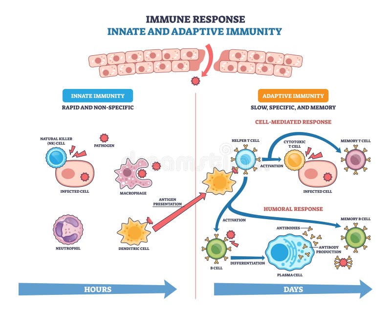
Free with trial Innate and adaptive immune response visual showing pathogen defense pathways, key elements, macrophage, T cell activation, antibody production. Outline diagram. T cell activation vectors Innate and adaptive immune response visual showing pathogen defense pathways, ... Innate and adaptive immune response visual showing pathogen defense pathways, key elements, macrophage, T cell activation, antibody production. Outline diagram
Free with trial An isometric illustration depicting advanced biotechnology research focusing on DNA sequencing, immune cell activation, and virus analysis. Scientists work in a modern lab environment, utilizing digital screens and advanced equipment to study cellular mechanisms and develop medical treatments. T cell activation illustrations Biotechnology Research DNA, Immune Cells, Virus Laboratory Science. An isometric illustration depicting advanced biotechnology research focusing on DNA sequencing, immune cell activation, and virus analysis. Scientists work in a modern lab environment, utilizing digital screens and advanced equipment to study cellular mechanisms and develop medical treatments.
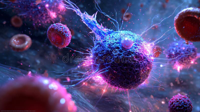
Free with trial A hyper-realistic 3D render depicts a futuristic, organic entity resembling a mutant cell or microorganism. The main spherical object, textured with intricate, fine details, appears bioluminescent with radiant blue and purple hues. It features extensions resembling tendrils or filaments, seemingly in an active state, emitting light. Surrounding smaller, similarly styled objects suggest a dynamic environment, with a vivid, energetic atmosphere. The scene conveys a science fiction theme, highlighting complexity and potential mutation. T cell activation illustrations Mutated immune cell attacks invading pathogens in sci-fi bloodstream. A hyper-realistic 3D render depicts a futuristic, organic entity resembling a mutant cell or microorganism. The main spherical object, textured with intricate, fine details, appears bioluminescent with radiant blue and purple hues. It features extensions resembling tendrils or filaments, seemingly in an active state, emitting light. Surrounding smaller, similarly styled objects suggest a dynamic environment, with a vivid, energetic atmosphere. The scene conveys a science fiction theme, highlighting complexity and potential mutation.
Free with trial A 3D render showing a Th1 immune cell releasing IFN-? cytokines that damage intestinal villi after activation by a gliadin-tTG complex, visualizing celiac pathogenesis. This illustration was created using AI Technology. T cell activation illustrations Celiac Disease: Th1 Immune Attack Causing Villous Atrophy. A 3D render showing a Th1 immune cell releasing IFN-? cytokines that damage intestinal villi after activation by a gliadin-tTG complex, visualizing celiac pathogenesis. This illustration was created using AI Technology
Free with trial This image presents an artistic representation of T and B lymphocytes, highlighting their pivotal roles in the immune response. The illustration showcases these cells in various stages of activation and interaction with other immune system components, emphasizing their importance in recognizing and combating pathogens. T cell activation illustrations T and B Lymphocytes: Pivotal Components of Immune Response. This image presents an artistic representation of T and B lymphocytes, highlighting their pivotal roles in the immune response. The illustration showcases these cells in various stages of activation and interaction with other immune system components, emphasizing their importance in recognizing and combating pathogens.
Free with trial Generative ai, The image depicts a complex biological scene with bright green and violet blue cells, likely showcasing cytotoxic T cell function, illuminated against a blended background. T cell activation illustrations The image shows cytotoxic T cells and targeted cells, exhibiting a detailed cellular interaction in vibrant. generative ai, The image depicts a complex biological scene with bright green and violet blue cells, likely showcasing cytotoxic T cell function, illuminated against a blended background
Free with trial Concept T cells, Cancer cells, Immune system, Cell recognition, Immunotherapy Distinguishing T Cells from Cancer Cells in the Immune System. T cell activation illustrations Distinguishing T Cells from Cancer Cells in the Immune System. Concept T cells, Cancer cells. Concept T cells, Cancer cells, Immune system, Cell recognition, Immunotherapy Distinguishing T Cells from Cancer Cells in the Immune System
Free with trial Contrast between T cells and cancer cells in the immune system. Concept T cells, Cancer Cells, Immune Response, Cell Recognition, Defense Mechanisms. T cell activation illustrations Concept T cells, Cancer Cells, Contrast between T cells and cancer cells in the immune system. Contrast between T cells and cancer cells in the immune system. Concept T cells, Cancer Cells, Immune Response, Cell Recognition, Defense Mechanisms
Free with trial 'T-2 Toxin in Poultry Feed: A Tiny Troublemaker with Big Consequences! ' 'Mycotoxins and Immunity: The Hidden Impact on Animal Health'. T cell activation illustrations T-2 Toxin in Poultry Feed: A Tiny Troublemaker with Big Consequences!
Free with trial This vector illustration portrays the cluster of differentiation factor 3 on a white background, highlighting its role as a cellular marker in immunology research. It illustrates the CD3 molecule, a crucial component of the T cell receptor complex, involved in antigen recognition and lymphocyte activation. Ideal for scientific and educational purposes, this high-quality vector graphic serves as a valuable visual aid for understanding the function and significance of CD3 in the immune system. The detailed and precise depiction ensures clarity and accuracy, making it an indispensable resource for researchers, educators, and professionals in the field of immunology and molecular biology. T cell activation vectors Illustration of Cluster of Differentiation Factor 3 on White Background Demonstrating Cellular Marker in Immunology Research. This vector illustration portrays the cluster of differentiation factor 3 on a white background, highlighting its role as a cellular marker in immunology research. It illustrates the CD3 molecule, a crucial component of the T cell receptor complex, involved in antigen recognition and lymphocyte activation. Ideal for scientific and educational purposes, this high-quality vector graphic serves as a valuable visual aid for understanding the function and significance of CD3 in the immune system. The detailed and precise depiction ensures clarity and accuracy, making it an indispensable resource for researchers, educators, and professionals in the field of immunology and molecular biology.
Free with trial Strategies of tumor immune evasion, Antigenic escape. Immune escape. Immune evasion or escape mutation, labeled tumor cells, t cells, tregs, exosome, immune system. Biology education diagram. T cell activation vectors Strategies of tumor immune evasion, Antigenic escape. Immune escape. Immune evasion or escape mutation, labeled tumor cells, t