Free with trial Collagen Fibers 3D of tendon in longitudinal section, fibrous connective tissue, microscopic on Isolated Transparent png background. Generative ai. Tendon microscope illustrations Collagen Fibers 3D of tendon in longitudinal section, fibrous connective tissue, microscopic on Isolated Transparent png
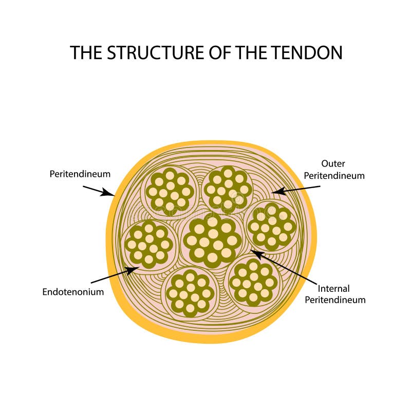
Free with trial Structure of tendon. Infographics. Vector illustration on isolated background. Tendon microscope vectors Structure of tendon. Infographics. Vector illustration on isolated background
Free with trial Types of human muscle tissue infographic. Cardiac, smooth, and skeletal muscle fibers. Walls of the heart, visceral organs and blood vessels. Skeleton with joints and and ligaments vector illustration. Tendon microscope vectors Human muscle tissues. Types of human muscle tissue infographic. Cardiac, smooth, and skeletal muscle fibers. Walls of the heart, visceral organs and blood vessels. Skeleton with joints and and ligaments vector illustration
Free with trial Smooth muscle tissue structure. Walls of internal organs and blood vessels in the human body. Spindle shaped myocyte. Intestine anatomy, digestive system diagram, medical flat vector illustration. Tendon microscope vectors Cells structure sketch. Smooth muscle tissue structure. Walls of internal organs and blood vessels in the human body. Spindle shaped myocyte. Intestine anatomy, digestive system diagram, medical flat vector illustration.
Free with trial Smooth muscle tissue structure. Walls of internal organs and blood vessels in the human body. Spindle shaped myocyte. Intestine anatomy, digestive system diagram, medical flat vector illustration. Tendon microscope vectors Cells structure sketch. Smooth muscle tissue structure. Walls of internal organs and blood vessels in the human body. Spindle shaped myocyte. Intestine anatomy, digestive system diagram, medical flat vector illustration.
Free with trial Types of human muscle tissue infographic. Cardiac, smooth, and skeletal muscle fibers. Walls of the heart, visceral organs and blood vessels. Skeleton with joints and and ligaments vector illustration. Tendon microscope vectors Cells structure sketch. Types of human muscle tissue infographic. Cardiac, smooth, and skeletal muscle fibers. Walls of the heart, visceral organs and blood vessels. Skeleton with joints and and ligaments vector illustration
Free with trial Types of human muscle tissue infographic. Cardiac, smooth, and skeletal muscle fibers. Walls of the heart, visceral organs and blood vessels. Skeleton with joints and and ligaments vector illustration. Tendon microscope vectors Cells structure sketch. Types of human muscle tissue infographic. Cardiac, smooth, and skeletal muscle fibers. Walls of the heart, visceral organs and blood vessels. Skeleton with joints and and ligaments vector illustration
Free with trial Types of human muscle tissue infographic. Cardiac, smooth, and skeletal muscle fibers. Walls of the heart, visceral organs and blood vessels. Skeleton with joints and and ligaments vector illustration. Tendon microscope vectors Cells structure sketch. Types of human muscle tissue infographic. Cardiac, smooth, and skeletal muscle fibers. Walls of the heart, visceral organs and blood vessels. Skeleton with joints and and ligaments vector illustration
Free with trial Smooth muscle tissue structure. Walls of internal organs and blood vessels in the human body. Spindle shaped myocyte. Intestine anatomy, digestive system diagram, medical flat vector illustration. Tendon microscope vectors Cells structure sketch. Smooth muscle tissue structure. Walls of internal organs and blood vessels in the human body. Spindle shaped myocyte. Intestine anatomy, digestive system diagram, medical flat vector illustration.
Free with trial Smooth muscle tissue structure. Walls of internal organs and blood vessels in the human body. Spindle shaped myocyte. Intestine anatomy, digestive system diagram, medical flat vector illustration. Tendon microscope vectors Cells structure sketch. Smooth muscle tissue structure. Walls of internal organs and blood vessels in the human body. Spindle shaped myocyte. Intestine anatomy, digestive system diagram, medical flat vector illustration.
Free with trial Types of human muscle tissue infographic. Cardiac, smooth, and skeletal muscle fibers. Walls of the heart, visceral organs and blood vessels. Skeleton with joints and and ligaments vector illustration. Tendon microscope vectors Cells structure sketch. Types of human muscle tissue infographic. Cardiac, smooth, and skeletal muscle fibers. Walls of the heart, visceral organs and blood vessels. Skeleton with joints and and ligaments vector illustration
Free with trial Types of human muscle tissue infographic. Cardiac, smooth, and skeletal muscle fibers. Walls of the heart, visceral organs and blood vessels. Skeleton with joints and and ligaments vector illustration. Tendon microscope vectors Cells structure sketch. Types of human muscle tissue infographic. Cardiac, smooth, and skeletal muscle fibers. Walls of the heart, visceral organs and blood. Types of human muscle tissue infographic. Cardiac, smooth, and skeletal muscle fibers. Walls of the heart, visceral organs and blood vessels. Skeleton with joints and and ligaments vector illustration
Free with trial Smooth muscle tissue structure. Walls of internal organs and blood vessels in the human body. Spindle shaped myocyte. Intestine anatomy, digestive system diagram, medical flat vector illustration. Tendon microscope vectors Cells structure sketch. Smooth muscle tissue structure. Walls of internal organs and blood vessels in the human body. Spindle shaped myocyte. Intestine anatomy, digestive system diagram, medical flat vector illustration.
Free with trial Smooth muscle tissue structure. Walls of internal organs and blood vessels in the human body. Spindle shaped myocyte. Intestine anatomy, digestive system diagram, medical flat vector illustration. Tendon microscope vectors Cells structure sketch. Smooth muscle tissue structure. Walls of internal organs and blood vessels in the human body. Spindle shaped myocyte. Intestine anatomy, digestive system diagram, medical flat vector illustration.
Free with trial Medical examination and illness - set of line design style icons isolated on white background. Quality images of operating table, IV, defibrillator, ultrasound, x-ray, lungs, doctor and rehabilitation. Tendon microscope vectors Medical examination and illness - set of line design style icons
Free with trial Blue glass structure Collagen fibers move inside 3d render. Tendon microscope illustrations Blue glass structure Collagen fibers move inside 3d
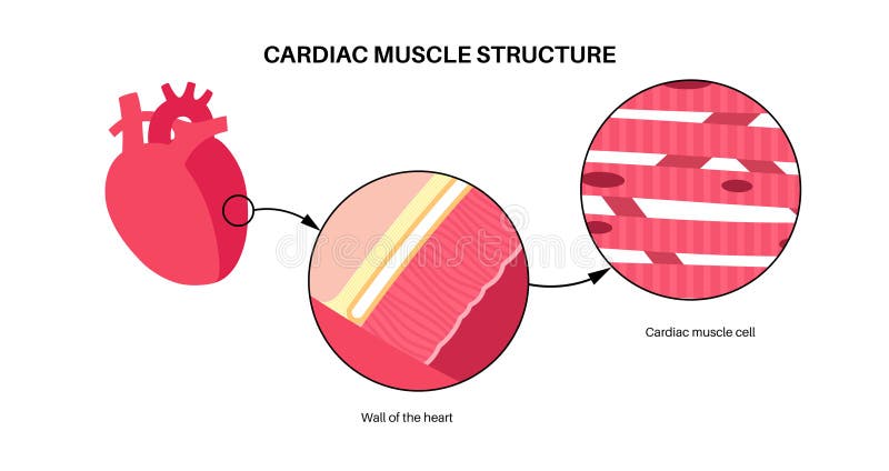
Free with trial Cardiac muscle tissue structure. Myocardium anatomical poster. Cardiomyocytes cells. Walls of the heart in the human body, relaxation and contraction of muscle fibers flat vector medical illustration. Tendon microscope vectors Cardiac muscle tissue
Free with trial Cardiac muscle tissue structure. Myocardium anatomical poster. Cardiomyocytes cells. Walls of the heart in the human body, relaxation and contraction of muscle fibers flat vector medical illustration. Tendon microscope vectors Cardiac muscle tissue
Free with trial Cardiac muscle tissue structure. Myocardium anatomical poster. Cardiomyocytes cells. Walls of the heart in the human body, relaxation and contraction of muscle fibers flat vector medical illustration. Tendon microscope vectors Cardiac muscle tissue
Free with trial Cardiac muscle tissue structure. Myocardium anatomical poster. Cardiomyocytes cells. Walls of the heart in the human body, relaxation and contraction of muscle fibers flat vector medical illustration. Tendon microscope vectors Cardiac muscle tissue
Free with trial Skeletal muscle tissue structure. Skeleton with joints, cartilages and ligaments in the human body. Muscle fibers and connective tissue sheaths. Musculoskeletal anatomy flat vector medical anatomy. Tendon microscope vectors Skeletal muscle tissue
Free with trial Skeletal muscle tissue structure. Skeleton with joints, cartilages and ligaments in the human body. Muscle fibers and connective tissue sheaths. Musculoskeletal anatomy flat vector medical anatomy. Tendon microscope vectors Skeletal muscle tissue
Free with trial Skeletal muscle tissue structure. Skeleton with joints, cartilages and ligaments in the human body. Muscle fibers and connective tissue sheaths. Musculoskeletal anatomy flat vector medical anatomy. Tendon microscope vectors Skeletal muscle tissue
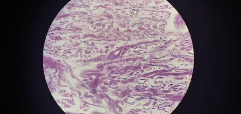
Free with trial This image appears to be a histological slide showing skeletal muscle tissue under a microscope. The elongated, multi-nucleated muscle fibers exhibit distinct striations, characteristic of muscle cells. The lighter connective tissue surrounds the muscle fibers, indicating the perimysium and endomysium. The image highlights the intricate organization and structure of muscle fibers, which are. Tendon microscope illustrations Microscopic view of muscle tissue with connective fibers and striations. This image appears to be a histological slide showing skeletal muscle tissue under a microscope. The elongated, multi-nucleated muscle fibers exhibit distinct striations, characteristic of muscle cells. The lighter connective tissue surrounds the muscle fibers, indicating the perimysium and endomysium. The image highlights the intricate organization and structure of muscle fibers, which are
Free with trial This image appears to be a microscopic cross-section of skeletal muscle tissue. The elongated, multi-nucleated muscle fibers are visible, running parallel to each other, with connective tissue surrounding them. The lighter, wavy lines likely represent perimysium and endomysium, while the pinkish background is indicative of the staining used to highlight cellular structures. Blood vessels and. Tendon microscope illustrations Microscopic view of muscle tissue fibers with connective tissue and blood vessels. This image appears to be a microscopic cross-section of skeletal muscle tissue. The elongated, multi-nucleated muscle fibers are visible, running parallel to each other, with connective tissue surrounding them. The lighter, wavy lines likely represent perimysium and endomysium, while the pinkish background is indicative of the staining used to highlight cellular structures. Blood vessels and
Free with trial Microscopic view of red muscle fibers intertwined. These bundles show striations and small particles on their surface suggesting biological detail. Useful for health or science visuals. Tendon microscope illustrations Microscopic view of red muscle fibers intertwined. These bundles show striations and small particles on their surface, suggesting. Microscopic view of red muscle fibers intertwined. These bundles show striations and small particles on their surface suggesting biological detail. Useful for health or science visuals.
Free with trial Smooth muscle tissue structure. Walls of internal organs and blood vessels in the human body. Spindle shaped myocyte. Intestine anatomy, digestive system diagram, medical flat vector illustration. Tendon microscope vectors Smooth muscle tissue
Free with trial Types of human muscle tissue infographic. Cardiac, smooth, and skeletal muscle fibers. Walls of the heart, visceral organs and blood vessels. Skeleton with joints and and ligaments vector illustration. Tendon microscope vectors Human muscle tissues. Types of human muscle tissue infographic. Cardiac, smooth, and skeletal muscle fibers. Walls of the heart, visceral organs and blood vessels. Skeleton with joints and and ligaments vector illustration
Free with trial Types of human muscle tissue infographic. Cardiac, smooth, and skeletal muscle fibers. Walls of the heart, visceral organs and blood vessels. Skeleton with joints and and ligaments vector illustration. Tendon microscope vectors Human muscle tissues. Types of human muscle tissue infographic. Cardiac, smooth, and skeletal muscle fibers. Walls of the heart, visceral organs and blood vessels. Skeleton with joints and and ligaments vector illustration
Free with trial Types of human muscle tissue infographic. Cardiac, smooth, and skeletal muscle fibers. Walls of the heart, visceral organs and blood vessels. Skeleton with joints and and ligaments vector illustration. Tendon microscope vectors Human muscle tissues. Types of human muscle tissue infographic. Cardiac, smooth, and skeletal muscle fibers. Walls of the heart, visceral organs and blood vessels. Skeleton with joints and and ligaments vector illustration
Free with trial Smooth muscle tissue structure. Walls of internal organs and blood vessels in the human body. Spindle shaped myocyte. Intestine anatomy, digestive system diagram, medical flat vector illustration. Tendon microscope vectors Smooth muscle tissue
Free with trial Smooth muscle tissue structure. Walls of internal organs and blood vessels in the human body. Spindle shaped myocyte. Intestine anatomy, digestive system diagram, medical flat vector illustration. Tendon microscope vectors Smooth muscle tissue
Free with trial Skeletal muscle tissue structure. Skeleton with joints, cartilages and ligaments in the human body. Muscle fibers and connective tissue sheaths. Musculoskeletal anatomy flat vector medical anatomy. Tendon microscope vectors Skeletal muscle tissue
Free with trial Detailed illustration of damaged or severed fibers (nerve or muscle), representing tissue injury, degeneration, or rupture. Ideal for communications on neuromuscular diseases, sports injuries, tissue repair, or research on nerve fiber regeneration and muscle atrophy. Tendon microscope illustrations Neuromuscular Tissue Injury and Repair. Detailed illustration of damaged or severed fibers (nerve or muscle), representing tissue injury, degeneration, or rupture. Ideal for communications on neuromuscular diseases, sports injuries, tissue repair, or research on nerve fiber regeneration and muscle atrophy.
Free with trial A 3d illustration of smooth muscle cells, common arrangement in tissue layers within organ walls. Tendon microscope illustrations A 3d illustration smooth muscle tissue layers. A 3d illustration of smooth muscle cells, common arrangement in tissue layers within organ walls
Free with trial A 3d illustration of smooth muscle cells, common arrangement in tissue layers within organ walls. Tendon microscope illustrations A 3d illustration smooth muscle tissue layers. A 3d illustration of smooth muscle cells, common arrangement in tissue layers within organ walls
Free with trial Structure of tendon. Infographics. Vector illustration on isolated background. Tendon microscope vectors Structure of tendon. Infographics. Vector illustration on isolated background
Free with trial Histology of human tissue, show epithelium cell, connective tissue and muscle tissue with microscope view. Tendon microscope illustrations Picture of histology human tissue with microscope from laboratory (not Illustration Designation). Histology of human tissue, show epithelium cell, connective tissue and muscle tissue with microscope view
Free with trial Histology of human tissue, show epithelium cell, connective tissue and muscle tissue with microscope view. Tendon microscope illustrations Picture of histology human tissue with microscope from laboratory (not Illustration Designation). Histology of human tissue, show epithelium cell, connective tissue and muscle tissue with microscope view
Free with trial Histology of human tissue, show epithelium cell, connective tissue and muscle tissue with microscope view. Tendon microscope illustrations Picture of histology human tissue with microscope from laboratory (not Illustration Designation). Histology of human tissue, show epithelium cell, connective tissue and muscle tissue with microscope view
Free with trial Histology of human tissue, show epithelium cell, connective tissue and muscle tissue with microscope view. Tendon microscope illustrations Picture of histology human tissue with microscope from laboratory (not Illustration Designation). Histology of human tissue, show epithelium cell, connective tissue and muscle tissue with microscope view
Free with trial Histology of human tissue, show epithelium cell, connective tissue and muscle tissue with microscope view. Tendon microscope illustrations Picture of histology human tissue with microscope from laboratory (not Illustration Designation). Histology of human tissue, show epithelium cell, connective tissue and muscle tissue with microscope view
Free with trial Ofloxacin molecular structure, 3d model molecule, quinolones, structural chemical formula view from a microscope. Tendon microscope illustrations Ofloxacin molecular structure, 3d model molecule, quinolones, structural chemical formula view from a microscope
Free with trial Beautiful picture full of colors and love. Tendon microscope illustrations Connective tissue under a microscope. beautiful picture full of colors and love
Free with trial Vector flat illustration, big human foot on white background, medical research. Scientists and doctors examine shin and foot, look through a microscope and monitor. Bottles and Medication Packages. Tendon microscope vectors Vector flat illustrations, large human foot on a white background. Vector flat illustration, big human foot on white background, medical research. Scientists and doctors examine shin and foot, look through a microscope and monitor. Bottles and Medication Packages
Free with trial Vector flat illustration, big human foot on white background, medical research. Scientists and doctors examine shin and foot, look through a microscope and monitor. Bottles and Medication Packages. Tendon microscope vectors Vector flat illustrations, large human foot on a white background. Vector flat illustration, big human foot on white background, medical research. Scientists and doctors examine shin and foot, look through a microscope and monitor. Bottles and Medication Packages
Free with trial Vector flat illustration, big human foot on white background, medical research. Scientists and doctors examine shin and foot, look through a microscope and monitor. Bottles and Medication Packages. Tendon microscope vectors Vector flat illustrations, large human foot on a white background. Vector flat illustration, big human foot on white background, medical research. Scientists and doctors examine shin and foot, look through a microscope and monitor. Bottles and Medication Packages
Free with trial Vector flat illustration, big human foot on white background, medical research. Scientists and doctors examine shin and foot, look through a microscope and monitor. Bottles and Medication Packages. Tendon microscope vectors Vector flat illustrations, large human foot on a white background. Vector flat illustration, big human foot on white background, medical research. Scientists and doctors examine shin and foot, look through a microscope and monitor. Bottles and Medication Packages
Free with trial Vector flat illustration, big human foot on white background, medical research. Scientists and doctors examine shin and foot, look through a microscope and monitor. Bottles and Medication Packages. Tendon microscope vectors Vector flat illustrations, large human foot on a white background. Vector flat illustration, big human foot on white background, medical research. Scientists and doctors examine shin and foot, look through a microscope and monitor. Bottles and Medication Packages
Free with trial Vector flat illustration, big human foot on white background, medical research. Scientists and doctors examine shin and foot, look through a microscope and monitor. Bottles and Medication Packages. Tendon microscope vectors Vector flat illustrations, large human foot on a white background. Vector flat illustration, big human foot on white background, medical research. Scientists and doctors examine shin and foot, look through a microscope and monitor. Bottles and Medication Packages