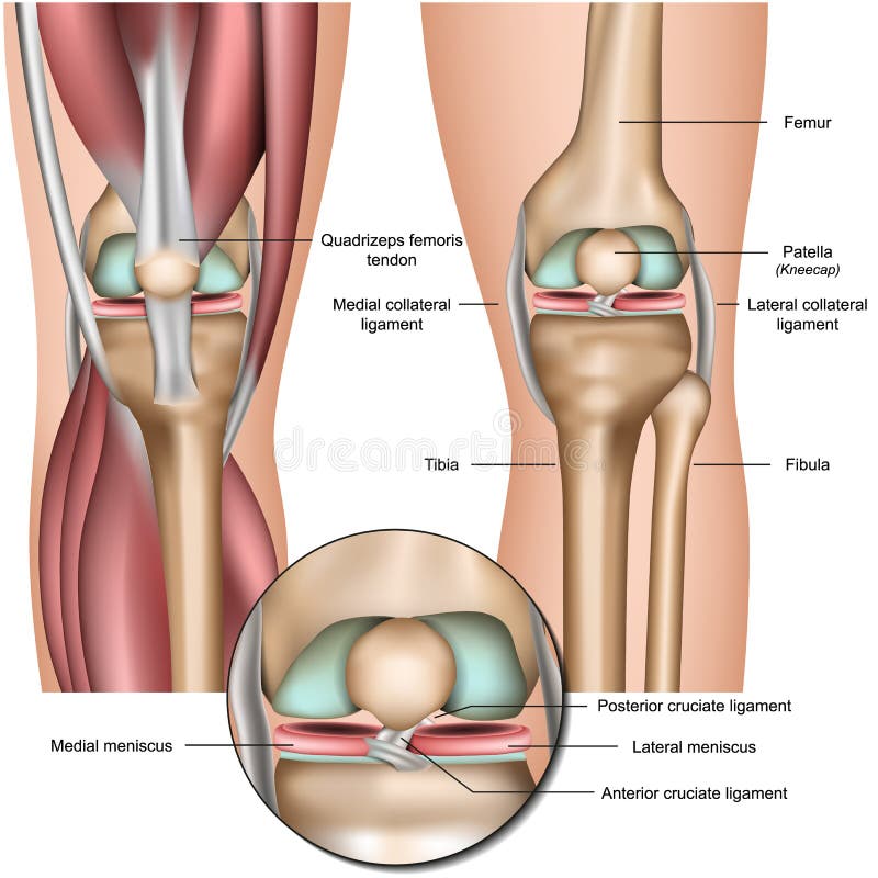
Free with trial Anatomy of human Knee Joint, eps8. Thigh muscle anatomy vectors Anatomy of the Knee Joint. Anatomy of human Knee Joint, eps8
Free with trial Anatomy of the posterior of the right knee in extension. Thigh muscle anatomy vectors Posterior view of the right knee in extension. Anatomy of the posterior of the right knee in extension
Free with trial Human heart anatomy from a healthy body isolated on white background. Medical health care symbol of an inner cardiovascular organ. illustration. Thigh muscle anatomy vectors Heart structure anatomy. Heart cross section. Human heart anatomy from a healthy body isolated on white background. Medical health care symbol of an inner cardiovascular organ. illustration
Free with trial Anatomy. Knee Joint Cross Section Showing the major parts which made the knee joint For Basic Medical Education Also for clinics. Thigh muscle anatomy vectors Anatomy. Structure knee joint vector. Anatomy. Knee Joint Cross Section Showing the major parts which made the knee joint For Basic Medical Education Also for clinics
Free with trial Muscle Strain degree and Treatment isolated on white background. Vector illustrations. Thigh muscle anatomy vectors Pulled Muscle. Muscle Strain degree and Treatment isolated on white background.Vector illustrations
Free with trial Anatomy of the knee joint side view, template for training a medical surgical poster, traumatology pages, vector illustration. Thigh muscle anatomy vectors Anatomy of the knee joint side view, template for training a medical surgical poster, traumatology page. Vector illustration.
Free with trial Anatomy. Knee Joint Cross Section Showing the major parts which made the knee joint For Basic Medical Education Also for clinics. Thigh muscle anatomy vectors Anatomy. Structure knee joint vector. Anatomy. Knee Joint Cross Section Showing the major parts which made the knee joint For Basic Medical Education Also for clinics
Free with trial Anatomy. Knee Joint Cross Section Showing the major parts which made the knee joint For Basic Medical Education Also for clinics. Thigh muscle anatomy vectors Anatomy. Structure knee joint vector. Anatomy. Knee Joint Cross Section Showing the major parts which made the knee joint For Basic Medical Education Also for clinics
Free with trial Synovial Joint. Human anatomy. vector. Thigh muscle anatomy vectors Synovial bursa. Synovial Joint. Human anatomy. vector
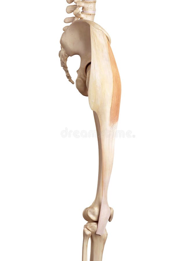
Free with trial Medical accurate illustration of the tensor fascia lata. Thigh muscle anatomy illustrations The tensor fascia lata
Free with trial Medical accurate illustration of the biceps femoris longus. Thigh muscle anatomy illustrations The biceps femoris longus
Free with trial Medical accurate illustration of the vastus intermedius. Thigh muscle anatomy illustrations The vastus intermedius
Free with trial Medical accurate illustration of the vastus lateralis. Thigh muscle anatomy illustrations The vastus lateralis
Free with trial Medical accurate illustration of the adductor magnus. Thigh muscle anatomy illustrations The adductor magnus
Free with trial Sciatic nerve pain in the lower back through hip, thigh, knee to leg. Educational or informational poster. Flat vector medical illustration isolated on white background. Thigh muscle anatomy vectors Sciatic nerve pain
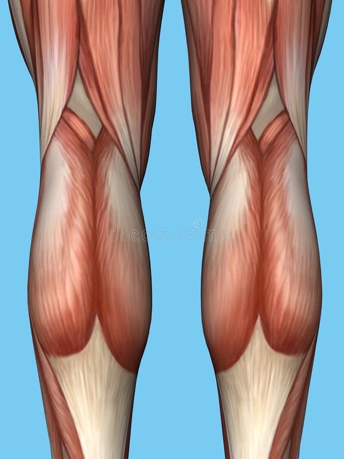
Free with trial Anatomy of back of legs featuring quadriceps, hamstrings, achilles tendon, gastrocnemius, peroneus longus muscle, biceps femoris, semimembranosus and soleus muscles. Thigh muscle anatomy illustrations Anatomy of back of legs. Anatomy of back of legs featuring quadriceps, hamstrings, achilles tendon, gastrocnemius, peroneus longus muscle, biceps femoris, semimembranosus and soleus muscles.
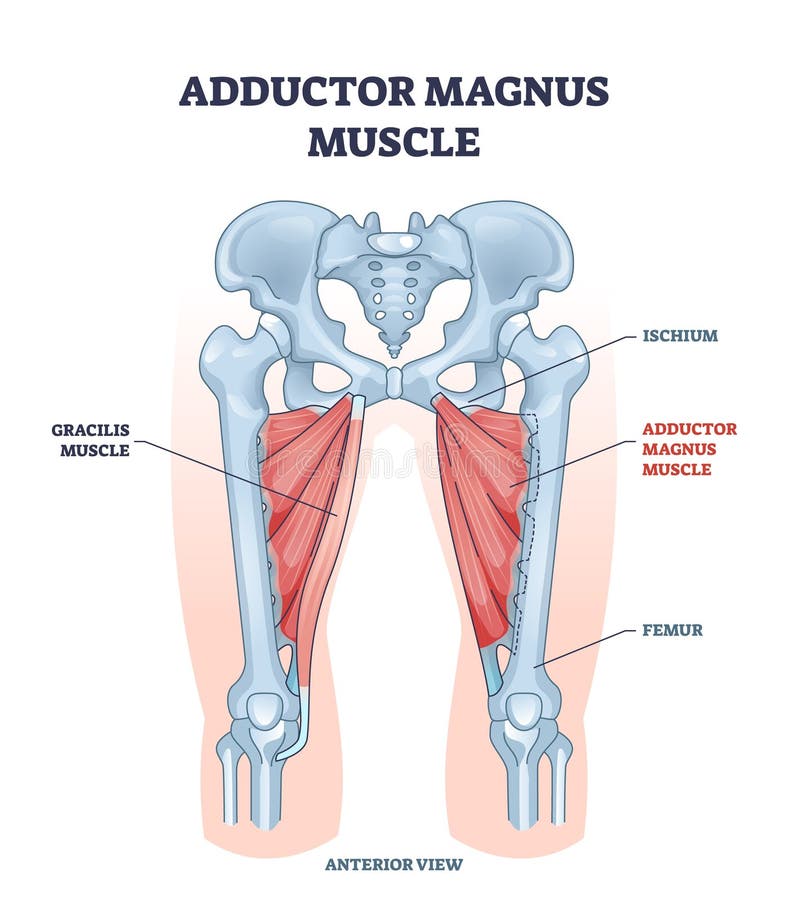
Free with trial Adductor magnus muscle with ischium and femur skeleton outline diagram. Labeled educational gracilis muscular system from anterior view vector illustration. Human body hips and legs inner structure. Thigh muscle anatomy vectors Adductor magnus muscle with ischium and femur skeleton outline diagram
Free with trial Adductor longus muscle location with hips and leg, ischium and femur, bones outline diagram. Labeled educational medical scheme with pectineus, magnus and gracilis muscular system vector illustration. Thigh muscle anatomy vectors Adductor longus muscle location with hips and leg bones outline diagram. Adductor longus muscle location with hips and leg, ischium and femur, bones outline diagram. Labeled educational medical scheme with pectineus, magnus and gracilis muscular system vector illustration.
Free with trial Vector illustration of a healthy knee joint. Anatomy of the human knee, side view. For advertising, medical publications. EPS 10. Thigh muscle anatomy vectors Anatomy of the knee. Vector illustration of a healthy knee joint . Anatomy of the human knee, side view. For advertising, medical publications. EPS 10.
Free with trial Meniscus knee anatomy medical vector illustration isolated on white background infographic eps 10. Thigh muscle anatomy vectors Meniscus knee anatomy medical vector illustration isolated on white background infographic
Free with trial Vector illustration of a healthy human knee joint and unhealthy knee with Hoffa`s fat pad impingement syndrome. Knee anatomy, sagittal section. For advertising and medical publications. EPS 10. Thigh muscle anatomy vectors Anatomy of the knee_Fat pad impingement due to hyperextension. Vector illustration of a healthy human knee joint and unhealthy knee with Hoffa`s fat pad impingement syndrome. Knee anatomy, sagittal section. For advertising and medical publications. EPS 10.
Free with trial 3D medical rendering of sciatica showing posterior full leg from lower back to heel, sciatic nerve in yellow with red pain path brightest at gluteal exit fading down thigh, muted pink muscles. Thigh muscle anatomy illustrations 3D medical rendering of sciatica showing posterior full leg from lower back to heel, sciatic nerve in yellow with red pain path
Free with trial Medical accurate illustration of the upper leg muscles. Thigh muscle anatomy illustrations The upper leg muscles
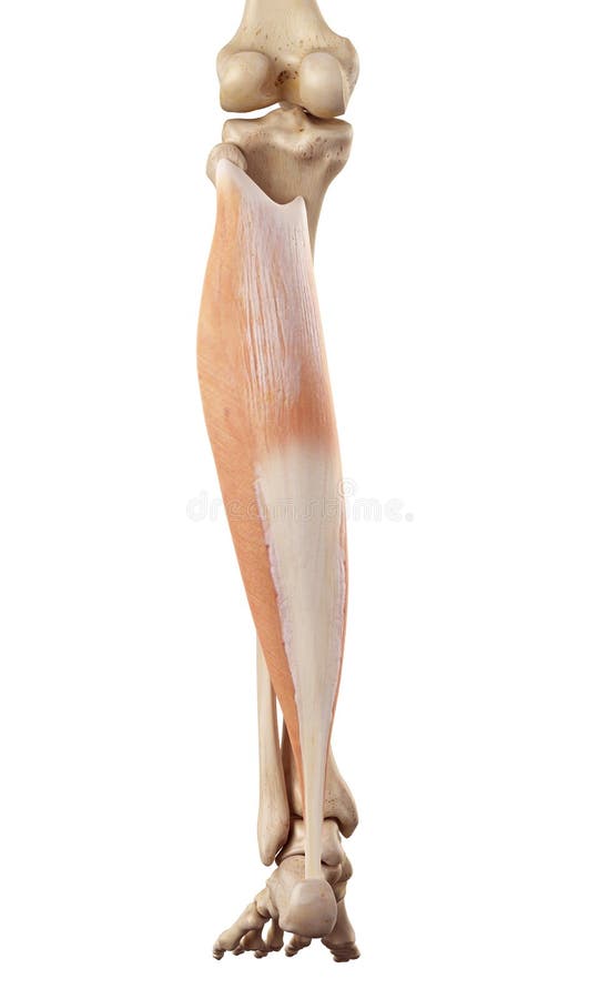
Free with trial Medical accurate illustration of the soleus. Thigh muscle anatomy illustrations The soleus
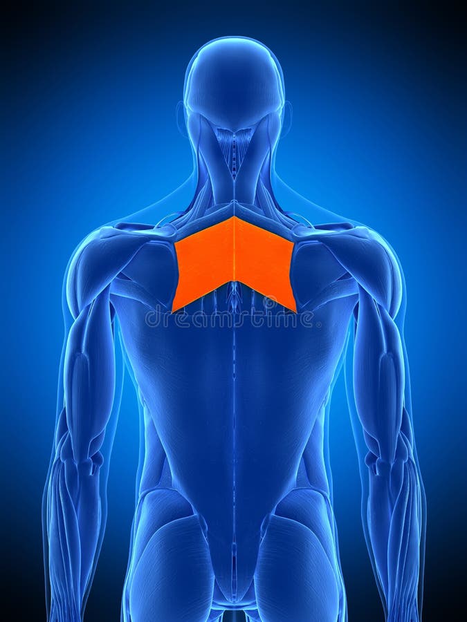
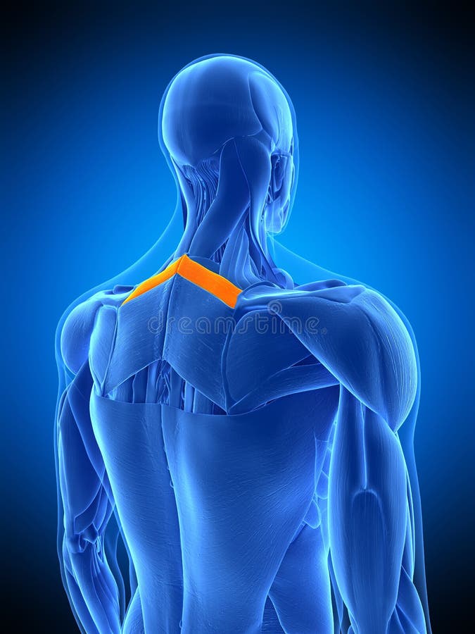
Free with trial Medically accurate illustration of the rhomboid major. Thigh muscle anatomy illustrations The rhomboid major
Free with trial Medical accurate illustration of the adductor longus. Thigh muscle anatomy illustrations The adductor longus
Free with trial Medical accurate illustration of the tibialis posterior. Thigh muscle anatomy illustrations The tibialis posterior
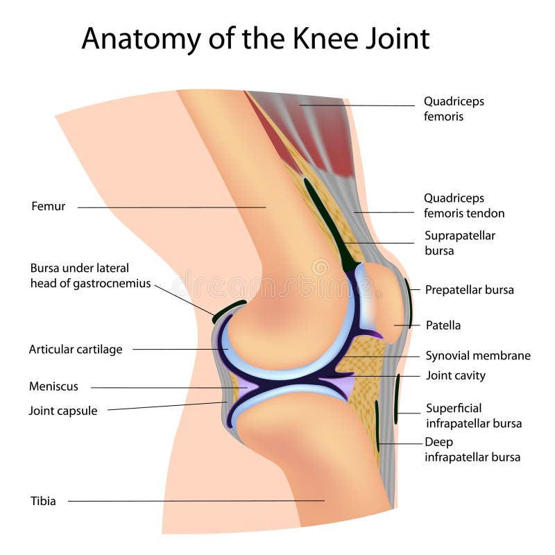
Free with trial The bursae of the knee are the fluid sacs and synovial pockets that surround and sometimes communicate with the joint cavity. Thin-walled and filled with synovial fluid, they represent the weak point of the joint, but also produce enlargements to the joint space. Thigh muscle anatomy illustrations Frontal section. The bursae of the knee are the fluid sacs and synovial pockets that surround and sometimes communicate with the joint cavity. Thin-walled and filled with synovial fluid, they represent the weak point of the joint, but also produce enlargements to the joint space.
Free with trial Medically accurate illustration of the rhomboid major. Thigh muscle anatomy illustrations The rhomboid major
Free with trial Medical accurate illustration of the tensor fascia lata. Thigh muscle anatomy illustrations The tensor fascia lata
Free with trial Medical accurate illustration of the tibialis posterior. Thigh muscle anatomy illustrations The tibialis posterior
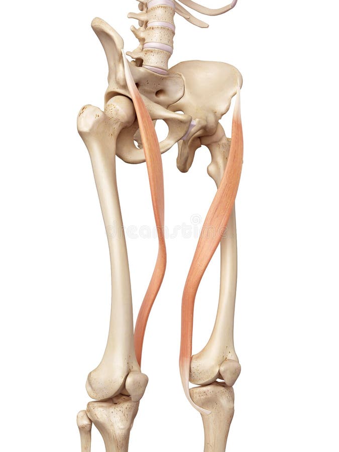
Free with trial Medical accurate illustration of the gracilis. Thigh muscle anatomy illustrations The gracilis
Free with trial Medical accurate illustration of the sartorius. Thigh muscle anatomy illustrations The sartorius
Free with trial Medically accurate illustration of the rhomboid minor. Thigh muscle anatomy illustrations The rhomboid minor
Free with trial Medical accurate illustration of the adductor magnus. Thigh muscle anatomy illustrations The adductor magnus
Free with trial Medical accurate illustration of the semimembranosus. Thigh muscle anatomy illustrations The semimembranosus
Free with trial Medically accurate illustration of the latissimus dorsi. Thigh muscle anatomy illustrations The latissimus dorsi
Free with trial Medical accurate illustration of the biceps femoris short. Thigh muscle anatomy illustrations The biceps femoris short
Free with trial Vector illustration of a healthy knee joint and an unhealthy knee with a quadriceps tendon rupture problem. Anatomy of the human knee, side view of the bent knee. Thigh muscle anatomy vectors Knee problem_Quadriceps tendon rupture. Vector illustration of a healthy knee joint and an unhealthy knee with a quadriceps tendon rupture problem. Anatomy of the human knee, side view of the bent knee.
Free with trial Vector illustration of a human pelvis, hip and a knee joint with iliotibial band tract syndrome. Front view. For advertising and medical publications. EPS 10. Thigh muscle anatomy vectors Pain in the hip joint iliotibial band syndrome. Vector illustration of a human pelvis, hip and a knee joint with iliotibial band tract syndrome. Front view. For advertising and medical publications. EPS 10.
Free with trial Frontal thigh anatomy with red glow on rectus femoris fiber tear, ideal for illustrating quadriceps muscle tear or sports sprint injury in clean medical, sports, and rehabilitation visuals, Generative AI. Thigh muscle anatomy illustrations Frontal thigh anatomy with red glow on rectus femoris fiber tear, ideal for illustrating quadriceps muscle tear or sports sprint
Free with trial Infographic of healthy and sarcopenia thigh muscle vector illustration isolated on white background. Cross section of strength and losing muscle mass. Anatomy and health care concept illustration. Thigh muscle anatomy illustrations Healthy and sarcopenia thigh muscle vector illustration on white background. Infographic of healthy and sarcopenia thigh muscle vector illustration isolated on white background. Cross section of strength and losing muscle mass. Anatomy and health care concept illustration.
Free with trial Anatomy of human thigh muscles in vintage style. Hand drawn engraved monochrome sketch. Vector illustration. Poster or banner for biology lesson. Description of the body part. Thigh muscle anatomy vectors Anatomy of human thigh muscles in vintage style. Hand drawn engraved monochrome sketch. Vector illustration. Poster or
Free with trial Thigh anatomy. Didactic scheme of structure of bone structure, muscular and circulatory system with anatomical captions. Thigh muscular, circulation system and skeleton. Flat vector illustration. Thigh muscle anatomy vectors Thigh anatomy. Didactic scheme of structure of bone structure
Free with trial Calf muscle. Trigger points in the leg, reflected pain on the back of the thigh and in the foot. Leg pain. Thigh muscle anatomy illustrations Calf muscle. Trigger points in the leg, reflected pain on the back of the thigh and in the foot. Leg pain
Free with trial Knee joint capsule anatomy infographic diagram. structure and components including bone femur tibia patella muscle ligament cartilage cavity and bursa. lateral view vector drawing. illustration for medical science. Thigh muscle anatomy vectors Knee joint capsule anatomy infographic diagram
Free with trial Gracilis muscle. Structure, trigger points, and reflected pain in the thigh muscle. Isolate on black background. Thigh muscle anatomy illustrations Gracilis muscle. Structure, trigger points, and reflected pain in the thigh muscle
Free with trial Calf muscle. Trigger points in the leg, reflected pain on the back of the thigh and in the foot. Leg pain. Thigh muscle anatomy illustrations Calf muscle. Trigger points in the leg, reflected pain on the back of the thigh and in the foot. Leg pain
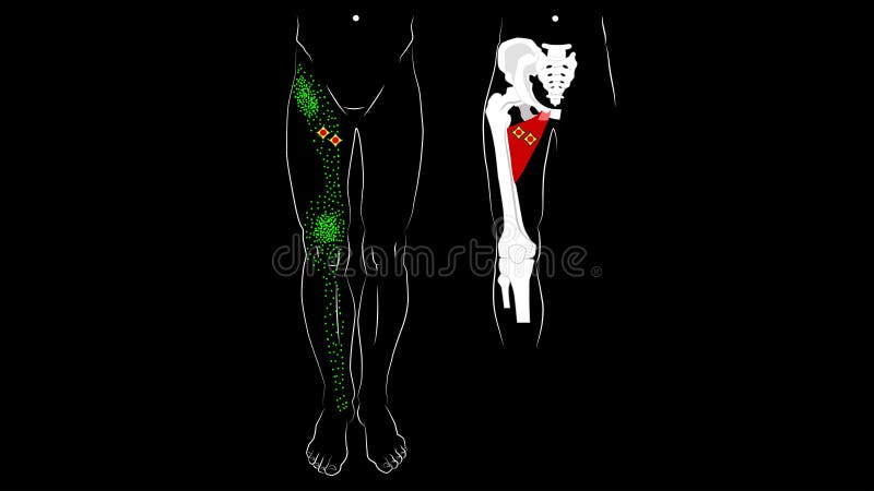
Free with trial Adductor longus muscle. Adductor brevis muscle. Trigger points and reflected pain on the inner thigh. Isolate on black background. Thigh muscle anatomy illustrations Adductor longus muscle. Adductor brevis muscle. Trigger points and reflected pain on the inner thigh.
Free with trial Front view of leg muscle anatomy featuring quadriceps, calf and knees. Thigh muscle anatomy illustrations Frontal View Leg Anatomy. Front view of leg muscle anatomy featuring quadriceps, calf and knees.
Free with trial Muscles of the anterior thigh Human structure diagram hand drawn schematic vector illustration. Medical science educational illustration. Thigh muscle anatomy illustrations Anatomy of anterior thigh muscles medical science. muscles of the anterior thigh Human structure diagram hand drawn schematic vector illustration. Medical science educational illustration
Free with trial Hip muscles. Didactic scheme of anatomy of human muscular system with anatomical captions. Hip and thigh gluteus maximus, inferior gemellus, quadratus femoris. Flat vector illustration. Thigh muscle anatomy vectors Hip muscles. Didactic scheme of anatomy of human muscular system
Free with trial Intramuscular injection areas. Guide to injecting medication into muscle. Sites with large, easy-to-locate muscles and little fatty tissue, upper arm, thigh, hip and buttock. Flat vector illustration. Thigh muscle anatomy vectors Intramuscular injection areas. Guide to injecting medication into muscle
Free with trial Adductor brevis muscle with hips and leg skeletal system outline diagram. Labeled educational scheme with medical magnus and gracilis muscular location and ischium or femur bones vector illustration. Thigh muscle anatomy vectors Adductor brevis muscle with hips and leg skeletal system outline diagram
Free with trial Vector illustration of a healthy human knee joint. Anatomy of the sagittal section of a knee, side view. For advertising, medical publications. EPS 8. Thigh muscle anatomy vectors Anatomy of the knee_Sagittal section. Vector illustration of a healthy human knee joint . Anatomy of the sagittal section of a knee, side view. For advertising, medical publications. EPS 8.
Free with trial Knee and meniscus anatomy medical illustration isolated on white background eps 10. Thigh muscle anatomy vectors Knee and meniscus anatomy medical illustration isolated on white background
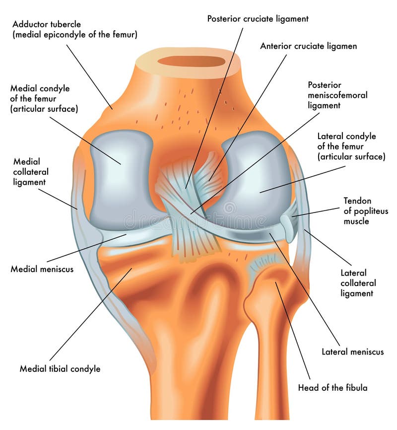
Free with trial Anatomy of the knee joint front view, template for training a medical surgical poster, traumatology page. Vector illustration. Thigh muscle anatomy vectors Anatomy of the knee joint front view, template for training a medical surgical poster
Free with trial Medical illustration showing the Tensor Fasciae Latae muscle, IT band, and iliac crest, diagram. Thigh muscle anatomy vectors Medical illustration showing the Tensor Fasciae Latae muscle, IT band,... Medical illustration showing the Tensor Fasciae Latae muscle, IT band, and iliac crest, diagram.
Free with trial Sciatica pain and inflammation in pelvis, leg and hip. Piriformis muscle syndrome concept. Human nervous system and skeleton anatomical poster. Medical flat vector illustration for clinic. X ray image. Thigh muscle anatomy vectors Pelvis bones and muscle. sciatica pain and inflammation in pelvis, leg and hip. Piriformis muscle syndrome concept. Human nervous system and skeleton anatomical poster. Medical flat vector illustration for clinic. X ray image
Free with trial Anatomy. Knee Joint Cross Section Showing the major parts which made the knee joint For Basic Medical Education Also for clinics set text. Thigh muscle anatomy illustrations Anatomy. Structure knee joint raster set text. Anatomy. Knee Joint Cross Section Showing the major parts which made the knee joint For Basic Medical Education Also for clinics set text
Free with trial Anatomy. Subscribe. Knee Joint Cross Section Showing the major parts which made the knee joint For Basic Medical Education Also for clinics. Thigh muscle anatomy illustrations Anatomy. Subscribe. Structure knee joint raster. Anatomy. Subscribe. Knee Joint Cross Section Showing the major parts which made the knee joint For Basic Medical Education Also for clinics
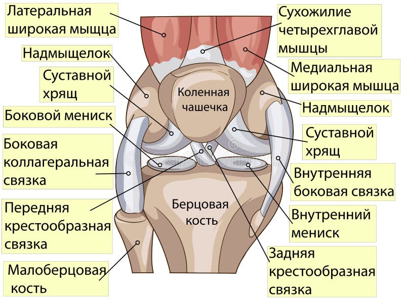
Free with trial Anatomy. Knee Joint Cross Section Showing the major parts which made the knee joint For Basic Medical Education Also for clinics. Inscriptions in Russian. Thigh muscle anatomy vectors Anatomy. Structure knee joint vector. Anatomy. Knee Joint Cross Section Showing the major parts which made the knee joint For Basic Medical Education Also for clinics. Inscriptions in Russian
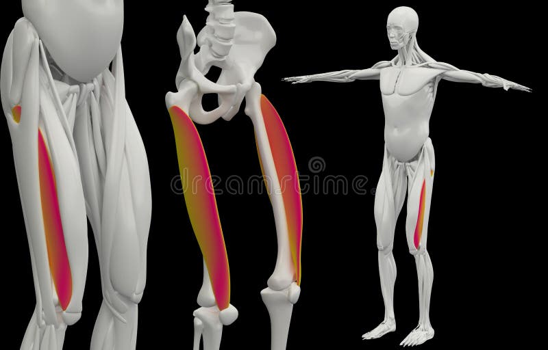
Free with trial 3d rendering medically accurate muscle illustration of the vastus lateralis. Thigh muscle anatomy illustrations Medically accurate muscle illustration of the vastus lateralis
Free with trial Anatomy. Subscribe. Knee Joint Cross Section Showing the major parts which made the knee joint For Basic Medical Education Also for clinics. Thigh muscle anatomy illustrations Anatomy. Subscribe. Structure knee joint raster Basic Medical Education. Anatomy. Subscribe. Knee Joint Cross Section Showing the major parts which made the knee joint For Basic Medical Education Also for clinics
Free with trial Anatomy pop art design. Knee Joint Cross Section Showing the major parts which made the knee joint For Basic Medical Education Also for clinics. Thigh muscle anatomy vectors Anatomy. Structure knee joint vector pop art design. Anatomy pop art design. Knee Joint Cross Section Showing the major parts which made the knee joint For Basic Medical Education Also for clinics
Free with trial Anatomy. Knee Joint Cross Section Showing the major parts which made the knee joint For Basic Medical Education Also for clinics set text. Thigh muscle anatomy vectors Anatomy. Structure knee joint vector set text. Anatomy. Knee Joint Cross Section Showing the major parts which made the knee joint For Basic Medical Education Also for clinics set text
Free with trial Hamstring muscle pain against a serene blue backdrop. This stock image highlights the physical strain and injury that athletes and fitness enthusiasts may experience, emphasizing the importance of proper training and injury prevention techniques. Thigh muscle anatomy illustrations Hamstring Muscle Pain with blue background. Hamstring muscle pain against a serene blue backdrop. This stock image highlights the physical strain and injury that athletes and fitness enthusiasts may experience, emphasizing the importance of proper training and injury prevention techniques.
Free with trial Anatomy. Knee Joint Cross Section Showing the major parts which made the knee joint For Basic Medical Education Also for clinics. Thigh muscle anatomy illustrations Anatomy. Knee Joint Cross Section Showing the major parts which made the knee joint For Basic Medical Education
Free with trial Lower limb muscles diagram showing anterior and posterior views with labeled quadriceps, hamstrings, and gastrocnemius for study and anatomy reference. Outline diagram. Thigh muscle anatomy vectors Lower limb muscles diagram showing anterior and posterior views with ... Lower limb muscles diagram showing anterior and posterior views with labeled quadriceps. Lower limb muscles diagram showing anterior and posterior views with labeled quadriceps, hamstrings, and gastrocnemius for study and anatomy reference. Outline diagram
Free with trial Medically accurate muscle illustration of the rectus femoris. Thigh muscle anatomy illustrations The rectus femoris
Free with trial Medically accurate muscle illustration of the semitendinosus. Thigh muscle anatomy illustrations The semitendinosus
Free with trial Medically accurate muscle illustration of the semimembranosus. Thigh muscle anatomy illustrations The semimembranosus
Free with trial Medically accurate muscle illustration of the biceps femoris longus. Thigh muscle anatomy illustrations The biceps femoris longus
Free with trial Medically accurate muscle illustration of the vastus intermedius. Thigh muscle anatomy illustrations The vastus intermedius
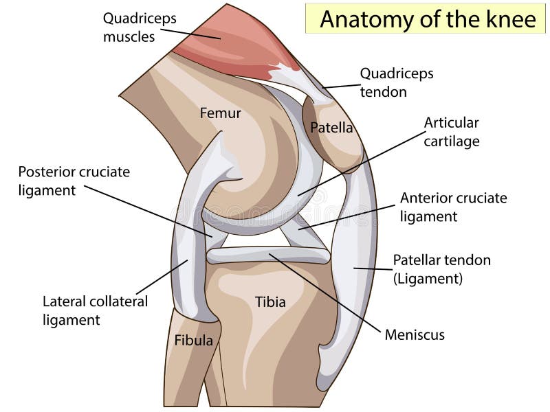
Free with trial The knee joint joins the thigh with the leg and consists of two articulations: one between the femur and tibia, and one between the femur and patella. Thigh muscle anatomy illustrations Knee joint
Free with trial Medically accurate muscle illustration of the pectineus. Thigh muscle anatomy illustrations The pectineus
Free with trial The knee joint joins the thigh with the leg and consists of two articulations: one between the femur and tibia, and one between the femur and patella. Thigh muscle anatomy illustrations Knees. The knee joint joins the thigh with the leg and consists of two articulations: one between the femur and tibia, and one between the femur and patella.
Free with trial The knee joint joins the thigh with the leg and consists of two articulations: one between the femur and tibia, and one between the femur and patella. Thigh muscle anatomy illustrations Knee joint
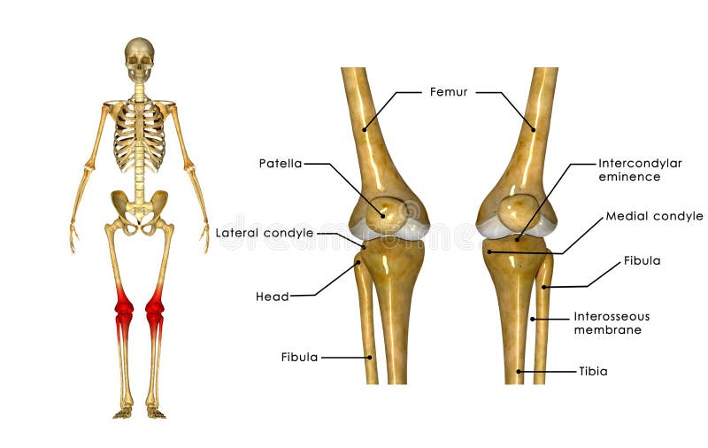
Free with trial The knee joint is one of the strongest and most important joints in the human body. It allows the lower leg to move relative to the thigh while supporting the body’s weight. Movements at the knee joint are essential to many everyday activities, including walking, running, sitting and standing. The knee, also known as the tibiofemoral joint, is a synovial hinge joint formed between three bones: the femur, tibia, and patella. Two rounded, convex processes (known as condyles) on the distal end of the femur meet two rounded, concave condyles at the proximal end of the tibia. Thigh muscle anatomy illustrations Knee joints. The knee joint is one of the strongest and most important joints in the human body. It allows the lower leg to move relative to the thigh while supporting the body’s weight. Movements at the knee joint are essential to many everyday activities, including walking, running, sitting and standing. The knee, also known as the tibiofemoral joint, is a synovial hinge joint formed between three bones: the femur, tibia, and patella. Two rounded, convex processes (known as condyles) on the distal end of the femur meet two rounded, concave condyles at the proximal end of the tibia
Free with trial Medically accurate muscle illustration of the adductor brevis. Thigh muscle anatomy illustrations The adductor brevis