Free with trial VECTOR İLLUSTRATİON OF A BONE MARROW ASPIRATION. Tissue samples vectors VECTOR İLLUSTRATİON OF A BONE MARROW ASPIRATION
Free with trial An abstract texture background pattern resembling green and blue tissue paper samples casually arranged. Tissue samples illustrations Blue Tissue Paper Texture. An abstract texture background pattern resembling green and blue tissue paper samples casually arranged
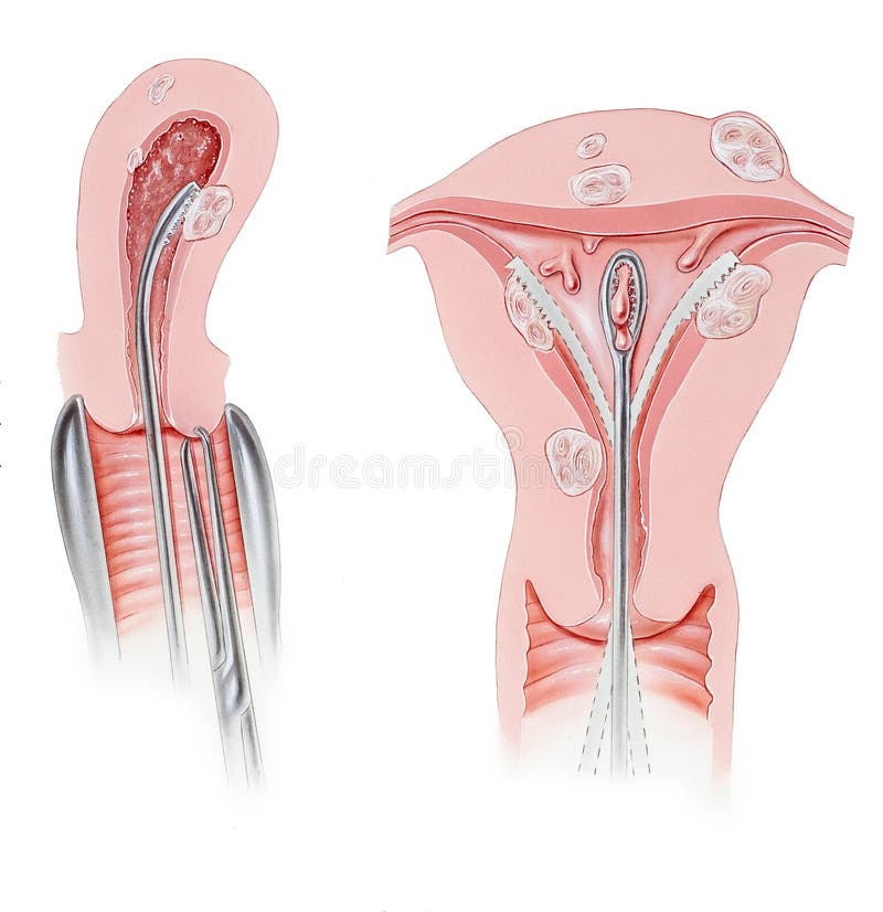
Free with trial A dilation and curettage D&C procedure, in which the cervix is dilated so that an instrument can be inserted to scrape or suction the lining of the uterus and take tissue samples. Shown on this abnormal uterus are intramural and submucous fibroids and polyps. Tissue samples illustrations Cervix - Dilation and Curettage D&C Procedure. A dilation and curettage D&C procedure, in which the cervix is dilated so that an instrument can be inserted to scrape or suction the lining of the uterus and take tissue samples. Shown on this abnormal uterus are intramural and submucous fibroids and polyps.
Free with trial An abstract texture background pattern resembling green and blue tissue paper samples casually arranged. Tissue samples illustrations Green Tissue Paper Texture. An abstract texture background pattern resembling green and blue tissue paper samples casually arranged
Free with trial Set of nine samples checkered cloth for a picnic. Seamless texture. Tablecloth, fabric, material, textile. Tissue samples vectors Set of tissue samples for a picnic. Set of nine samples checkered cloth for a picnic. Seamless texture. Tablecloth, fabric, material, textile
Free with trial Set of nine samples checkered cloth for a picnic. Seamless texture. Tablecloth, fabric, material, textile. Tissue samples vectors Set of tissue samples for a picnic. Set of nine samples checkered cloth for a picnic. Seamless texture. Tablecloth, fabric, material, textile
Free with trial This visual representation illustrates the process of transrectal ultrasound-guided core needle biopsy, a crucial procedure for diagnosing prostate cancer. The image depicts the various stages involved, from initial ultrasound imaging to the precise extraction of tissue samples. This minimally invasive technique allows for the collection of tissue samples for pathological examination, providing. Tissue samples illustrations Understanding Prostate Cancer Treatment A Visual Guide to Transrectal UltrasoundGuided Core Needle Biopsy. This visual representation illustrates the process of transrectal ultrasound-guided core needle biopsy, a crucial procedure for diagnosing prostate cancer. The image depicts the various stages involved, from initial ultrasound imaging to the precise extraction of tissue samples. This minimally invasive technique allows for the collection of tissue samples for pathological examination, providing
Free with trial Cotton variegated fabric texture sampler. Tissue samples illustrations Fabric texture sampler.
Free with trial Leather variegated fabric texture sampler. Tissue samples illustrations Leather variegated fabric texture. Leather variegated fabric texture sampler.
Free with trial DNA banking. Stock vector illustration of a building with columns formed by double helix. Storage for cryopreservation of body samples for regenerative medicine and scientific purpose. Tissue samples vectors Stem cell banking. DNA banking. Stock vector illustration of a building with columns formed by double helix. Storage for cryopreservation of body samples for regenerative medicine and scientific purpose.
Free with trial Stem cell banking. Stock vector illustration of a doctor`s hand manipulation and lab equipment used for cryopreservation of human body samples for regenerative medicine purpose. Tissue samples vectors Stem cell banking
Free with trial Stem cell banking. Stock vector illustration for cryopreservation of human body samples for regenerative medicine purpose. Tissue samples vectors Stem cell banking
Free with trial A laboratory setup featuring a microscope, computer screen showing cell analysis, and lab equipment on a desk, 150 chars exactly. Tissue samples illustrations Digital pathology lab with microscope and computer displaying cell samples. A laboratory setup featuring a microscope, computer screen showing cell analysis, and lab equipment on a desk, 150 chars exactly
Free with trial Black denim cloth texture with pockets with stitches and rivets. Vector realistic background of dark gray jean fabric with samples of different pockets for pants back. Tissue samples vectors Black denim cloth texture with pockets
Free with trial Variations in Design and multicolored wavy background in separate samples by white stripes. Tissue samples illustrations Wavy Design and Multicolored. Variations in Design and multicolored wavy background in separate samples by white stripes
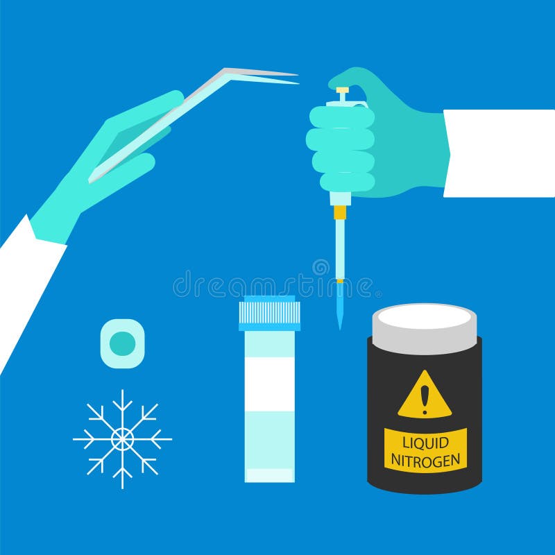
Free with trial Cryopreservation uses liquid nitrogen to freeze biological materials for research. Tissue samples illustrations Cryopreservation uses liquid nitrogen to freeze biological materials for research
Free with trial 3D Bioprinter A futuristic 3D bioprinter creating organic tissue samples with a closeup view of the printing process happening. Tissue samples illustrations 3D Bioprinter A futuristic 3D bioprinter creating organic tissue samples with a closeup view of the printing process
Free with trial Close up of plant tissue samples in a sterile lab setting sterile samples Vector illustration. Tissue samples illustrations Close up of plant tissue samples in a sterile lab setting , sterile, samples. Close up of plant tissue samples in a sterile lab setting sterile samples Vector illustration.
Free with trial Artificial skin tissue samples in laboratory containers for medical transplant research. Synthetic bioengineered membrane development with various cellular structure patterns. Tissue samples illustrations Artificial skin tissue samples in laboratory containers for medical transplant research. Synthetic bioengineered membrane
Free with trial Futuristic lab bench with layered 3D-printed tissue samples, sterile white setting, 16:9 digital. Tissue samples illustrations Futuristic lab bench with layered 3D-printed tissue samples, sterile
Free with trial Bright and colorful cellular tissue samples arranged on glass slides, showcasing high fidelity and intricate details, ideal for scientific and medical themes. Tissue samples illustrations Bright Cellular Tissue Samples on Glass Slides. Bright and colorful cellular tissue samples arranged on glass slides, showcasing high fidelity and intricate details, ideal for scientific and medical themes
Free with trial The image shows four microscopic views of tissue samples arranged in a 2x2 grid. The top-left sample appears to have a distinct coloration compared to the other three, which are more uniformly shaded in blue tones. The differences in texture and density suggest variations in cellular structure, potentially due to different treatments, staining techniques, or pathological conditions. The scale bars. Tissue samples illustrations Microscopic comparison of tissue samples under varying conditions or treatments. The image shows four microscopic views of tissue samples arranged in a 2x2 grid. The top-left sample appears to have a distinct coloration compared to the other three, which are more uniformly shaded in blue tones. The differences in texture and density suggest variations in cellular structure, potentially due to different treatments, staining techniques, or pathological conditions. The scale bars
Free with trial A futuristic diagnostic laboratory featuring AI-driven machines that analyze blood and tissue samples. The machines process samples with advanced technology, and detailed reports and medical insights are displayed on large digital screens. This scene showcases the integration of artificial intelligence in medical diagnostics, enhancing precision and efficiency in healthcare. Tissue samples illustrations AI-Driven Diagnostic Laboratory Analyzing Blood and Tissue Samples with Digital Reports. A futuristic diagnostic laboratory featuring AI-driven machines that analyze blood and tissue samples. The machines process samples with advanced technology, and detailed reports and medical insights are displayed on large digital screens. This scene showcases the integration of artificial intelligence in medical diagnostics, enhancing precision and efficiency in healthcare
Free with trial This captivating image showcases the intricate details of 3D bioprinted tissue samples, a crucial advancement in regenerative medicine. The vibrant pink hues highlight the engineered tissue structures within clear glass vessels, ideal for observation under low-light microscopy. The technique employed in this bioprinting process allows for the precise layering and deposition of biological. Tissue samples illustrations Advanced 3D Bioprinting Techniques Showcase Pink Tissue Samples in Glass Vessels A LowLight Microscopy Study of. This captivating image showcases the intricate details of 3D bioprinted tissue samples, a crucial advancement in regenerative medicine. The vibrant pink hues highlight the engineered tissue structures within clear glass vessels, ideal for observation under low-light microscopy. The technique employed in this bioprinting process allows for the precise layering and deposition of biological
Free with trial A vibrant display of glass slides featuring colorful tissue samples. Ideal for illustrating microscopy, laboratory settings, and scientific research concepts. Generative AI Illustration. Tissue samples illustrations Colorful Glass Slides with Tissue Samples on Display. A vibrant display of glass slides featuring colorful tissue samples. Ideal for illustrating microscopy, laboratory settings, and scientific research concepts. Generative AI Illustration
Free with trial Colorful tissue samples on glass slides showcase intricate cellular details, ideal for medical and scientific visualization, emphasizing microscopic research. Generative AI Illustration. Tissue samples illustrations Tissue Samples on Glass Slides with Cellular Details. Colorful tissue samples on glass slides showcase intricate cellular details, ideal for medical and scientific visualization, emphasizing microscopic research. Generative AI Illustration
Free with trial A vibrant arrangement of colorful cellular tissue samples on a glass surface, showcasing high fidelity and intricate details. Ideal for scientific and medical visuals. Tissue samples illustrations High Fidelity Cellular Tissue Samples on Glass Surface. A vibrant arrangement of colorful cellular tissue samples on a glass surface, showcasing high fidelity and intricate details. Ideal for scientific and medical visuals
Free with trial A futuristic diagnostic laboratory featuring AI-driven machines that analyze blood and tissue samples. The machines process samples with advanced technology, and detailed reports and medical insights are displayed on large digital screens. This scene showcases the integration of artificial intelligence in medical diagnostics, enhancing precision and efficiency in healthcare. Tissue samples illustrations Advanced AI Diagnostic Lab Processing Blood and Tissue Samples with Automated Digital Reports. A futuristic diagnostic laboratory featuring AI-driven machines that analyze blood and tissue samples. The machines process samples with advanced technology, and detailed reports and medical insights are displayed on large digital screens. This scene showcases the integration of artificial intelligence in medical diagnostics, enhancing precision and efficiency in healthcare
Free with trial Microscopic tissue samples are submerged in vibrant pink fluid within clear glass laboratory vials, showcasing precise scientific research and analysis, perfect for medical and educational contexts. Tissue samples illustrations Microscopic Tissue Samples in Pink Fluid Within Laboratory Vials for Scientific Research and Analysis. Microscopic tissue samples are submerged in vibrant pink fluid within clear glass laboratory vials, showcasing precise scientific research and analysis, perfect for medical and educational contexts
Free with trial Micro preparation samples section tissue for microscope examination. Tissue samples illustrations Micro preparation samples section tissue for microscope examination
Free with trial Micro preparation samples section tissue for microscope examination. Tissue samples illustrations Micro preparation samples section tissue for microscope examination
Free with trial Micro preparation samples section tissue for microscope examination. Tissue samples illustrations Micro preparation samples section tissue for microscope examination
Free with trial Micro preparation samples section tissue for microscope examination. Tissue samples illustrations Micro preparation samples section tissue for microscope examination
Free with trial Plant tissue culture, a revolutionary method in horticulture and agricultural science, is showcased here. These meticulously prepared lab samples in sterile test tubes represent a crucial step in cultivating high-yielding, disease-resistant plant varieties. The process involves isolating small pieces of plant tissue, such as leaves or stems, and nurturing them in a controlled environment. This. Tissue samples illustrations Cultivating the Future Plant Tissue Culture Lab Samples Support Vegetarian Research and Sustainable Agriculture. Plant tissue culture, a revolutionary method in horticulture and agricultural science, is showcased here. These meticulously prepared lab samples in sterile test tubes represent a crucial step in cultivating high-yielding, disease-resistant plant varieties. The process involves isolating small pieces of plant tissue, such as leaves or stems, and nurturing them in a controlled environment. This
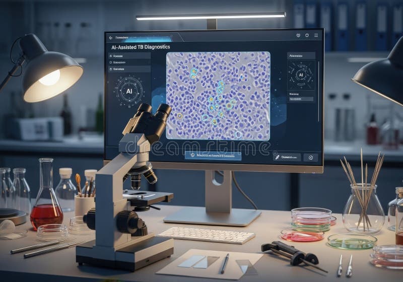
Free with trial Explore the advanced technology of an AI powered microscope used for analyzing tissue samples in a modern laboratory. Ideal for research and healthcare applications. Tissue samples illustrations Advanced AI Powered Microscope Analyzing Tissue Samples in Lab Setting. Explore the advanced technology of an AI powered microscope used for analyzing tissue samples in a modern laboratory. Ideal for research and healthcare applications
Free with trial A medical lab technician preparing a microscope slide with tissue samples for examination under magnification. Tissue samples illustrations A medical lab technician preparing a microscope slide with tissue samples
Free with trial Scientist Works with Petri Dishes with Various Bacteria, Tissue and Blood Samples. Tissue samples illustrations Scientist Works with Petri Dishes with Various Bacteria, Tissue and Blood Samples
Free with trial Futuristic lab bench with layered 3D-printed tissue samples, sterile white setting, 16:9 digital. Tissue samples illustrations Futuristic lab bench with layered 3D-printed tissue samples, sterile white setting, 16:9 .
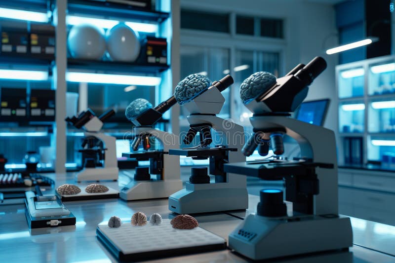
Free with trial A high-tech laboratory with brain tissue samples under microscopes, illustrating the meticulous work in brain tumor research. Tissue samples illustrations A high-tech laboratory with brain tissue samples under microscopes, illustrating the meticulous work in brain tumor research
Free with trial A high-tech laboratory with brain tissue samples under microscopes, illustrating the meticulous work in brain tumor research. Tissue samples illustrations A high-tech laboratory with brain tissue samples under microscopes, illustrating the meticulous work in brain tumor research
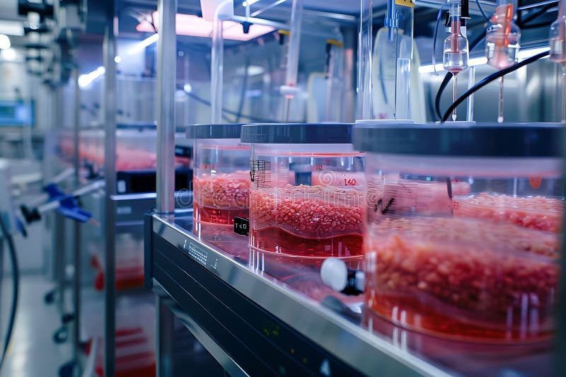
Free with trial The image shows a close-up view of a laboratory bioreactor system. Several cylindrical glass containers are visible, each filled with a reddish-pink substance that appears to be cultivated tissue samples submerged in a liquid medium. The containers are neatly arranged on a stainless steel rack, suggesting a controlled environment for cellular growth. The overall setup suggests a highly technological and sterile environment indicative of advanced biological research or tissue engineering. A digital display on the rack is partially visible. The lighting and cleanliness of the laboratory suggest a meticulously maintained facility. Tissue samples illustrations Laboratory Bioreactor System with Tissue Samples. The image shows a close-up view of a laboratory bioreactor system. Several cylindrical glass containers are visible, each filled with a reddish-pink substance that appears to be cultivated tissue samples submerged in a liquid medium. The containers are neatly arranged on a stainless steel rack, suggesting a controlled environment for cellular growth. The overall setup suggests a highly technological and sterile environment indicative of advanced biological research or tissue engineering. A digital display on the rack is partially visible. The lighting and cleanliness of the laboratory suggest a meticulously maintained facility.
Free with trial Close-up of a surgical tray with medical instruments, tissue samples, and a measuring cup with a substance, all in a sterile environment, ready for analysis. Tissue samples illustrations Medical Instruments and Samples on a Tray in a Sterile Environment. Close-up of a surgical tray with medical instruments, tissue samples, and a measuring cup with a substance, all in a sterile environment, ready for analysis
Free with trial A scientist examines lab-grown human tissue samples under a microscope, showcasing the intricate details of cellular structures in a modern laboratory setting. Tissue samples illustrations Examination of Lab Grown Human Tissue Samples Under Microscope. A scientist examines lab-grown human tissue samples under a microscope, showcasing the intricate details of cellular structures in a modern laboratory setting
Free with trial Microscopic view of blood cells and tissue samples. Close-up microscopic images showing blood cells, tissue layers, and cellular structures under magnification. Tissue samples illustrations Microscopic view of blood cells and tissue samples. Close-up microscopic images showing blood cells, tissue layers, and cellular structures under magnification
Free with trial Modern automated digital pathology slide scanner in a medical laboratory, used for digitizing tissue samples for ai-assisted diagnosis. Tissue samples illustrations Modern automated digital pathology slide scanner in a medical laboratory, used for digitizing tissue samples
Free with trial Modern automated digital pathology slide scanner in a medical laboratory, used for digitizing tissue samples for ai-assisted diagnosis. Tissue samples illustrations Modern automated digital pathology slide scanner in a medical laboratory, used for digitizing tissue samples
Free with trial This image showcases vibrant tissue samples on glass slides with illuminated colors, highlighting cellular structures in a laboratory setting for scientific research. Generative AI Illustration. Tissue samples illustrations Vibrant Cellular Samples on Glass Slides. This image showcases vibrant tissue samples on glass slides with illuminated colors, highlighting cellular structures in a laboratory setting for scientific research. Generative AI Illustration
Free with trial Artificial skin samples stored in laboratory dish show cellular structure for medical transplant research. Bioengineered tissue displays microscopic patterns in clinical setting. Tissue samples illustrations Artificial skin samples stored in laboratory dish show cellular structure for medical transplant research. Bioengineered tissue
Free with trial Artificial skin tissue samples showing cellular structure and smooth surface for medical transplantation. Synthetic biomaterial comparison. Biotechnology healthcare innovation research. Tissue samples illustrations Artificial skin tissue samples showing cellular structure and smooth surface for medical transplantation. Synthetic biomaterial
Free with trial Scientific laboratory equipment shows a petri dish with trabecular bone structure and tissue samples forming text for medical research and biotechnology studies. Tissue samples illustrations Microscopic view of bone marrow tissue samples in laboratory petri dish. Scientific laboratory equipment shows a petri dish with trabecular bone structure and tissue samples forming text for medical research and biotechnology studies
Free with trial Biobanks are vital repositories for biological samples, including DNA, RNA, tissue, and other biosamples, enabling groundbreaking research and advancements in personalized medicine. These collections, often referred to as biorepositories, meticulously manage the storage and cryopreservation of samples in specialized freezers. Rigorous quality control procedures and standardization protocols are. Tissue samples illustrations Biobanks Secure Storage and Management of Biological Samples for Advanced Research and Personalized Medicine. Biobanks are vital repositories for biological samples, including DNA, RNA, tissue, and other biosamples, enabling groundbreaking research and advancements in personalized medicine. These collections, often referred to as biorepositories, meticulously manage the storage and cryopreservation of samples in specialized freezers. Rigorous quality control procedures and standardization protocols are
Free with trial Close-up of gloved hands holding glass histology slides with purple stained tissue samples. Medical research involves microscopic analysis, of biological specimens. Scientists examine. Tissue samples illustrations Close-up of gloved hands holding glass histology slides with purple stained tissue samples. Medical research involves microscopic
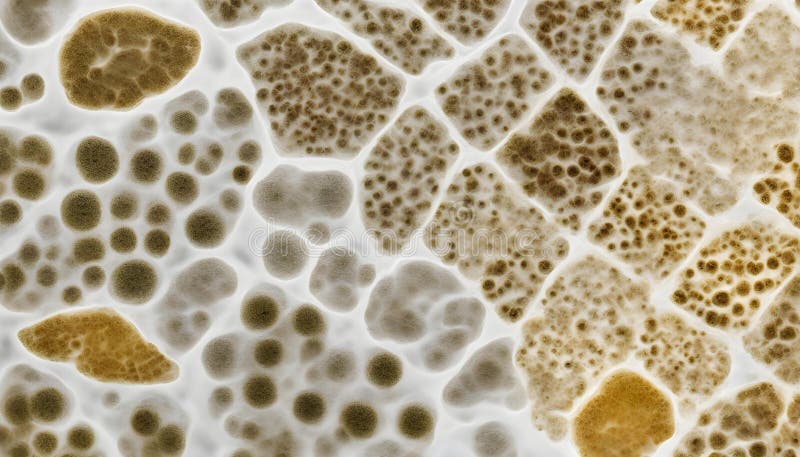
Free with trial This image presents a microscopic view of three distinct biological tissue samples, each exhibiting unique cell structures and densities. The samples are arranged side by side, allowing for a comparative analysis of their morphological characteristics. The first sample on the left displays a densely packed arrangement of cells with prominent, radiating lines, suggesting a highly organized tissue. Tissue samples illustrations Microscopic view of three biological tissue samples with different cell structures. This image presents a microscopic view of three distinct biological tissue samples, each exhibiting unique cell structures and densities. The samples are arranged side by side, allowing for a comparative analysis of their morphological characteristics. The first sample on the left displays a densely packed arrangement of cells with prominent, radiating lines, suggesting a highly organized tissue
Free with trial This captivating image showcases the meticulous process of plant tissue culture within a controlled laboratory environment. Glass vials, each containing vibrant green plant tissue samples, are meticulously arranged, highlighting the precision and care involved in scientific research. The controlled laboratory setting, complete with specialized equipment and sterile conditions, emphasizes the. Tissue samples illustrations Cultivating the Future Controlled Plant Tissue Culture in a Scientific Laboratory Setting. This captivating image showcases the meticulous process of plant tissue culture within a controlled laboratory environment. Glass vials, each containing vibrant green plant tissue samples, are meticulously arranged, highlighting the precision and care involved in scientific research. The controlled laboratory setting, complete with specialized equipment and sterile conditions, emphasizes the
Free with trial Clinical pathologist examines tissue samples with microscope in laboratory. Doctor in gloves, white lab coat studies histological slides. Medical equipment and research work in the lab. Tissue samples illustrations Clinical pathologist examines tissue samples with microscope in laboratory. Doctor in gloves, white lab coat, studies. Clinical pathologist examines tissue samples with microscope in laboratory. Doctor in gloves, white lab coat studies histological slides. Medical equipment and research work in the lab.
Free with trial Floating Petri Dishes with Biological Cell Culture and Microscopic Tissue Samples in High Tech Laboratory for Stem Cell Research Genetic Engineering, and Biotechnology Science Innovation Concepts. Tissue samples illustrations Floating Petri Dishes with Biological Cell Culture and Microscopic Tissue Samples in High Tech Laboratory for Stem Cell Research
Free with trial This image presents a detailed close up of human skin tissue highlighting a vibrant red substance, indicative of collagen within the cellular structure. The visualization is captured in a fluorescence image, emphasizing the advanced techniques employed in medical laboratory experiments to study tissue samples. Such images are vital for understanding skin biology and pathology. Tissue samples illustrations Exploring the Intricacies of Cellular Structures through Close Up Imaging of Skin Tissue with Red Collagen and Medical Laboratory. This image presents a detailed close up of human skin tissue highlighting a vibrant red substance, indicative of collagen within the cellular structure. The visualization is captured in a fluorescence image, emphasizing the advanced techniques employed in medical laboratory experiments to study tissue samples. Such images are vital for understanding skin biology and pathology
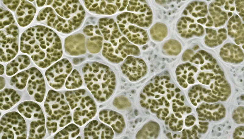
Free with trial The image shows six circular cross-sections of plant stems arranged in a grid pattern under a microscope. The sections reveal detailed internal cellular structures with visible vascular bundles and surrounding parenchyma cells, exhibiting variations in tissue density and arrangement. The samples appear to be stained, enhancing contrast and highlighting different cell types and tissue layers. Tissue samples illustrations Microscopic view of plant tissue revealing intricate cellular structures isolated on transparent background, isolated on white. The image shows six circular cross-sections of plant stems arranged in a grid pattern under a microscope. The sections reveal detailed internal cellular structures with visible vascular bundles and surrounding parenchyma cells, exhibiting variations in tissue density and arrangement. The samples appear to be stained, enhancing contrast and highlighting different cell types and tissue layers
Free with trial Harnessing the power of nature's genetic code, this captivating image showcases the cutting-edge field of green biotechnology. The meticulously prepared plant samples, nestled within laboratory flasks, represent a crucial stage in research and development. These samples are likely undergoing various analyses, including genetic modification, tissue culture, and metabolic profiling, to unlock. Tissue samples illustrations Exploring the Future of Agriculture Green Biotechnology Research with Plant Samples in Lab Flasks. Harnessing the power of nature's genetic code, this captivating image showcases the cutting-edge field of green biotechnology. The meticulously prepared plant samples, nestled within laboratory flasks, represent a crucial stage in research and development. These samples are likely undergoing various analyses, including genetic modification, tissue culture, and metabolic profiling, to unlock
Free with trial Histology lab photo shows microscope slides stained with hematoxylin, eosin. Medical research includes samples of tissue analyzed under microscope for cancer diagnosis. Laboratory. Tissue samples illustrations Histology lab photo shows microscope slides stained with hematoxylin, eosin. Medical research includes samples of tissue analyzed
Free with trial Rows of petri dishes filled with vibrant green plant tissue culture are arranged neatly in a laboratory setting. The controlled environment promotes growth and research in plant biology, showcasing the intricate details of the cultures. Tissue samples illustrations Green plant tissue culture in petri dishes under laboratory conditions. Rows of petri dishes filled with vibrant green plant tissue culture are arranged neatly in a laboratory setting. The controlled environment promotes growth and research in plant biology, showcasing the intricate details of the cultures
Free with trial A detailed view of a microscope in a laboratory setting, highlighting vibrant gel samples under observation. This imagery captures the essence of scientific research and innovation. Tissue samples illustrations Close-Up of Laboratory Microscopy with Bright Gel Samples in Focus. A detailed view of a microscope in a laboratory setting, highlighting vibrant gel samples under observation. This imagery captures the essence of scientific research and innovation
Free with trial The image depicts a sterile, high-tech laboratory meticulously organized with rows of large glass containers and numerous smaller dishes. The containers are likely holding cell cultures or organ cultures, suggesting biological research or tissue engineering. A central monitoring system is visible, overseeing the process, and the overall scene suggests a controlled environment for advanced scientific experimentation, potentially in the field of regenerative medicine or similar. The rows of samples on trays indicate a potentially large-scale operation, emphasizing precision and organization within the experiment. Tissue samples illustrations Scientific Laboratory with Cellular Growth and Organ Culture. The image depicts a sterile, high-tech laboratory meticulously organized with rows of large glass containers and numerous smaller dishes. The containers are likely holding cell cultures or organ cultures, suggesting biological research or tissue engineering. A central monitoring system is visible, overseeing the process, and the overall scene suggests a controlled environment for advanced scientific experimentation, potentially in the field of regenerative medicine or similar. The rows of samples on trays indicate a potentially large-scale operation, emphasizing precision and organization within the experiment.
Free with trial Several laboratory bottles filled with green plant tissue cultures are arranged on a wet surface, representing science and agriculture innovation. Tissue samples illustrations Plant Tissue Culture in Glass Bottles for Scientific Research and Development. Several laboratory bottles filled with green plant tissue cultures are arranged on a wet surface, representing science and agriculture innovation.
Free with trial A detailed illustration of a classic microscope examining various biological samples on glass slides, including cells, tissues, and microorganisms, set against a warm, softly lit background that highlights the scientific and analytical nature of the scene. The image conveys a sense of precision and discovery, with the microscope as the central focus and the slides arranged dynamically around it. Tissue samples illustrations Microscope Analyzing Biological Samples on Glass Slides Under Warm Light. A detailed illustration of a classic microscope examining various biological samples on glass slides, including cells, tissues, and microorganisms, set against a warm, softly lit background that highlights the scientific and analytical nature of the scene. The image conveys a sense of precision and discovery, with the microscope as the central focus and the slides arranged dynamically around it
Free with trial Generative AI : Plant Tissue Culture Young Plants Growing in Test Tubes for Biotechnology and Research business concept. Tissue samples illustrations Generative AI Plant Tissue Culture Young Plants Growing in Test Tubes for Biotechnology and Research business conc
Free with trial Layers of human skin and tissue structure with detailed cross-section highlighting multiple cell types, collagen fibers, and extracellular matrix components in realistic anatomical illustration. Tissue samples vectors Layers of human skin and tissue structure with detailed cross-section highlighting
Free with trial A biopsy jar isolated on a white background, often used in medical laboratories for tissue samples. Tissue samples illustrations Biopsy jar isolated on white background. A biopsy jar isolated on a white background, often used in medical laboratories for tissue samples
Free with trial A biopsy jar isolated on a white background, used in medical practices for tissue samples. Tissue samples illustrations Biopsy jar isolated on white background. A biopsy jar isolated on a white background, used in medical practices for tissue samples
Free with trial Colorful immunofluorescent photomicrograph of breast tissue with ductal carcinoma. The fluorescent staining reveals intricate cellular structures, aiding in histopathological examination. Tissue samples illustrations Immunofluorescent photomicrograph of breast tissue showing ductal carcinoma under the microscope, highlighting cancerous cells. Colorful immunofluorescent photomicrograph of breast tissue with ductal carcinoma. The fluorescent staining reveals intricate cellular structures, aiding in histopathological examination.
Free with trial This striking image reveals the complexity of microscopic anatomy, likely renal or glandular tissue, stained using immunohistochemical techniques. It is ideal for scientific publications, medical textbooks, research presentations in pathology or cell biology, and to illustrate articles on medical diagnostics or laboratory innovations. Use it to highlight the precision of research and the importance of detail in life sciences. Tissue samples illustrations Tissue Micrography: Exploring Cellular Health. This striking image reveals the complexity of microscopic anatomy, likely renal or glandular tissue, stained using immunohistochemical techniques. It is ideal for scientific publications, medical textbooks, research presentations in pathology or cell biology, and to illustrate articles on medical diagnostics or laboratory innovations. Use it to highlight the precision of research and the importance of detail in life sciences.
Free with trial A scientist's hand holding a petri dish with samples in a laboratory. The image highlights microbiological research and experimentation, showcasing precision and safety. Tissue samples illustrations Laboratory Hand Holding Petri Dish with Samples. A scientist's hand holding a petri dish with samples in a laboratory. The image highlights microbiological research and experimentation, showcasing precision and safety