Free with trial Inside the brain. Concept of neurons and nervous system. Two neurons transmitting information. Two neurons illustrations Neuron Energy. Inside the brain. Concept of neurons and nervous system. Two neurons transmitting information.
Free with trial Two neurons connecting by using electrochemical transmissions. Digital illustration. Two neurons illustrations Neuron synapse. Two neurons connecting by using electrochemical transmissions. Digital illustration.
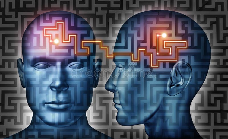
Free with trial Communication solutions and mind control with a group of communicating human heads on a labyrinth or maze pattern with a laser light connection the thinking network of two brains. Two neurons illustrations Communication Solutions
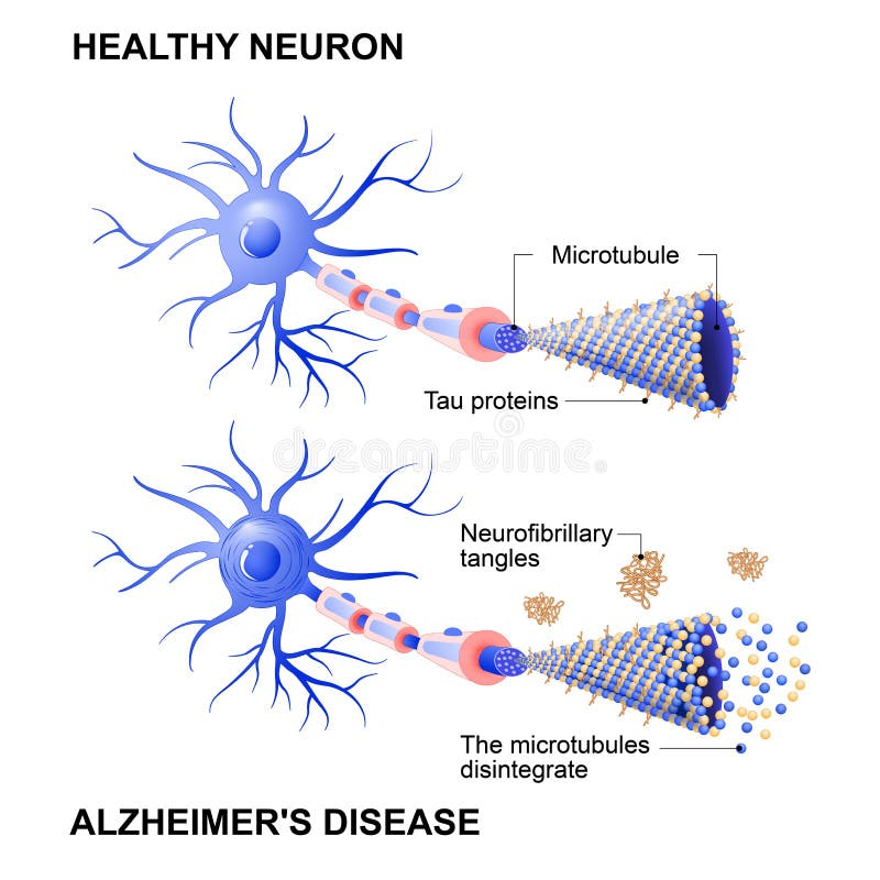
Free with trial Alzheimer`s disease is the change in tau protein that results in the breakdown of microtubules in brain cells. Mechanism of disease. Diagram shows two neurons: healthy cell and neuron with Alzheimer`s disease. Tau hypothesis. Neurofibrillary tangles. Two neurons vectors Healthy cell and neurons with Alzheimer`s disease. Tau hypothesi. Alzheimer`s disease is the change in tau protein that results in the breakdown of microtubules in brain cells. Mechanism of disease. Diagram shows two neurons: healthy cell and neuron with Alzheimer`s disease. Tau hypothesis. Neurofibrillary tangles
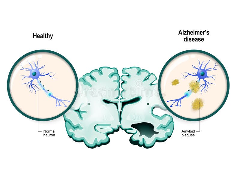
Free with trial Human brain, in two halves: healthy and Alzheimer`s disease. Healthy neuron and neuron with amyloid plaques. in comparison. Two neurons vectors Alzheimer`s disease. Neurons and brain. Human brain, in two halves: healthy and Alzheimer`s disease. Healthy neuron and neuron with amyloid plaques. in comparison
Free with trial Neural communication. Transmission of the nerve signal between two neurons with axon and synapse. Close-up of a chemical synapse. vector diagram for education, medical, science use. Two neurons vectors Transmission of the nerve signal between two neurons with axon and synapse. Close-up of a chemical synapse
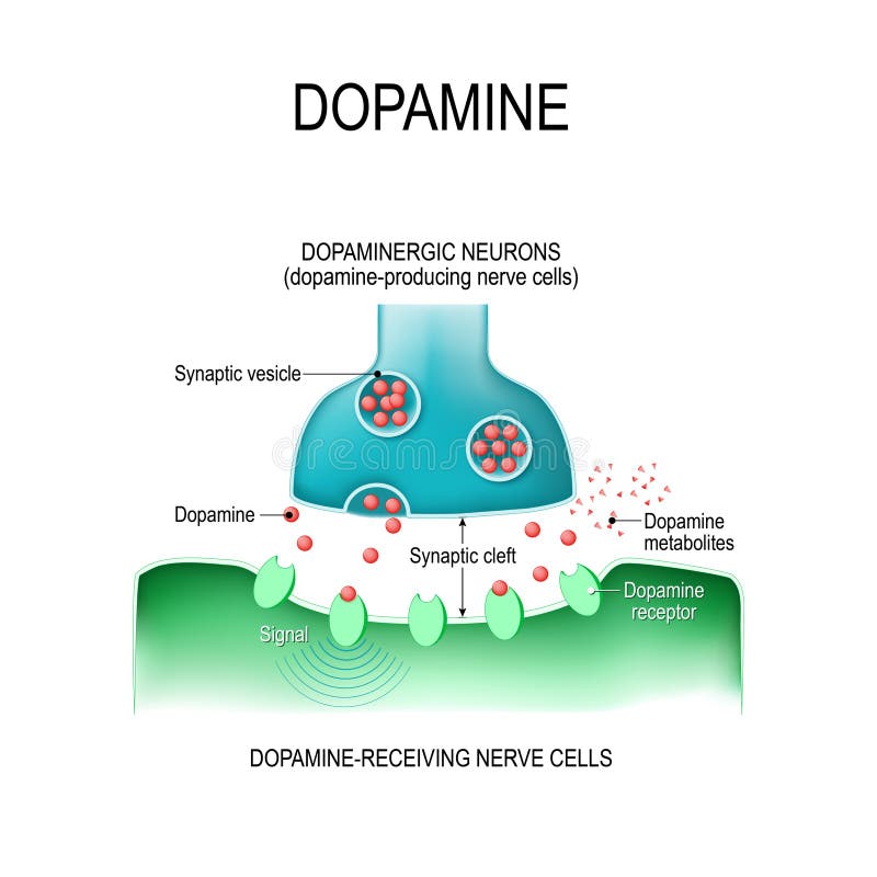
Free with trial Dopamine. two neurons dopamine-producing and dopamine-receiving nerve cells, receptors, and synaptic cleft with dopamine. Two neurons vectors Dopamine. two neurons with receptors, and synaptic cleft with d. Dopamine. two neurons dopamine-producing and dopamine-receiving nerve cells, receptors, and synaptic cleft with dopamine.
Free with trial Three dimensional illustration of synapse between two neurons. Two neurons illustrations Neuronal synapse. Three dimensional illustration of synapse between two neurons.
Free with trial Nervous tissue is the main component of the two parts of the nervous system; the brain and spinal cord of the central nervous system (CNS), and the branching peripheral nerves of the peripheral nervous system (PNS), which regulates and controls bodily functions and activity. Two neurons illustrations Neural Tissue. Nervous tissue is the main component of the two parts of the nervous system; the brain and spinal cord of the central nervous system (CNS), and the branching peripheral nerves of the peripheral nervous system (PNS), which regulates and controls bodily functions and activity.
Free with trial Synapse between two neurons neural synapse receptors neuron link neural network 3d render. Two neurons illustrations Synapse between two neurons neural synapse receptors neuron link neural network
Free with trial Neural synapse, electric and chemical signals between two neurons, human nervous system. Futuristic low polygonal design vector illustration. Two neurons vectors Neural synapse, electric and chemical signals between two neurons, human nervous system.
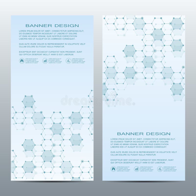
Free with trial Two scientific banners. Molecular structure of DNA and neurons. Geometric abstract background. Medicine, science, technology, business and website templates. Vector illustration. Two neurons vectors Two scientific banners. Molecular structure of DNA and neurons. Geometric abstract background. Medicine, science
Free with trial Nervous tissue is the main component of the two parts of the nervous system; the brain and spinal cord of the central nervous system (CNS), and the branching peripheral nerves of the peripheral nervous system (PNS),. Two neurons illustrations Neural Tissue. Nervous tissue is the main component of the two parts of the nervous system; the brain and spinal cord of the central nervous system (CNS), and the branching peripheral nerves of the peripheral nervous system (PNS),
Free with trial Dopamine and Antipsychotics. Neuroleptics or tranquilizers are medications that use to manage psychosis hallucinations, paranoia, schizophrenia, bipolar disorder. Neuroleptics blocks the ability of dopamine to activate receptor. two neurons, receptors, and synaptic cleft with dopamine and Neuroleptic. Two neurons vectors Dopamine and Antipsychotic
Free with trial Synapse between two neurons neural synapse receptors neuron link neural network 3d render. Two neurons illustrations Synapse between two neurons neural synapse receptors neuron link neural network
Free with trial Synapse between two neurons neural synapse receptors neuron link neural network 3d render. Two neurons illustrations Synapse between two neurons neural synapse receptors neuron link neural network
Free with trial Synapse between two neurons neural synapse receptors neuron link neural network 3d render. Two neurons illustrations Synapse between two neurons neural synapse receptors neuron link neural network
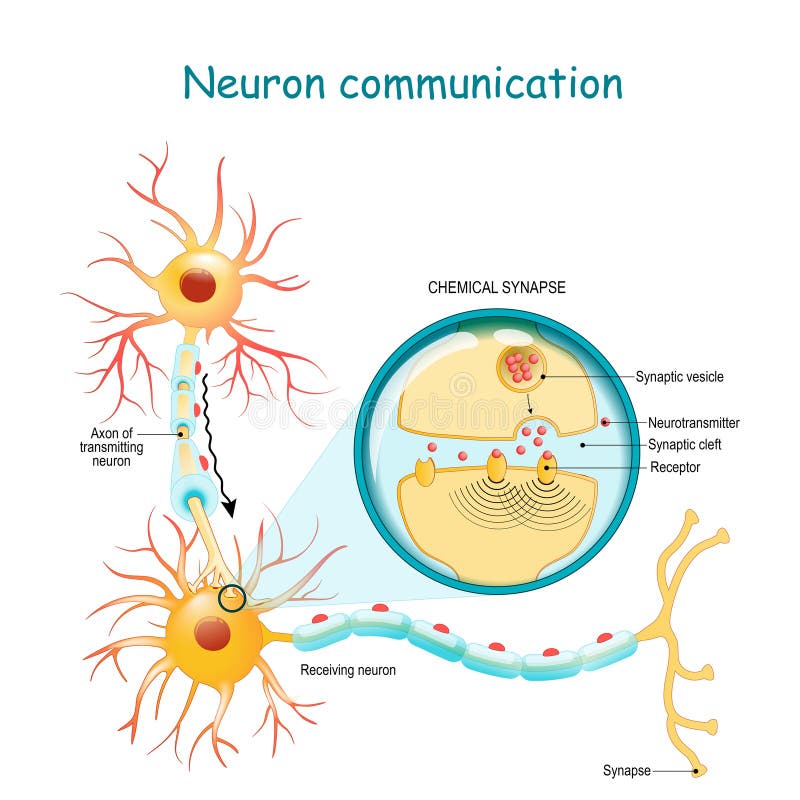
Free with trial Neuronal communication. Transmission of the nerve signal between two neurons with axon and synapse. Neural network. Neuron anatomy. Two neurons vectors Transmission of the nerve signal between two neurons with axon and synapse
Free with trial Abstract image of two pink and white vertical neurons over blue background with nervous cells. Concept of science and medicine. 3d rendering. Two neurons illustrations Two neurons close up. Abstract image of two pink and white vertical neurons over blue background with nervous cells. Concept of science and medicine. 3d rendering
Free with trial Cannabinoid. and Endocannabinoid system. pharmacological effects of cannabis. Two neurons with receptors for keys cannabinoid and endocannabinoid. neurotransmitter in synaptic cleft. Two neurons vectors Effects of cannabis. Two neurons with receptors and neurotransmitter. Cannabinoid. and Endocannabinoid system. pharmacological effects of cannabis. Two neurons with receptors for keys cannabinoid and endocannabinoid. neurotransmitter in synaptic cleft
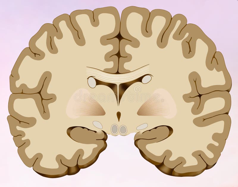
Free with trial Coronal section of the human brain in which we can see the brain composed of two halves, one right and one left, in this illustration we can distinguish the main commissure that unites both hemispheres: the Corpus Callosum, we see the Inter-hemispheric Cisura, the Ventricles Laterals, drawing the typical image on butterfly wings. Caudate Nucleus, laterally, to the Lenticular Nucleus, Internal Capsule, Silvio Cisura, Lobe of the insula. Two neurons illustrations Coronal cut of the human brain in which we can see the brain composed of two halves, one right and one left, in this illustration. Coronal section of the human brain in which we can see the brain composed of two halves, one right and one left, in this illustration we can distinguish the main commissure that unites both hemispheres: the Corpus Callosum, we see the Inter-hemispheric Cisura, the Ventricles Laterals, drawing the typical image on butterfly wings. Caudate Nucleus, laterally, to the Lenticular Nucleus, Internal Capsule, Silvio Cisura, Lobe of the insula.
Free with trial A detailed illustration showcasing the connection between the human brain and the digestive system, highlighting the neural network that links the two, set against a soft gradient background to emphasize the intricate relationship. The brain is depicted at the top with branching neurons extending downward, connecting to the intestines below, symbolizing the gut-brain axis and its role in overall. Two neurons illustrations Illustration of Brain and Gut Connection with Neural Network. A detailed illustration showcasing the connection between the human brain and the digestive system, highlighting the neural network that links the two, set against a soft gradient background to emphasize the intricate relationship. The brain is depicted at the top with branching neurons extending downward, connecting to the intestines below, symbolizing the gut-brain axis and its role in overall
Free with trial Two scientific banners. Molecular structure of DNA and neurons. Geometric abstract background. Medicine, science, technology, business and website templates. Vector illustration. Two neurons vectors Two scientific banners. Molecular structure of DNA and neurons. Geometric abstract background. Medicine, science
Free with trial Two scientific banner. Molecular structure of DNA and neurons. Geometric abstract background. Medicine, science, technology, business and website templates. Vector illustration. Two neurons vectors Two scientific banner. Molecular structure of DNA and neurons. Geometric abstract background. Medicine, science
Free with trial Two brain cells communicating. Neuroscience, medical concept digital render. Two neurons illustrations Two brain cells communicating. Neuroscience, medical concept digital render
Free with trial Two of modern vertical scientific banners. Molecular structure of DNA and neurons. Geometric abstract background. Medicine, science, technology, business and website templates. Vector illustration. Two neurons vectors Two of modern vertical scientific banners. Molecular structure of DNA and neurons. Geometric abstract background
Free with trial Nervous tissue is the main component of the two parts of the nervous system; the brain and spinal cord of the central nervous system (CNS), and the branching peripheral nerves of the peripheral nervous system (PNS),. Two neurons illustrations Neural Tissue. Nervous tissue is the main component of the two parts of the nervous system; the brain and spinal cord of the central nervous system (CNS), and the branching peripheral nerves of the peripheral nervous system (PNS),
Free with trial Neurons communication. Transmission of the nerve signal between two neurons. Close-up of a Synaptic cleft, chemical synapse, synaptic vesicle, Neurotransmitter. Vector illustration. Two neurons vectors Close-up of a Synaptic cleft, chemical synapse, synaptic vesicle, Neurotransmitter
Free with trial Artistic yellow gold colored neurons in the brain illustration on black background. Selective focus used. Two neurons illustrations Artistic neurons in the brain on black background. Artistic yellow gold colored neurons in the brain illustration on black background. Selective focus used.
Free with trial Amygdala in the brain, and closeup view of amygdala neurons, 3D illustration. Two almond-shaped clusters of nuclei within temporal lobes, part of the limbic system, play role in memory and emotions. Two neurons illustrations Amygdala in the brain, and closeup view of amygdala neurons, 3D illustration
Free with trial Amygdala in the brain, and closeup view of amygdala neurons, 3D illustration. Two almond-shaped clusters of nuclei within temporal lobes, part of the limbic system, play role in memory and emotions. Two neurons illustrations Amygdala in the brain, and closeup view of amygdala neurons, 3D illustration
Free with trial Illustration of two neurons communicating across a synapse. Neurotransmitters carry signals between the nerve cells. This image represents the nervous system and neural communication in the brain. Two neurons illustrations Neurons communicating with neurotransmitters across a synapse connection. Illustration of two neurons communicating across a synapse. Neurotransmitters carry signals between the nerve cells. This image represents the nervous system and neural communication in the brain
Free with trial Illustration of a red brain with two neurons connected to it, set against a light blue background. Simple, clean, and modern design. Two neurons vectors Brain and Neurons Illustration in Red on Light Blue Background. Illustration of a red brain with two neurons connected to it, set against a light blue background. Simple, clean, and modern design
Free with trial Two spiritual human bodies on dark background. Souls connection. Created with Generative AI. Two neurons illustrations Two human soul silhouettes on dark background. Generative AI. Two spiritual human bodies on dark background. Souls connection. Created with Generative AI
Free with trial The cell bodies of the three neurons in a typical somatosensory pathway are located in the dorsal root ganglion, the spinal cord, and the thalamus. A major target of somatosensory pathways is the postcentral gyrus in the parietal lobe of the cerebral cortex. Two neurons illustrations Somatosensory association area of human brain. The cell bodies of the three neurons in a typical somatosensory pathway are located in the dorsal root ganglion, the spinal cord, and the thalamus. A major target of somatosensory pathways is the postcentral gyrus in the parietal lobe of the cerebral cortex.
Free with trial Dendritic spine. Neuron anatomy. Learning and memory. Neuronal plasticity. Neural communication. Transmission of the nerve signal between two neurons with axon and synapse. Vector illustration. Two neurons vectors Dendritic spine. Neuron anatomy. Learning and memory. Neuronal plasticity
Free with trial Amygdala in the brain, and closeup view of amygdala neurons, 3D illustration. Two almond-shaped clusters of nuclei within temporal lobes, part of the limbic system, play role in memory and emotions. Two neurons illustrations Amygdala, also known as corpus amygdaloideum, in the brain. Amygdala in the brain, and closeup view of amygdala neurons, 3D illustration. Two almond-shaped clusters of nuclei within temporal lobes, part of the limbic system, play role in memory and emotions
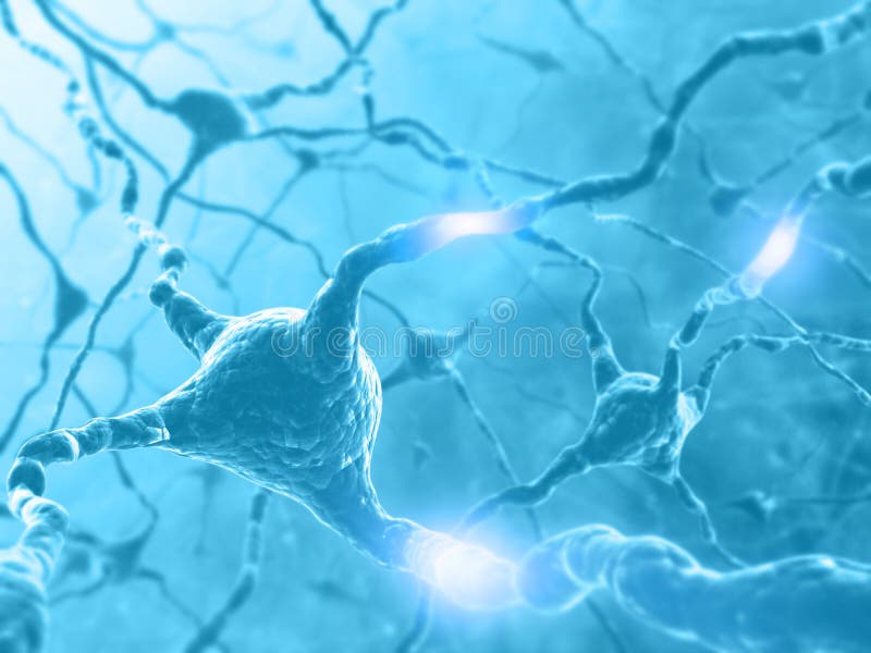
Free with trial Gray neuron with signals impulses 3d render. Two neurons illustrations Two neurons with impulses 3d illustration. Gray neuron with signals impulses 3d render
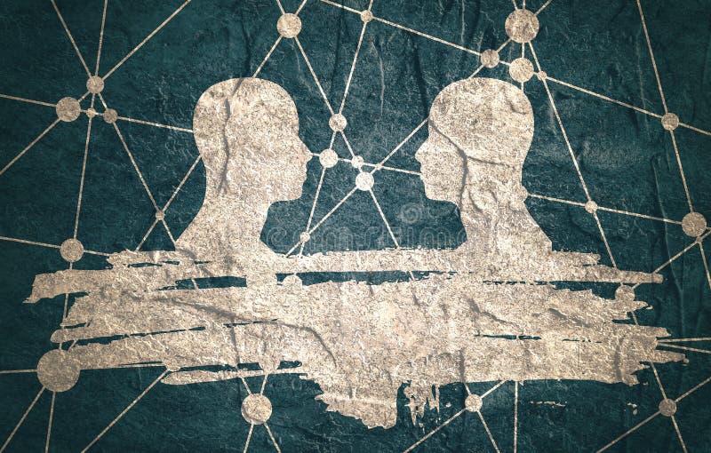
Free with trial Human Communication Background. Connected circles with dots. Two man silhouettes looking at each other. Grunge brush stroke. Abstract business meeting. Social network. Concrete textured. Two neurons illustrations Molecule And Communication Background. Illustration. Human Communication Background.Connected circles with dots. Two man silhouettes looking at each other. Grunge brush stroke. Abstract business meeting. Social network. Concrete textured.
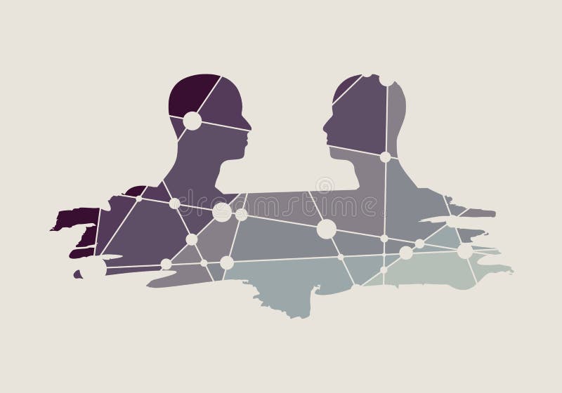
Free with trial Human communication background. Brochure or web banner template. Connected circles with dots. Two man silhouettes looking at each other. Grunge brush stroke. Abstract business meeting. Social network. Two neurons vectors Molecule And Communication Background. Human communication background. Brochure or web banner template. Connected circles with dots. Two man silhouettes looking at each other. Grunge brush stroke. Abstract business meeting. Social network
Free with trial Amygdala in the brain, and closeup view of amygdala neurons, 3D illustration. Two almond-shaped clusters of nuclei within temporal lobes, part of the limbic system, play role in memory and emotions. Two neurons illustrations Amygdala, also known as corpus amygdaloideum, in the human brain. Amygdala in the brain, and closeup view of amygdala neurons, 3D illustration. Two almond-shaped clusters of nuclei within temporal lobes, part of the limbic system, play role in memory and emotions
Free with trial The human brain and telepathy. AI generated. Two neurons illustrations The human brain and telepathy. AI generated
Free with trial Antique engraved illustration of the earthworm. Vintage illustration of the dew worm. Old engraved picture. Book illustration published 1907. A: worm, B: front end (view from above), C: front end (side view), D: earthworm (view from below), E: the seta/cheata of the dew worm. An earthworm is a terrestrial invertebrate that belongs to the phylum Annelida. They exhibit a tube-within-a-tube body plan they are externally segmented with corresponding internal segmentation and they usually have setae on all segments. They occur worldwide where soil, water, and temperature allow. Earthworms are commonly found in soil, eating a wide variety of organic matter. This organic matter includes plant matter, living protozoa, rotifers, nematodes, bacteria, fungi, and other microorganisms. An earthworm's digestive system runs the length of its body. An earthworm respires through its skin. It has a double transport system made of coelomic fluid that moves within the fluid-filled coelom and a simple, closed circulatory system. It has a central and peripheral nervous system. Its central nervous system consists of two ganglia above the mouth, one on either side, connected to a nerve running along its length to motor neurons and sensory cells in each segment. Large numbers of chemoreceptors concentrate near its mouth. Two neurons illustrations Antique engraved illustration of the earthworm. Vintage illustration of the dew worm. Old engraved picture. Book
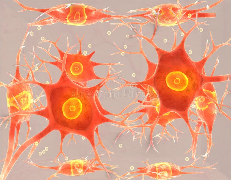
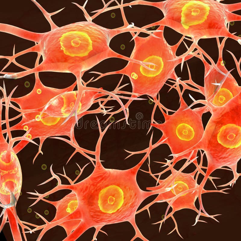
Free with trial This image shows a close-up view of two neurons, which are the basic working units of the brain, connected by a network of fibers. The neurons are depicted in a vibrant pink color, with numerous thin, hair-like structures extending from their surfaces. Two neurons illustrations Microscopic view of two neurons connected by a network of fibers. This image shows a close-up view of two neurons, which are the basic working units of the brain, connected by a network of fibers. The neurons are depicted in a vibrant pink color, with numerous thin, hair-like structures extending from their surfaces
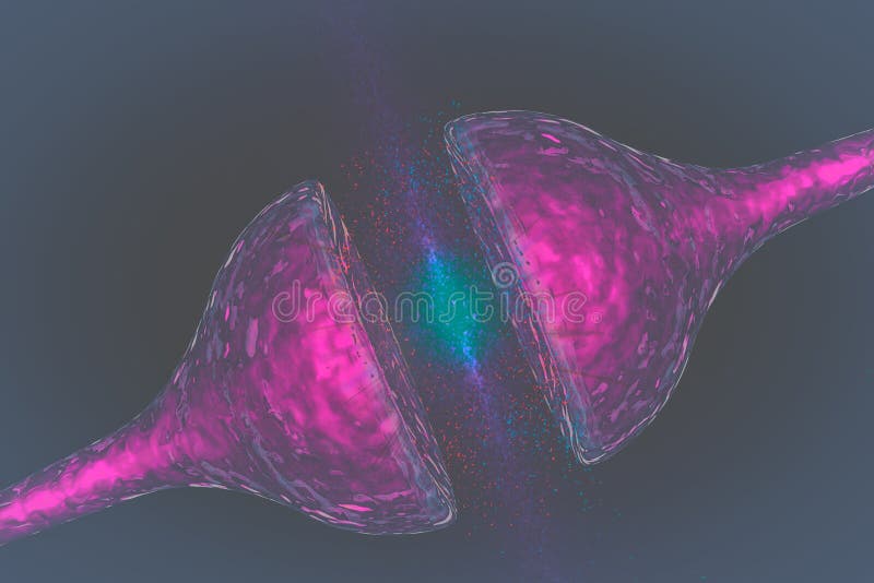
Free with trial This image depicts two neurons connected by a synapse, illustrating the junction where nerve impulses are transmitted from one neuron to another. The orange and yellow structures represent the synaptic terminal and vesicles containing neurotransmitters, while the branching structures are dendrites and axons. This visualization emphasizes the complexity and connectivity of the nervous system at a. Two neurons illustrations Two interconnected neurons highlighting the synapse between them. This image depicts two neurons connected by a synapse, illustrating the junction where nerve impulses are transmitted from one neuron to another. The orange and yellow structures represent the synaptic terminal and vesicles containing neurotransmitters, while the branching structures are dendrites and axons. This visualization emphasizes the complexity and connectivity of the nervous system at a
Free with trial This image shows a detailed scientific illustration of two neurons. The neurons are depicted with their soma (cell body), dendrites (branched projections), and axon (long projection extending from the soma). The intricate network of dendrites is visible, highlighting the complexity of neuronal connections in the nervous system. The image is likely used for educational or anatomical reference. Two neurons illustrations Illustration of two neurons with detailed dendritic and axonal structures. This image shows a detailed scientific illustration of two neurons. The neurons are depicted with their soma (cell body), dendrites (branched projections), and axon (long projection extending from the soma). The intricate network of dendrites is visible, highlighting the complexity of neuronal connections in the nervous system. The image is likely used for educational or anatomical reference
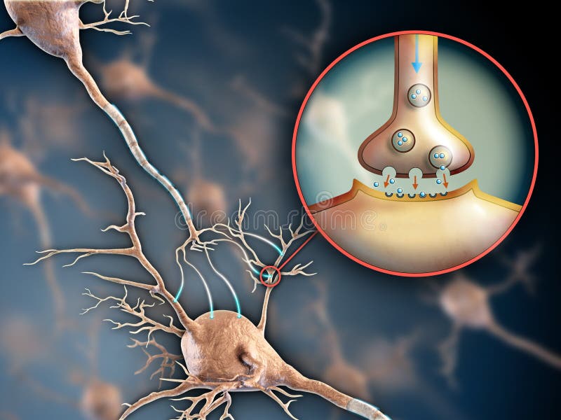
Free with trial The image illustrates the process of synaptic transmission between two neurons, highlighting the presynaptic neuron, postsynaptic neuron, and the synaptic cleft. It shows the release of neurotransmitters from synaptic vesicles into the synaptic cleft, which then bind to receptors on the postsynaptic neuron. The diagram also labels key components such as the nucleus, cell body, dendrites, axon,. Two neurons illustrations Diagram of synapse communication between two neurons in the nervous system. The image illustrates the process of synaptic transmission between two neurons, highlighting the presynaptic neuron, postsynaptic neuron, and the synaptic cleft. It shows the release of neurotransmitters from synaptic vesicles into the synaptic cleft, which then bind to receptors on the postsynaptic neuron. The diagram also labels key components such as the nucleus, cell body, dendrites, axon,
Free with trial Vibrant illustration of two neurons with extensive dendrites interconnected against a dark backdrop, symbolizing neural communication and connection in the human brain for educational or medical purposes. Two neurons illustrations Colorful illustration of two interconnected neurons on black background in medical concept. Vibrant illustration of two neurons with extensive dendrites. Vibrant illustration of two neurons with extensive dendrites interconnected against a dark backdrop, symbolizing neural communication and connection in the human brain for educational or medical purposes
Free with trial Degenerative Brain Cell Concept. a brain cell and the loss of synaptic communication. a synapse between two neurons, showing the plaques typical of Alzheimer that block signals in the brain. Two neurons illustrations Degenerative Brain Cell Concept. a brain cell and the loss of synaptic communication. a synapse between two neurons, showing the
Free with trial Two neurons connecting at a synapse. Electricity flows between them, illustrating how signals travel in the brain. The design highlights neural communication. Two neurons illustrations High tech visualization shows synaptic transmission between two neurons meeting at a synapse in a digital representation. Two neurons connecting at a synapse. Electricity flows between them, illustrating how signals travel in the brain. The design highlights neural communication
Free with trial Two abstract neurons with vibrant purple and blue glowing branches and bodies are depicted against a soft pink and blue gradient background. Two neurons illustrations Two interconnected abstract neurons with purple and blue glowing dendrites and cell bodies against a gradient background. Two abstract neurons with vibrant purple and blue glowing branches and bodies are depicted against a soft pink and blue gradient background
Free with trial Microscopic image of two neurons connecting, on black background. Concept of biology, science and research. Vertical. Two neurons illustrations Microscopic image of two neurons connecting, on black background. Concept of biology, science and research
Free with trial Stylized illustration of two interconnected neurons, showcasing elaborate dendritic branches. The neurons feature large, circular cell bodies with prominent, abstract patterns within. The black and white color scheme emphasizes neural complexity and structure. Branches extend outward, intertwining to form an intricate network, resembling a biological neural pathway. The image captures the complex architecture of a neural network, highlighting the interconnectedness essential for signal transmission in the nervous system. Two neurons illustrations Stylized illustration of two interconnected neurons, showcasing elaborate dendri
Free with trial Two glowing neurons connected by a glowing axon. stock photo concept. Two neurons illustrations Two glowing neurons connected by a glowing axon
Free with trial Two neurons in a blue and green color scheme Design Background for Instagram, Facebook wall painting AI Generative green, background art, background image, AI Generative, background ai, ai wallpaper, color, ai, wallpaper, background photo, wall painting, Generative, scheme, Two, blue, ai background, and, background, neurons, backgrounds. Two neurons illustrations Two neurons in a blue and green color scheme AI Generative. Two neurons in a blue and green color scheme Design Background for Instagram, Facebook wall painting AI Generative green, background art, background image, AI Generative, background ai, ai wallpaper, color, ai, wallpaper, background photo, wall painting, Generative, scheme, Two, blue, ai background, and, background, neurons, backgrounds
Free with trial Two neurons in a blue and green color scheme Design Background for Instagram, Facebook wall painting AI Generative blue, neurons, Generative, wallpaper, background, background image, background photo, backgrounds, background ai, green, and, color, AI Generative, ai background, wall painting, ai wallpaper, scheme, background art, Two, ai. Two neurons illustrations Two neurons in a blue and green color scheme AI Generative. Two neurons in a blue and green color scheme Design Background for Instagram, Facebook wall painting AI Generative blue, neurons, Generative, wallpaper, background, background image, background photo, backgrounds, background ai, green, and, color, AI Generative, ai background, wall painting, ai wallpaper, scheme, background art, Two, ai
Free with trial Two glowing red cells with pink tendrils, resembling neurons or a biological network, in a dark blue background. stock photo concept. Two neurons illustrations Two glowing red cells with pink tendrils, resembling neurons or a biological network, in a dark blue background
Free with trial Detailed close-up of two red neurons with intricate synaptic connections, showcasing their structure and network on a dark background. Two neurons illustrations Close-up of two red neurons with interconnected synapses on a dark background. Detailed close-up of two red neurons with intricate synaptic connections, showcasing their structure and network on a dark background
Free with trial Two Glowing Neurons Illustrating Brain Activity and Connectivity in Neuroscience Concepts, Generated by AI. Two neurons illustrations Two Glowing Neurons Illustrating Brain Activity and Connectivity in Neuroscience Concepts
Free with trial Illustration depicting a synaptic transmission between neurons. Two neuron endings (synaptic knobs) are shown in close proximity, with a vivid, bright blue electric-like discharge representing the synaptic transmission process. The background displays a dark, space-like environment with scattered points of light, highlighting the dynamic exchange. The synaptic knobs are colored in shades of red and blue, emphasizing their activity. This visual represents the intricate process of communication within the nervous system. Two neurons illustrations Illustration depicting a synaptic transmission between neurons