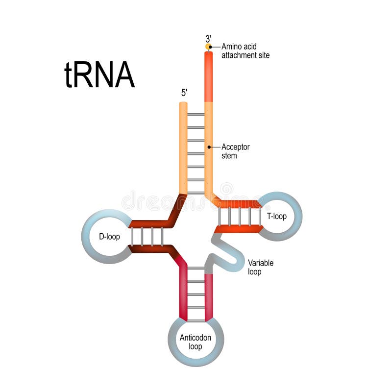
Free with trial Transfer RNA tRNA. Structure of Transfer RNA tRNA. tRNAs are a component of synthesis of new proteins in accordance with the genetic code. Variable binding vectors Transfer RNA tRNA. Structure of Transfer RNA tRNA. tRNAs are a component of synthesis of new proteins in accordance with the genetic code
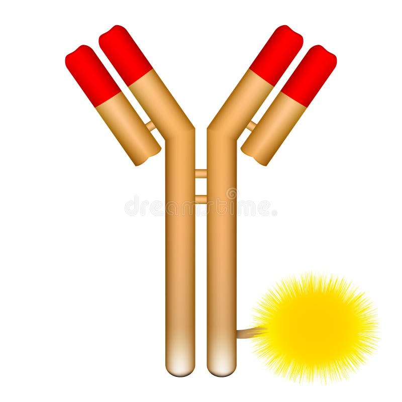
Free with trial Antibody molecule conjugated with fluorophore. Vector illustration of antibody molecule with conjugated fluorophore. Variable binding illustrations Antibody molecule conjugated with fluorophore. Vector illustration of antibody molecule with conjugated fluorophore
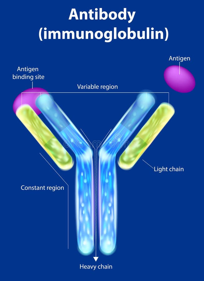
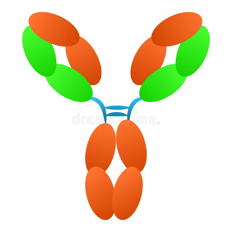
Free with trial The structure of the antibody. Immunoglobulin. Antibody binds to a specific antigen. Variable binding vectors The structure of the antibody. Immunoglobulin. Antibody binds to a specific antigen
Free with trial Antibody molecule cell vector / Antigen. On white. Variable binding vectors Antibody molecule cell vector / Antigen. On white
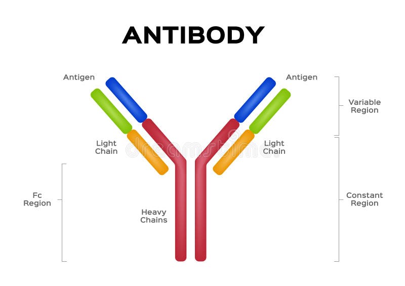
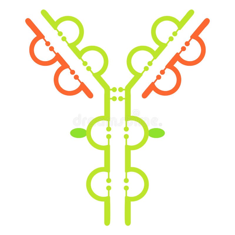
Free with trial Antibody immunoglobulin molecule structure. Simple scheme of structure of immunoglobulin IgG antibody molecule vector. Variable binding vectors Antibody immunoglobulin molecule structure. Simple scheme of structure of immunoglobulin IgG antibody molecule vector
Free with trial Antibody molecule floats in water. Binds only specific target. Variable binding vectors Antibody molecule floats in water. Binds only specific target
Free with trial Antibody logo. Antibody, immunoglobulin IgG antibody molecule logo vector. Variable binding vectors Antibody logo. Antibody, immunoglobulin IgG antibody molecule logo vector
Free with trial Antibody Structure. Immunoglobulin anatomy. Antibody anatomy. Immunoglobulin Structure. Adaptive immune response. Vector poster. Illustration in flat style. Variable binding vectors Antibody Structure. Immunoglobulin anatomy. Antibody anatomy. Immunoglobulin Structure. Adaptive immune response. Vector poster. Illustration in flat style
Free with trial Antibodies Word Cloud Concept with great terms such as genes, bacteria, system immunie, plasma, proteins, genetics and more. Variable binding illustrations Antibodies Word Cloud Concept. With great terms such as genes, bacteria, system immunie, plasma, proteins, genetics and more
Free with trial Antibody immunoglobulin molecule structure. Structure of immunoglobulin IgG antibody molecule vector. Variable binding vectors Antibody immunoglobulin molecule structure. Structure of immunoglobulin IgG antibody molecule vector
Free with trial Antibodies Word Cloud Concept on a Chalkboard with great terms such as bacteria, system immunie, plasma, proteins, genetics and more. Variable binding illustrations Antibodies Word Cloud Concept on a Blackboard. Antibodies Word Cloud Concept on a Chalkboard with great terms such as bacteria, system immunie, plasma, proteins. Antibodies Word Cloud Concept on a Chalkboard with great terms such as bacteria, system immunie, plasma, proteins, genetics and more.
Free with trial Mortgage Word Cloud. On White Background. Variable binding illustrations Mortgage Word Cloud. On White Background
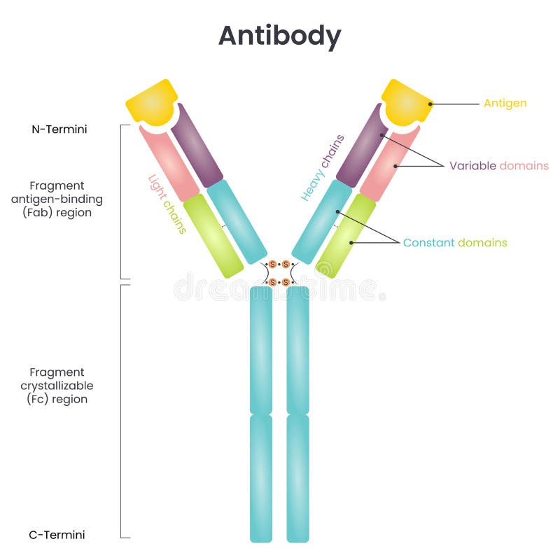
Free with trial Antibody structure vector diagram. Vector illustration diagram infographic an the typical structure of an antibody. Variable binding vectors Antibody structure vector diagram. Vector illustration diagram infographic an the typical structure of an antibody
Free with trial Antibody Fragment scFv, labeled diagram, educational immunology poster design, cloning of single-chain variable fragment, IgG comparison, Fab region, VH, VL, CH1, CL, linker, recombinant protein. Variable binding vectors Antibody Fragment scFv, labeled diagram, educational immunology poster design, cloning of single-chain variable fragment, IgG. Comparison, Fab region, VH, VL. Antibody Fragment scFv, labeled diagram, educational immunology poster design, cloning of single-chain variable fragment, IgG comparison, Fab region, VH, VL, CH1, CL, linker, recombinant protein
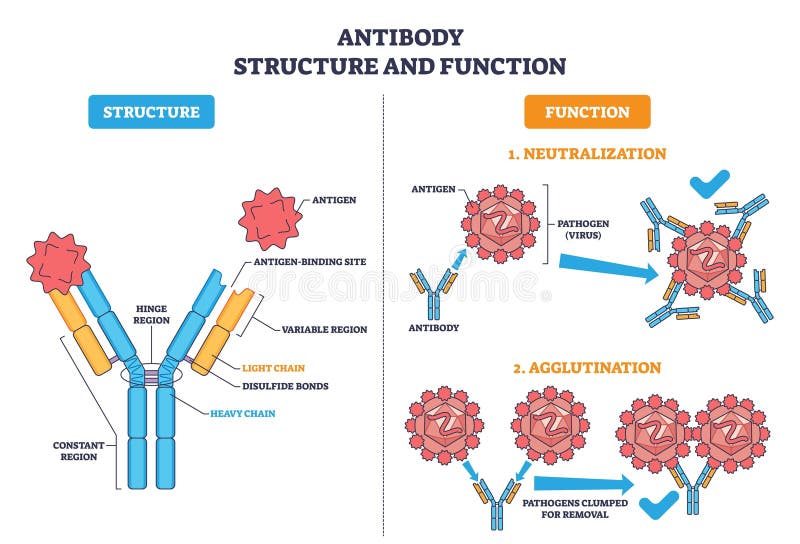
Free with trial Antibody structure and function, Y shaped immunoglobulin binds antigens to neutralize and agglutinate pathogens, key parts include heavy-light chains and variable regions. Outline diagram. Variable binding vectors Antibody structure and function, Y shaped immunoglobulin binds antigens to ... Antibody structure and function, Y shaped immunoglobulin binds antigens to. Antibody structure and function, Y shaped immunoglobulin binds antigens to neutralize and agglutinate pathogens, key parts include heavy-light chains and variable regions. Outline diagram
Free with trial Antibody explained, Y-shaped protein binds antigen to neutralize a virus, showing structure and binding sites. Key objects, antibody, antigen, pathogen. Outline diagram. Variable binding vectors Antibody explained, Y-shaped protein binds antigen to neutralize a. Virus, showing structure and binding sites. Key objects, antibody, antigen, pathogen
Free with trial Scientific educational showing the basic structure of an antibody, including heavy chains, light chains, variable regions, and constant regions. Clear molecular diagram suitable for immunology, microbiology, molecular biology, medical education, biomedical research, healthcare publications, academic textbooks, and scientific presentations. Variable binding vectors Antibody Structure Scientific Medical Illustration. Scientific educational showing the basic structure of an antibody, including heavy chains, light chains. Scientific educational showing the basic structure of an antibody, including heavy chains, light chains, variable regions, and constant regions. Clear molecular diagram suitable for immunology, microbiology, molecular biology, medical education, biomedical research, healthcare publications, academic textbooks, and scientific presentations.
Free with trial Scientific educational showing the basic structure of an antibody, including heavy chains, light chains, variable regions, and constant regions. Clear molecular diagram suitable for immunology, microbiology, molecular biology, medical education, biomedical research, healthcare publications, academic textbooks, and scientific presentations. Variable binding vectors Antibody Structure Scientific Medical Illustration. Scientific educational showing the basic structure of an antibody, including heavy chains, light chains. Scientific educational showing the basic structure of an antibody, including heavy chains, light chains, variable regions, and constant regions. Clear molecular diagram suitable for immunology, microbiology, molecular biology, medical education, biomedical research, healthcare publications, academic textbooks, and scientific presentations.
Free with trial Scientific educational showing the basic structure of an antibody, including heavy chains, light chains, variable regions, and constant regions. Clear molecular diagram suitable for immunology, microbiology, molecular biology, medical education, biomedical research, healthcare publications, academic textbooks, and scientific presentations. Variable binding vectors Antibody Structure Scientific Medical Illustration. Scientific educational showing the basic structure of an antibody, including heavy chains, light chains. Scientific educational showing the basic structure of an antibody, including heavy chains, light chains, variable regions, and constant regions. Clear molecular diagram suitable for immunology, microbiology, molecular biology, medical education, biomedical research, healthcare publications, academic textbooks, and scientific presentations.
Free with trial Scientific educational showing the basic structure of an antibody, including heavy chains, light chains, variable regions, and constant regions. Clear molecular diagram suitable for immunology, microbiology, molecular biology, medical education, biomedical research, healthcare publications, academic textbooks, and scientific presentations. Variable binding vectors Antibody Structure Scientific Medical Illustration. Scientific educational showing the basic structure of an antibody, including heavy chains, light chains. Scientific educational showing the basic structure of an antibody, including heavy chains, light chains, variable regions, and constant regions. Clear molecular diagram suitable for immunology, microbiology, molecular biology, medical education, biomedical research, healthcare publications, academic textbooks, and scientific presentations.
Free with trial This visual explains function application in lambda calculus through structure rather than text. The upper left box represents an abstraction: a lambda marker points to a dashed input slot, and the body beside it holds two dashed placeholders joined by a single arc, showing they are the same bound variable, with a square standing for a constant term. To the right, a separate box contains the argument, a solid hexagon matching those shapes exactly. Placing the two boxes side by side is the notation for applying one to the other, and a bracket underneath groups them into a single expression. A downward arrow marks the substitution step known as beta reduction. In the result box below, every bound position has been filled with the argument while the constant stays untouched, and the binder, arc and marker have disappeared because the function has been consumed. Suitable for computer science courses, theory of computation lectures, functional programming tutorials, Haskell or Lisp explainers, logic and foundations slides, and edtech content on how programs evaluate expressions. Clean minimalist outline style, single stroke weight, no labels or numerals, scalable for print and screen. Variable binding vectors Lambda Calculus Visual - function box applied to an argument. This visual explains function application in lambda calculus through structure rather than text. This visual explains function application in lambda calculus through structure rather than text. The upper left box represents an abstraction: a lambda marker points to a dashed input slot, and the body beside it holds two dashed placeholders joined by a single arc, showing they are the same bound variable, with a square standing for a constant term. To the right, a separate box contains the argument, a solid hexagon matching those shapes exactly. Placing the two boxes side by side is the notation for applying one to the other, and a bracket underneath groups them into a single expression. A downward arrow marks the substitution step known as beta reduction. In the result box below, every bound position has been filled with the argument while the constant stays untouched, and the binder, arc and marker have disappeared because the function has been consumed. Suitable for computer science courses, theory of computation lectures, functional programming tutorials, Haskell or Lisp explainers, logic and foundations slides, and edtech content on how programs evaluate expressions. Clean minimalist outline style, single stroke weight, no labels or numerals, scalable for print and screen.
Free with trial Antibodies Affinity Reagents, labeled diagram, educational immunology poster design, IgG, Fab fragment, scFv, nanobody, antibody fragments, antigen binding, recombinant protein illustration. Variable binding vectors Antibodies Affinity Reagents, labeled diagram, educational immunology poster design, IgG, Fab fragment, scFv, nanobody, antibody. Fragments, antigen binding. Antibodies Affinity Reagents, labeled diagram, educational immunology poster design, IgG, Fab fragment, scFv, nanobody, antibody fragments, antigen binding, recombinant protein illustration
Free with trial Visualization medical hud style cross section of an antibody cell illustration structure protein, immune antigen, binding epitope visualization medical hud style cross section of an antibody cell. Variable binding illustrations Visualization medical hud style cross section of an antibody cell. Illustration structure protein, immune antigen, binding epitope AI generated. visualization medical hud style cross section of an antibody cell illustration structure protein, immune antigen, binding epitope visualization medical hud style cross section of an antibody cell
Free with trial Cell high contrast cross section of an antibody cell illustration structure epitope, paratope immunoglobulin, binding affinity cell high contrast cross section of an antibody cell. Variable binding illustrations Cell high contrast cross section of an antibody cell. Illustration structure epitope, paratope immunoglobulin, binding affinity AI generated. cell high contrast cross section of an antibody cell illustration structure epitope, paratope immunoglobulin, binding affinity cell high contrast cross section of an antibody cell
Free with trial Antibody structure diagram illustrating antigen binding site and constant heavy chains. Scientific illustration detailing the structure of an antibody molecule. Variable binding illustrations Antibody structure diagram illustrating antigen binding site and constant heavy chains. Scientific illustration detailing the structure of an antibody molecule
Free with trial This minimalist graphic presents a bold symbol featuring a knot tied around a prominent 'X' shape, rendered in stark black and white. Shown against both light and dark split backgrounds, the design is clean, simple, and impactful. The knot symbolizes connection, binding, or complexity, while the 'X' can represent intersection, a challenge, or a variable. This abstract mark is versatile and can be used for branding, logos, or conceptual representation in business, technology, or creative fields. Its strong, clear execution makes it memorable and professional, suitable for conveying ideas of linkage, solutions, or overcoming obstacles. Variable binding vectors A minimalist black and white graphic depicts a knot tied around an 'X' shape, shown on split backgrounds. This minimalist graphic presents a bold symbol. This minimalist graphic presents a bold symbol featuring a knot tied around a prominent 'X' shape, rendered in stark black and white. Shown against both light and dark split backgrounds, the design is clean, simple, and impactful. The knot symbolizes connection, binding, or complexity, while the 'X' can represent intersection, a challenge, or a variable. This abstract mark is versatile and can be used for branding, logos, or conceptual representation in business, technology, or creative fields. Its strong, clear execution makes it memorable and professional, suitable for conveying ideas of linkage, solutions, or overcoming obstacles.
Free with trial This image depicts the intricate structure of an antibody, showcasing its Y-shaped form. The antibody is color-coded to highlight its different regions: the variable domains are shown in green and pink, while the constant domains are depicted in blue. The image provides a detailed view of the antibody's molecular composition, emphasizing the complexity and specificity of its structure. Variable binding illustrations A detailed molecular structure of an antibody with colored regions representing different domains. This image depicts the intricate structure of an antibody. This image depicts the intricate structure of an antibody, showcasing its Y-shaped form. The antibody is color-coded to highlight its different regions: the variable domains are shown in green and pink, while the constant domains are depicted in blue. The image provides a detailed view of the antibody's molecular composition, emphasizing the complexity and specificity of its structure
Free with trial This captivating 3D molecular model showcases the intricate structure of an immunoglobulin antibody. The sharp, bright, and colorful rendering highlights the protein's key components, including the variable and constant regions, heavy and light chains, and the antigen-binding site. The precise molecular structure is clearly depicted, emphasizing the critical role of antibodies in the body's. Variable binding illustrations Detailed 3D Molecular Model of an Antibody Immunoglobulin A Visual Exploration of Immune System Structure and Function. This captivating 3D molecular model. This captivating 3D molecular model showcases the intricate structure of an immunoglobulin antibody. The sharp, bright, and colorful rendering highlights the protein's key components, including the variable and constant regions, heavy and light chains, and the antigen-binding site. The precise molecular structure is clearly depicted, emphasizing the critical role of antibodies in the body's
Free with trial Antigens vs antibody structure comparison diagram. Illustration of antigens vs antibody structure comparison diagram. Variable binding vectors Antigens vs antibody structure comparison diagram. Illustration of antigens vs antibody structure comparison diagram
Free with trial Vector Illustration of the Molecular Structure of Immunoglobulin Antibody with Fc Fragment. Variable binding vectors Molecular Structure of Immunoglobulin Antibody with Fc Fragment. Vector Illustration of the Molecular Structure of Immunoglobulin Antibody with Fc Fragment. Vector Illustration of the Molecular Structure of Immunoglobulin Antibody with Fc Fragment.
Free with trial Molecular diagram showing hiv or hepatitis c virus envelope glycoprotein quasispecies interacting with neutralizing antibodies, representing viral immune evasion and vaccine development. Variable binding illustrations Molecular diagram showing hiv or hepatitis c virus envelope glycoprotein quasispecies interacting with neutralizing antibodies. Representing viral immune. Molecular diagram showing hiv or hepatitis c virus envelope glycoprotein quasispecies interacting with neutralizing antibodies, representing viral immune evasion and vaccine development
Free with trial First order logic post it on blue. Background. Variable binding illustrations First order logic post it on blue. Background
Free with trial An antibody molecule, also known as immunoglobulin, is a key component of the immune system that recognizes and neutralizes foreign invaders like viruses and bacteria. generative ai. Variable binding vectors Antibody molecule isolated on white background, a key immune component, generative ai. An antibody molecule, also known as immunoglobulin, is a key component of. An antibody molecule, also known as immunoglobulin, is a key component of the immune system that recognizes and neutralizes foreign invaders like viruses and bacteria.generative ai
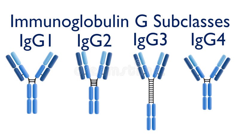
Free with trial 3D rendering of the four human IgG subclasses (IgG1, IgG2, IgG3, IgG4). Their distinct structures lead to varying immune response functions. Variable binding illustrations 3D rendering of the four human IgG subclasses (IgG1, IgG2, IgG3, IgG4). Their distinct structures lead to varying immune response functions
Free with trial Protein exploding view of antibody cell illustration antigen epitope, receptor immunology, monoclonal b protein exploding view of antibody cell. Variable binding illustrations Protein exploding view of antibody cell. Illustration antigen epitope, receptor immunology, monoclonal b AI generated. protein exploding view of antibody cell illustration antigen epitope, receptor immunology, monoclonal b protein exploding view of antibody cell
Free with trial 3D rendering of the four human IgG subclasses (IgG1, IgG2, IgG3, IgG4). Their distinct structures lead to varying immune response functions. Variable binding illustrations 3D rendering of the four human IgG subclasses (IgG1, IgG2, IgG3, IgG4). Their distinct structures lead to varying immune response functions
Free with trial 3D rendering of the four human IgG subclasses (IgG1, IgG2, IgG3, IgG4). Their distinct structures lead to varying immune response functions. Variable binding illustrations 3D rendering of the four human IgG subclasses (IgG1, IgG2, IgG3, IgG4). Their distinct structures lead to varying immune response functions
Free with trial 3D rendering of the four human IgG subclasses (IgG1, IgG2, IgG3, IgG4). Their distinct structures lead to varying immune response functions. Variable binding illustrations 3D rendering of the four human IgG subclasses (IgG1, IgG2, IgG3, IgG4). Their distinct structures lead to varying immune response functions
Free with trial 3D rendering of the four human IgG subclasses (IgG1, IgG2, IgG3, IgG4). Their distinct structures lead to varying immune response functions. Variable binding illustrations 3D rendering of the four human IgG subclasses (IgG1, IgG2, IgG3, IgG4). Their distinct structures lead to varying immune response functions
Free with trial A scientific diagram illustrating the structural components of an antibody molecule including light chains. Variable binding illustrations Antibody structure y-shaped immunoglobulin molecular biology diagram with y shaped, antibody. A scientific diagram illustrating the structural components of an. A scientific diagram illustrating the structural components of an antibody molecule including light chains
Free with trial Adult hands set a plain unmarked straightedge alongside the naturally wavy border of a loose rug on an inspection table. The tool rests on bare tabletop parallel to the bound edge with a narrow gap that varies along its length. The textile stays relaxed and flat. Even work light makes this top down comparison easy to read. The view keeps the existing curve and reference line visible together during an intake assessment that can inform suitable handling. This practical condition check precedes selection of an appropriate cleaning process. Variable binding illustrations Technician sets a straightedge beside a naturally wavy rug border during assessment before professional cleaning. Adult hands set a plain unmarked straightedge. Adult hands set a plain unmarked straightedge alongside the naturally wavy border of a loose rug on an inspection table. The tool rests on bare tabletop parallel to the bound edge with a narrow gap that varies along its length. The textile stays relaxed and flat. Even work light makes this top down comparison easy to read. The view keeps the existing curve and reference line visible together during an intake assessment that can inform suitable handling. This practical condition check precedes selection of an appropriate cleaning process.
Free with trial Scheme outline icons collection. Syntax, Evaluation, Procedure, Recursion, Macro, Closure, Variable vector and. Variable binding vectors Scheme line icons collection. Syntax, Evaluation, Procedure, Recursion, Macro, Closure, Variable vector and linear. Scheme outline icons collection. Syntax. Scheme outline icons collection. Syntax, Evaluation, Procedure, Recursion, Macro, Closure, Variable vector and
Free with trial Antibodies Word Cloud Concept in red caps with great terms such as bacteria, system immunie, plasma, proteins, genetics and more. Variable binding illustrations Antibodies Word Cloud Concept in Red Caps. With great terms such as bacteria, system immunie, plasma, proteins, genetics and more
Free with trial Mortgage Word Cloud. On Blue Background. Variable binding illustrations Mortgage Word Cloud. On Blue Background
Free with trial Inclined grid vector composition of small circles in variable sizes and color hues. Small circles are organized into inclined grid vector mosaic. Abstract vector illustration. Variable binding vectors Vector Inclined Grid Composition of Small Circles. Inclined grid vector composition of small circles in variable sizes and color hues. Small circles are. Inclined grid vector composition of small circles in variable sizes and color hues. Small circles are organized into inclined grid vector mosaic. Abstract vector illustration
Free with trial Square grid centers vector collage of small circles in variable sizes and color hues. Small circles are organized into square grid centers vector collage. Abstract vector illustration. Variable binding vectors Vector Square Grid Centers Composition of Dots. Square grid centers vector collage of small circles in variable sizes and color hues. Small circles are organized. Square grid centers vector collage of small circles in variable sizes and color hues. Small circles are organized into square grid centers vector collage. Abstract vector illustration.
Free with trial Shifted grid nodes vector mosaic of round dots in variable sizes and blue color tints, and rubber Transparency stamp seal. Blue round sharp rosette seal has Transparency title inside it. Variable binding vectors Vector Shifted Grid Nodes Composition of Small Circles with Rubber Transparency Badge. Shifted grid nodes vector mosaic of round dots in variable sizes and blue. Shifted grid nodes vector mosaic of round dots in variable sizes and blue color tints, and rubber Transparency stamp seal. Blue round sharp rosette seal has Transparency title inside it.
 Sign in