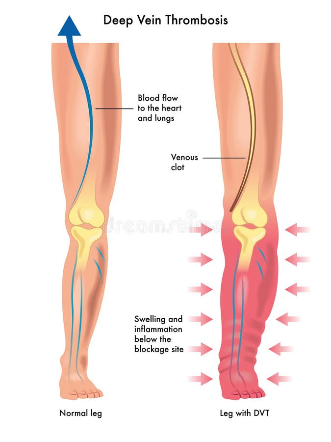
Free with trial Medical illustration of the symptoms of deep vein thrombosis. Vessel lumen vectors Deep vein thrombosis
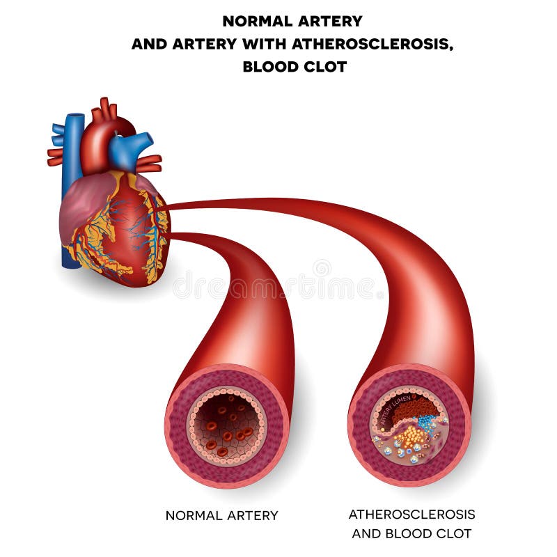
Free with trial Normal artery and unhealthy artery with blood clot. Plaque rupture detailed anatomy illustration. Artery lumen is narrowed and lead to thrombosis. Vessel lumen vectors Normal artery and unhealthy artery
Free with trial Fatty streak formation in the artery. It may lead to thrombosis, formation of a blood clot inside artery. Vessel lumen vectors Fatty streak formation in the artery
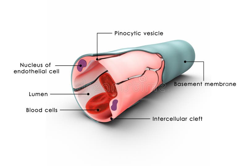
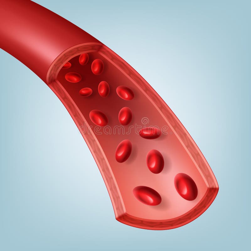
Free with trial Vector illustration of human blood vessel in section with red blood cells. Isolated on background. Vessel lumen vectors Blood vessel in section
Free with trial Blood vessels a tubular structure carrying blood through the tissues and organs; a vein, artery, or capillary. Blood enters the heart through two large veins, the inferior and superior vena cava, emptying oxygen-poor blood from the body into the right atrium of the heart. As the atrium contracts, blood flows from your right atrium into your right ventricle through the open tricuspid valve. Vessel lumen illustrations Blood vessels
Free with trial Lymphatic capillaries are tiny, thin-walled vessels located in the spaces between cells (except in the central nervous system and non-vascular tissues) which serve to drain and process extra-cellular fluid. Upon entering the lumen of a lymphatic capillary, the collected fluid and associated cells (notably white blood cells) is known as lymph. Vessel lumen illustrations Capillary labelled. Lymphatic capillaries are tiny, thin-walled vessels located in the spaces between cells (except in the central nervous system and non-vascular tissues) which serve to drain and process extra-cellular fluid. Upon entering the lumen of a lymphatic capillary, the collected fluid and associated cells (notably white blood cells) is known as lymph.
Free with trial There are two types of blood vessels in the circulatory system of the body - arteries that carry oxygenated blood from the heart to various parts of the body; and veins that carry blood towards the heart for purification. Vessel lumen illustrations Arteries and Veins. There are two types of blood vessels in the circulatory system of the body - arteries that carry oxygenated blood from the heart to various parts of the body; and veins that carry blood towards the heart for purification.
Free with trial Atherosclerosis detailed illustration, progression till Thrombus, blood clot, unstable plaque formation in the artery. Finnaly artery lumen is narrowed and lead to thrombosis and arterial occlusion. Vessel lumen vectors Atherosclerosis illustration. Atherosclerosis detailed illustration, progression till Thrombus, blood clot, unstable plaque formation in the artery. Finnaly artery lumen is narrowed and lead to thrombosis and arterial occlusion.
Free with trial Thrombus, blood clot, unstable plaque formation in the artery. Plaque rupture detailed anatomy illustration. Illustrative diagram how atherosclerosis is progressing till plaque rupture, artery lumen is narrowed and lead to thrombosis and arterial occlusion. Vessel lumen vectors Thrombus, blood clot
Free with trial Thrombus, blood clot, unstable plaque formation in the artery. Plaque rupture detailed anatomy illustration. Illustrative diagram how atherosclerosis is progressing till plaque rupture, artery lumen is narrowed and lead to thrombosis and arterial occlusion. Vessel lumen vectors Thrombus, blood clot, unstable plaque
Free with trial Small intestine anatomy. Digital illustration. Vessel lumen illustrations Bowel and villi cross-section. Small intestine anatomy. Digital illustration.
Free with trial Lymphatic capillaries are tiny, thin-walled vessels located in the spaces between cells (except in the central nervous system and non-vascular tissues) which serve to drain and process extra-cellular fluid. Upon entering the lumen of a lymphatic capillary, the collected fluid and associated cells (notably white blood cells) is known as lymph. Vessel lumen illustrations Capillary labelled. Lymphatic capillaries are tiny, thin-walled vessels located in the spaces between cells (except in the central nervous system and non-vascular tissues) which serve to drain and process extra-cellular fluid. Upon entering the lumen of a lymphatic capillary, the collected fluid and associated cells (notably white blood cells) is known as lymph.
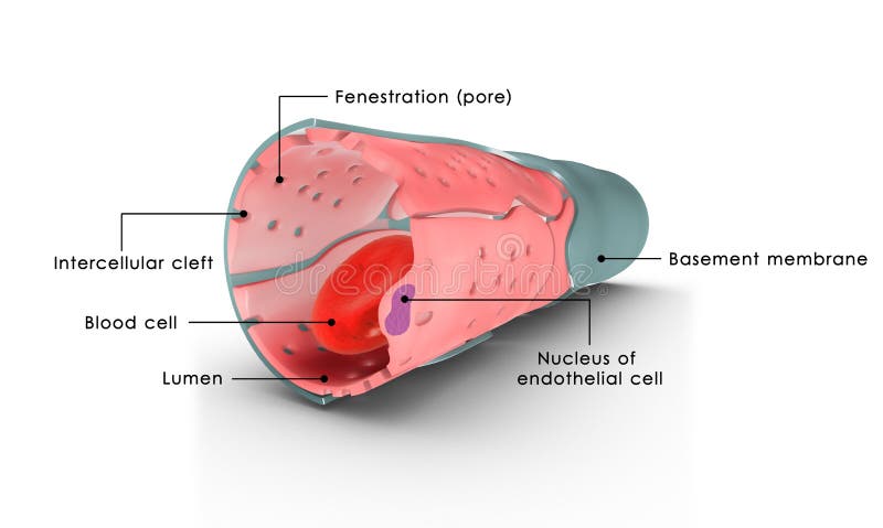
Free with trial Pericyte anatomy. Structure of Blood vessel. Cross section of capillary with Basement membrane, Capillary lumen, Endothelial cells, and Rouget cells. Pericytes wrap around the endothelial cells that line the capillaries and venules that embedded in basement membrane. Vessel lumen vectors Pericyte anatomy. Structure of Blood vessel. Cross section of capillary
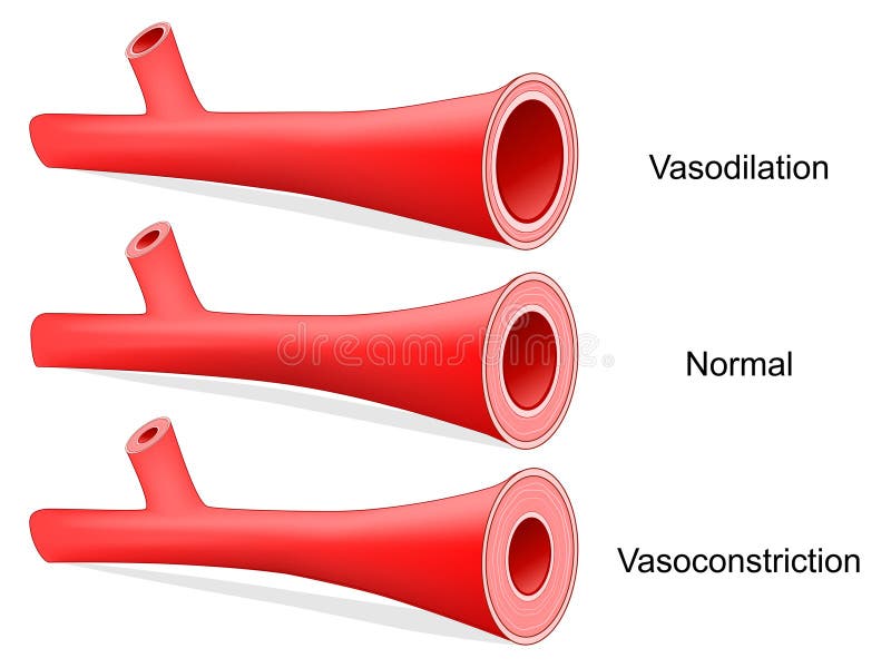
Free with trial Vasoconstriction and Vasodilation of an artery. Lumen of vein. Cross section of the blood vessel with red blood cells. comparison of normal, constricted, and dilated blood vessels. Vector illustration. Vessel lumen vectors Vasoconstriction and Vasodilation
Free with trial Blood vessels a tubular structure carrying blood through the tissues and organs; a vein, artery, or capillary. Blood enters the heart through two large veins, the inferior and superior vena cava, emptying oxygen-poor blood from the body into the right atrium of the heart. As the atrium contracts, blood flows from your right atrium into your right ventricle through the open tricuspid valve. Vessel lumen illustrations Blood vessel
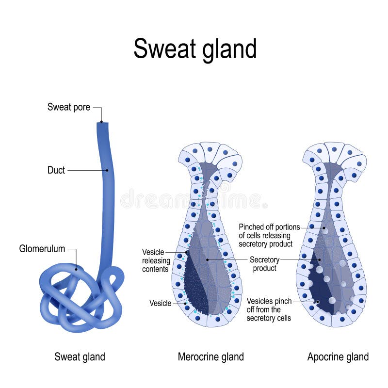
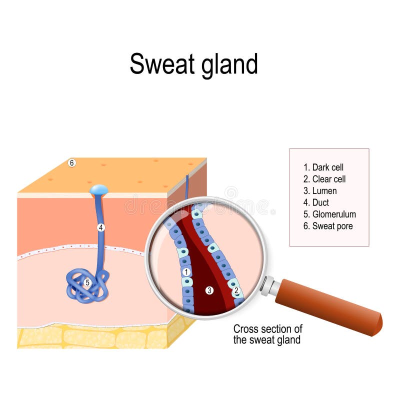
Free with trial Sweat gland. Merocrine and apocrine. different of manner of secretion. cross-section of the human skin, with the sweat gland. Close-up of dark and clear cells, lumen, sweat duct, glomerulum and pore. labeled Vector diagram for educational, medical, biological and science use. Vessel lumen vectors Sweat gland. cross-section. Sweat gland. Merocrine and apocrine. different of manner of secretion. cross-section of the human skin, with the sweat gland. Close-up of dark and clear cells, lumen, sweat duct, glomerulum and pore. labeled Vector diagram for educational, medical, biological and science use
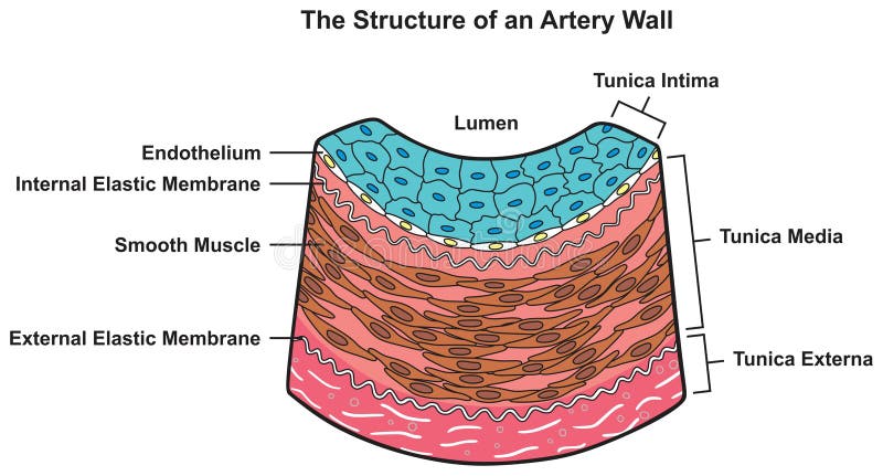
Free with trial Structure of artery wall infographic diagram. for human body biology physiology medical science education. cartoon vector drawing chart. tunica layers. illustration scheme. lumen muscle membrane. Vessel lumen vectors Structure of artery wall infographic diagram
Free with trial Lymphatic capillaries are tiny, thin-walled vessels located in the spaces between cells (except in the central nervous system and non-vascular tissues) which serve to drain and process extra-cellular fluid. Upon entering the lumen of a lymphatic capillary, the collected fluid and associated cells (notably white blood cells) is known as lymph. Vessel lumen illustrations Capillary labelled. Lymphatic capillaries are tiny, thin-walled vessels located in the spaces between cells (except in the central nervous system and non-vascular tissues) which serve to drain and process extra-cellular fluid. Upon entering the lumen of a lymphatic capillary, the collected fluid and associated cells (notably white blood cells) is known as lymph.
Free with trial Blood vessels a tubular structure carrying blood through the tissues and organs; a vein, artery, or capillary. Blood enters the heart through two large veins, the inferior and superior vena cava, emptying oxygen-poor blood from the body into the right atrium of the heart. As the atrium contracts, blood flows from your right atrium into your right ventricle through the open tricuspid valve. Vessel lumen illustrations Blood Cells. Blood vessels a tubular structure carrying blood through the tissues and organs; a vein, artery, or capillary. Blood enters the heart through two large veins, the inferior and superior vena cava, emptying oxygen-poor blood from the body into the right atrium of the heart. As the atrium contracts, blood flows from your right atrium into your right ventricle through the open tricuspid valve.
Free with trial The frog has three respiratory surfaces on its body that it uses to exchange gas with the surroundings: the skin, in the lungs and on the lining of the mouth. A frog may also breathe much like a human, by taking air in through their nostrils and down into their lungs. Vessel lumen illustrations Diagram of the respiratory system of an adult frog. The frog has three respiratory surfaces on its body that it uses to exchange gas with the surroundings: the skin, in the lungs and on the lining of the mouth. A frog may also breathe much like a human, by taking air in through their nostrils and down into their lungs.
Free with trial 3D anatomical render of a cross-section showing atherosclerosis artery, red circular vessel wall with lumen partially blocked by yellow plaque and cholesterol deposits, clear diseased contrast, Generative AI. Vessel lumen illustrations 3D anatomical render of a cross-section showing atherosclerosis artery, red circular vessel wall with lumen partially blocked by
Free with trial Blood vessel / text vector / vein นื ไ้ระำ. Vessel lumen vectors Blood vessel / text vector / vein
Free with trial Vector illustration of human blood vessel in section with red blood cells. Isolated on background. Vessel lumen vectors Blood vessel in section
Free with trial Each type of vessel has a lumen—a hollow passageway through which blood flows. Arteries have smaller lumens than veins, a characteristic that helps to maintain the pressure of blood moving through the system. Further, the walls of the larger vessels are too thick for nutrients to diffuse through to all of the cells. Vessel lumen illustrations Difference between artery and vein lumen. Each type of vessel has a lumen—a hollow passageway through which blood flows. Arteries have smaller lumens than veins, a characteristic that helps to maintain the pressure of blood moving through the system. Further, the walls of the larger vessels are too thick for nutrients to diffuse through to all of the cells.
Free with trial Stages of atherosclerosis vector illustration. Vessel lumen vectors Stages of atherosclerosis. Development of atherosclerosis in the lumen of the vessel. Stages of atherosclerosis vector illustration
Free with trial Blood vessel flat line icon. Vector thin pictogram of vein with molecules, outline illustration for hematology clinic. Vessel lumen vectors Blood vessel flat line icon. Vector thin pictogram of vein with molecules, outline illustration for hematology clinic
Free with trial Detailed 3D render, human head profile, carotid artery plaque, narrowed lumen, cholesterol plaque, red blood cells, medical illustration, copy space. Vessel lumen illustrations Carotid Artery Plaque Illustration - Human Head, Narrowed Lumen, Cholesterol Plaque. Detailed 3D render, human head profile, carotid artery plaque, narrowed lumen, cholesterol plaque, red blood cells, medical illustration, copy space
Free with trial Medical illustration showing carotid artery plaque, narrowed lumen, and cholesterol buildup in a human head silhouette, bright, informative, copy space. Vessel lumen illustrations Carotid Artery Plaque Illustration - Human Head, Narrowed Lumen, Cholesterol Buildup. Medical illustration showing carotid artery plaque, narrowed lumen, and cholesterol buildup in a human head silhouette, bright, informative, copy space
Free with trial A detailed 3D rendering illustrates atherosclerosis, a serious condition characterized by the accumulation of plaque within an artery. The image shows a cross-section of a blood vessel with significant yellow-white plaque deposits severely narrowing the lumen, impeding the normal flow of numerous red blood cells. A few white blood cells are also visible. This medical illustration effectively visualizes the dangerous obstruction caused by cholesterol and fatty substances, leading to reduced blood flow and increased risk of cardiovascular diseases like heart attack and stroke. Ideal for educational, scientific, and health-related content. Vessel lumen illustrations Atherosclerosis: Plaque Buildup in Artery with Red Blood Cells. A detailed 3D rendering illustrates atherosclerosis, a serious condition characterized by the accumulation of plaque within an artery. The image shows a cross-section of a blood vessel with significant yellow-white plaque deposits severely narrowing the lumen, impeding the normal flow of numerous red blood cells. A few white blood cells are also visible. This medical illustration effectively visualizes the dangerous obstruction caused by cholesterol and fatty substances, leading to reduced blood flow and increased risk of cardiovascular diseases like heart attack and stroke. Ideal for educational, scientific, and health-related content.
Free with trial A detailed, realistic 3D illustration of a cross-section of an artery showing significant plaque buildup (atherosclerosis). Yellowish, fatty deposits line the artery walls, narrowing the lumen and impeding blood flow. Numerous red blood cells are depicted flowing through the constricted vessel, highlighting the impact of the blockage. This image is suitable for medical education, scientific publications, and health awareness campaigns. Vessel lumen illustrations Atherosclerosis: Plaque Buildup in Artery with Red Blood Cells. A detailed, realistic 3D illustration of a cross-section of an artery showing significant plaque buildup (atherosclerosis). Yellowish, fatty deposits line the artery walls, narrowing the lumen and impeding blood flow. Numerous red blood cells are depicted flowing through the constricted vessel, highlighting the impact of the blockage. This image is suitable for medical education, scientific publications, and health awareness campaigns.
Free with trial Coronary stent in blood vessel - metal or plastic tube inserted into the lumen of vein to keep the passageway open. Vessel lumen vectors Coronary stent in vessel to keep the passageway. Coronary stent in blood vessel - metal or plastic tube inserted into the lumen of vein to keep the passageway open
Free with trial This medical illustration depicts a detailed internal structure of a human blood vessel. The illustration shows anatomical details. Vessel lumen illustrations Detailed anatomical illustration of a human blood vessel section. This medical illustration depicts a detailed internal structure of a human blood vessel. The illustration shows anatomical details
Free with trial Detailed medical illustration showing coronary artery spasm, highlighting contracted smooth muscle and narrowed lumen. Clear, informative composition with ample copy space. Vessel lumen illustrations Coronary Artery Spasm Illustration - Heart, Contracted Smooth Muscle, Narrowed Lumen. Detailed medical illustration showing coronary artery spasm, highlighting contracted smooth muscle and narrowed lumen. Clear, informative composition with ample copy space
Free with trial A detailed 3D rendering illustrates atherosclerosis, a serious cardiovascular condition. The image shows a cross-section of an artery with significant yellowish plaque buildup on its inner walls, severely narrowing the lumen. Red blood cells and white blood cells are depicted flowing through the constricted vessel. This visual represents the accumulation of cholesterol and fatty substances, leading to hardened and blocked arteries. It effectively conveys the concept of reduced blood flow and the increased risk of heart disease, stroke, and other circulatory problems, making it ideal for medical education and health awareness campaigns. Vessel lumen illustrations Atherosclerosis: 3D Illustration of Clogged Artery with Cholesterol Plaque. A detailed 3D rendering illustrates atherosclerosis, a serious cardiovascular condition. The image shows a cross-section of an artery with significant yellowish plaque buildup on its inner walls, severely narrowing the lumen. Red blood cells and white blood cells are depicted flowing through the constricted vessel. This visual represents the accumulation of cholesterol and fatty substances, leading to hardened and blocked arteries. It effectively conveys the concept of reduced blood flow and the increased risk of heart disease, stroke, and other circulatory problems, making it ideal for medical education and health awareness campaigns.
Free with trial Comparative 3D render of three vein segments: normal smooth lumen, moderately dilated with early bulging, and severely twisted varicose vein with incompetent valves and tortuous shape, Generative AI. Vessel lumen illustrations Comparative 3D render of three vein segments: normal smooth lumen, moderately dilated with early bulging, and severely twisted
Free with trial Photorealistic 3D render of dilated varicose vein with dark red clot partially obstructing lumen. Upstream section swollen, valves distorted, medical illustration vascular pathology and obstruction, Generative AI. Vessel lumen illustrations Photorealistic 3D render of dilated varicose vein with dark red clot partially obstructing lumen. Upstream section swollen, valves
Free with trial Sweat gland. cross-section of the human skin, with the sweat gland. Close-up of dark and clear cells, lumen, sweat duct, glomerulum and pore. labeled Vector diagram for educational, medical, biological and science use. Vessel lumen vectors Sweat gland
Free with trial Vein and Artery anatomy. comparison and difference. longitudinal and cross section human blood vessel. Poster for medical and education use. Vector diagram. Vessel lumen vectors Vein and Artery anatomy. comparison and difference
Free with trial Detailed medical illustration showing atherosclerosis, a condition where plaque accumulates inside an artery. The image highlights the fibrous cap, lipid core, and calcification within the artery wall, with red blood cells flowing through the narrowed lumen. Vessel lumen illustrations Atherosclerosis - Plaque Buildup in Artery Wall with Lipid Core and Calcification. Detailed medical illustration showing atherosclerosis, a condition where plaque accumulates inside an artery. The image highlights the fibrous cap, lipid core, and calcification within the artery wall, with red blood cells flowing through the narrowed lumen
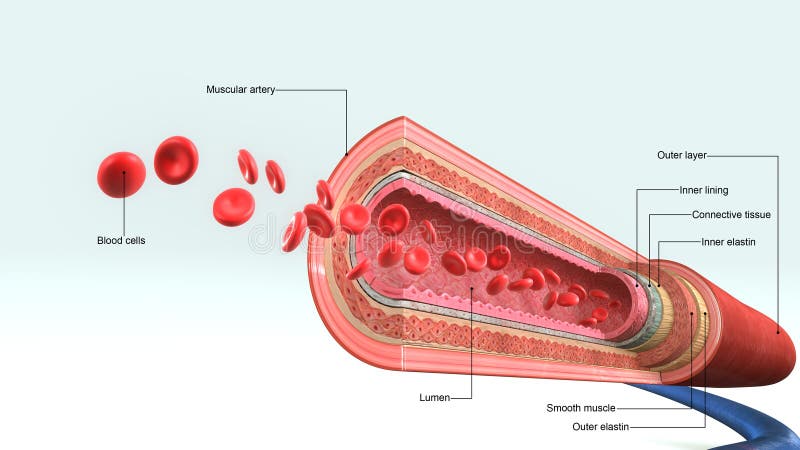
Free with trial Close-up view of an artery cross-section. The artery appears to be a vivid light pinkish-red color. Red blood cells fill the inside lumen. The image displays a 3D anatomical view of the artery and surrounding tissues. This medical illustration could be used in educational or scientific materials. Vessel lumen illustrations Blood vessels artery cross section. Close-up view of an artery cross-section. The artery appears to be a vivid light pinkish-red color. Red blood cells fill the inside lumen. The image displays a 3D anatomical view of the artery and surrounding tissues. This medical illustration could be used in educational or scientific materials
Free with trial 3D cutaway render of leg cross-section showing enlarged deep vein with dark red clot obstructing lumen, upstream dilation. Muscles and bones softly outlined for orientation, detailed medical realism, Generative AI. Vessel lumen illustrations 3D cutaway render of leg cross-section showing enlarged deep vein with dark red clot obstructing lumen, upstream dilation. Muscles
Free with trial During these procedures, doctor will insert a sheath, or soft plastic tube, into a large artery in your groin. Next a flexible wire will be guided through the sheath [pause] and gently up through your circulatory system into your narrowed coronary artery. Vessel lumen illustrations Angioplasty Stent. During these procedures, doctor will insert a sheath, or soft plastic tube, into a large artery in your groin. Next a flexible wire will be guided through the sheath [pause] and gently up through your circulatory system into your narrowed coronary artery.
Free with trial During these procedures, doctor will insert a sheath, or soft plastic tube, into a large artery in your groin. Next a flexible wire will be guided through the sheath [pause] and gently up through your circulatory system into your narrowed coronary artery. Vessel lumen illustrations Angioplasty Stent. During these procedures, doctor will insert a sheath, or soft plastic tube, into a large artery in your groin. Next a flexible wire will be guided through the sheath [pause] and gently up through your circulatory system into your narrowed coronary artery.
Free with trial During these procedures, doctor will insert a sheath, or soft plastic tube, into a large artery in your groin. Next a flexible wire will be guided through the sheath [pause] and gently up through your circulatory system into your narrowed coronary artery. Vessel lumen illustrations Angioplasty Stent. During these procedures, doctor will insert a sheath, or soft plastic tube, into a large artery in your groin. Next a flexible wire will be guided through the sheath [pause] and gently up through your circulatory system into your narrowed coronary artery.
Free with trial During these procedures, doctor will insert a sheath, or soft plastic tube, into a large artery in your groin. Next a flexible wire will be guided through the sheath [pause] and gently up through your circulatory system into your narrowed coronary artery. Vessel lumen illustrations Angioplasty Stent. During these procedures, doctor will insert a sheath, or soft plastic tube, into a large artery in your groin. Next a flexible wire will be guided through the sheath [pause] and gently up through your circulatory system into your narrowed coronary artery.
Free with trial The esophagus or oesophagus is an organ in vertebrates through which food passes, aided by peristaltic contractions, from the pharynx to the stomach. The esophagus is a fibromuscular tube, that travels behind the trachea and heart. During swallowing, the epiglottis tilts backwards to prevent food from going down the larynx and lungs. The trachea is the long tube that connects your larynx to your bronchi. Bronchi send air to your lungs. Trachea is a key part of respiratory system. The trachea is made of rings of cartilage. It is lined with cells that produce mucus. Vessel lumen illustrations Anatomy of Trachea and Esophagus. The esophagus or oesophagus is an organ in vertebrates through which food passes, aided by peristaltic contractions, from the pharynx to the stomach. The esophagus is a fibromuscular tube, that travels behind the trachea and heart. During swallowing, the epiglottis tilts backwards to prevent food from going down the larynx and lungs.The trachea is the long tube that connects your larynx to your bronchi. Bronchi send air to your lungs. Trachea is a key part of respiratory system. The trachea is made of rings of cartilage. It is lined with cells that produce mucus.
Free with trial The esophagus or oesophagus is an organ in vertebrates through which food passes, aided by peristaltic contractions, from the pharynx to the stomach. The esophagus is a fibromuscular tube, that travels behind the trachea and heart. During swallowing, the epiglottis tilts backwards to prevent food from going down the larynx and lungs. The trachea is the long tube that connects your larynx to your bronchi. Bronchi send air to your lungs. Trachea is a key part of respiratory system. The trachea is made of rings of cartilage. It is lined with cells that produce mucus. Vessel lumen illustrations Trachea and Esophagus cross section. The esophagus or oesophagus is an organ in vertebrates through which food passes, aided by peristaltic contractions, from the pharynx to the stomach. The esophagus is a fibromuscular tube, that travels behind the trachea and heart. During swallowing, the epiglottis tilts backwards to prevent food from going down the larynx and lungs.The trachea is the long tube that connects your larynx to your bronchi. Bronchi send air to your lungs. Trachea is a key part of respiratory system. The trachea is made of rings of cartilage. It is lined with cells that produce mucus
Free with trial Arteries carry blood away from the heart, and veins carry blood towards the heart. With the exception of pulmonary blood vessels, arteries carry oxygenated blood and veins carry deoxygenated blood. Arteries have thick walls with muscle tissue. Veins have thinner walls and use valves to keep your blood flowing. Vessel lumen illustrations Structure of arteries, veins and capillaries. Arteries carry blood away from the heart, and veins carry blood towards the heart. With the exception of pulmonary blood vessels, arteries carry oxygenated blood and veins carry deoxygenated blood. Arteries have thick walls with muscle tissue. Veins have thinner walls and use valves to keep your blood flowing
Free with trial Arteries have thick walls with muscle tissue. Veins have thinner walls and use valves to keep your blood flowing. Artery vs Capillary. Arteries carry blood from your heart to your organs. Vessel lumen illustrations Illustration of difference between arteries and veins. Arteries have thick walls with muscle tissue. Veins have thinner walls and use valves to keep your blood flowing. Artery vs Capillary. Arteries carry blood from your heart to your organs.
Free with trial Implantable venous access port. Under the skin central line access device for chemotherapy infusion, medication administration and blood drawing. Medical vector illustration. Vessel lumen vectors Implantable venous access port on male body. Implantable venous access port. Under the skin central line access device for chemotherapy infusion, medication administration and blood drawing. Medical vector illustration.
Free with trial System many small capillaries branch out of the large blood vessels into the circulatory system for the transportation of blood to different parts In the body. disease hemorrhagic stroke. 3D render. Vessel lumen illustrations 3D render. System many small capillaries branch out of the large blood. System many small capillaries branch out of the large blood vessels into the circulatory system for the transportation of blood to different parts In the body. disease hemorrhagic stroke. 3D render
Free with trial Vasoconstriction, Vasodilation, and normal artery. Comparison of normal, constricted, and dilated blood vessels. Vector illustration. Vessel lumen vectors Vasoconstriction, Vasodilation, and normal artery. Comparison of normal, constricted, and dilated blood vessels
Free with trial Atherosclerosis visualization showing coronary artery narrowing due to fatty plaque buildup and restricted blood flow inside ghostly chest silhouette for clinical cardiology illustration. Vessel lumen illustrations Atherosclerosis visualization showing coronary artery narrowing due to fatty plaque buildup and restricted blood flow inside
Free with trial System many small capillaries branch out of the large blood vessels into the circulatory system for the transportation of blood to different parts In the body. disease hemorrhagic stroke. 3D render. Vessel lumen illustrations 3D render. System many small capillaries branch out of the large blood. System many small capillaries branch out of the large blood vessels into the circulatory system for the transportation of blood to different parts In the body. disease hemorrhagic stroke. 3D render
Free with trial Tunneled central venous catheter placed in the subclavian vein. Patient with CVC long term access device for chemotherapy infusions and blood sampling. Central line tube close up. Vector illustration. Vessel lumen vectors Tunneled central line venous catheter medical diagram. Tunneled central venous catheter placed in the subclavian vein. Patient with CVC long term access device for chemotherapy infusions and blood sampling. Central line tube close up. Vector illustration.
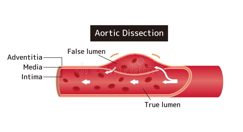
Free with trial Vector illustration of aortic dissection. Vessel lumen vectors Vector illustration of aortic dissection
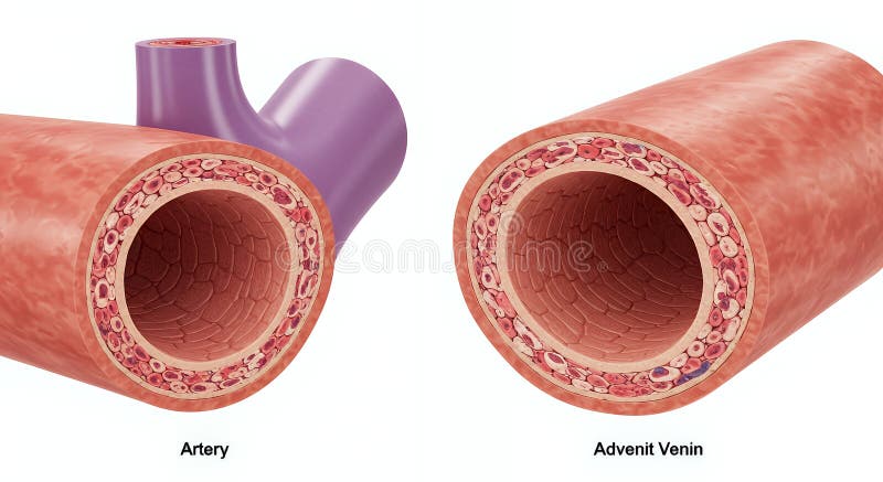
Free with trial The image shows a cross-sectional illustration comparing the layered structures of an artery and a vein. Both are depicted with their characteristic anatomy. Vessel lumen illustrations Illustration comparing the cross section of an artery and vein. The image shows a cross-sectional illustration comparing the layered structures of an artery and a vein. Both are depicted with their characteristic anatomy
Free with trial Microscopic View of Human Blood Vessels: Artery and Vein in Detail. Vessel lumen illustrations Microscopic View of Human Blood Vessels: Artery and Vein in Detail
Free with trial 3D medical illustration comparing healthy and diseased atherosclerosis artery, left vessel with smooth walls and open lumen, right artery narrowed with thick yellow plaque buildup, Generative AI. Vessel lumen illustrations 3D medical illustration comparing healthy and diseased atherosclerosis artery, left vessel with smooth walls and open lumen, right
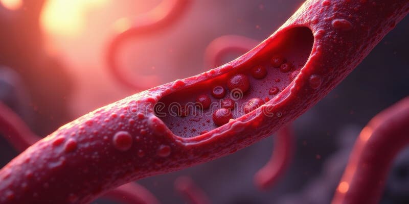
Free with trial Microscopic vessel with red cellular components flowing through lumen in warm light. Aesthetic image. Generative AI. Vessel lumen illustrations Microscopic vessel with red cellular components flowing through lumen in warm light. Generative AI
Free with trial Atherosclerosis illustration shows cholesterol plaque buildup in artery. Red blood cells navigate narrowed vessel lumen. Visualizes cardiovascular disease, risk factors prevention. Vessel lumen illustrations Atherosclerosis illustration shows cholesterol plaque buildup in artery. Red blood cells navigate narrowed vessel lumen.
Free with trial 3D anatomical render of aortic aneurysm cross-section. Circular vessel with normal red wall, one area thinned and outwardly bulging. Lumen widened at aneurysm site. minimal medical style, Generative AI. Vessel lumen illustrations 3D anatomical render of aortic aneurysm cross-section. Circular vessel with normal red wall, one area thinned and outwardly
Free with trial Cross section of clogged artery showing narrowed blood vessel lumen and blockage. Blood flow restricted to heart danger of heart attack shown with orange arrows. Vessel lumen illustrations Cross section of clogged artery showing narrowed blood vessel lumen and blockage. Blood flow restricted to heart, danger of heart. Cross section of clogged artery showing narrowed blood vessel lumen and blockage. Blood flow restricted to heart danger of heart attack shown with orange arrows.
Free with trial This detailed microscopic image reveals the intricate structure of a blood vessel showcasing red blood cells flowing smoothly through its lumen The artery's endothelium and surrounding tissue are clearly visible offering a clear visualization of cardiovascular anatomy Ideal for educational and medical purposes. Vessel lumen illustrations Microscopic View of Blood Vessel Red Blood Cells Flowing Through Artery AI generated. This detailed microscopic image reveals the intricate structure of a blood vessel showcasing red blood cells flowing smoothly through its lumen The artery's endothelium and surrounding tissue are clearly visible offering a clear visualization of cardiovascular anatomy Ideal for educational and medical purposes
Free with trial A close-up, microscopic view of a blood vessel interior, showing significant plaque buildup narrowing the lumen. Image. Vessel lumen illustrations Blood Vessel with Arterial Plaque. A close-up, microscopic view of a blood vessel interior, showing significant plaque buildup narrowing the lumen. Image
Free with trial This image displays a detailed cross-sectional view of a human blood vessel. The vessel wall shows significant plaque accumulation, which appears as a dense, irregular layer within the lumen. The plaque is likely composed of lipids, calcium, and cellular debris, indicative of atherosclerosis. The inner lining of the vessel, or endothelium, is partially visible, and the surrounding tissue appears. Vessel lumen vectors Cross-sectional view of a human blood vessel with plaque buildup. This image displays a detailed cross-sectional view of a human blood vessel. The vessel wall. This image displays a detailed cross-sectional view of a human blood vessel. The vessel wall shows significant plaque accumulation, which appears as a dense, irregular layer within the lumen. The plaque is likely composed of lipids, calcium, and cellular debris, indicative of atherosclerosis. The inner lining of the vessel, or endothelium, is partially visible, and the surrounding tissue appears
Free with trial This image displays a detailed cross-sectional view of a human blood vessel. The vessel wall shows significant plaque accumulation, which appears as a dense, irregular layer within the lumen. The plaque is likely composed of lipids, calcium, and cellular debris, indicative of atherosclerosis. The inner lining of the vessel, or endothelium, is partially visible, and the surrounding tissue appears. Vessel lumen illustrations Cross-sectional view of a human blood vessel with plaque buildup. This image displays a detailed cross-sectional view of a human blood vessel. The vessel wall shows significant plaque accumulation, which appears as a dense, irregular layer within the lumen. The plaque is likely composed of lipids, calcium, and cellular debris, indicative of atherosclerosis. The inner lining of the vessel, or endothelium, is partially visible, and the surrounding tissue appears
Free with trial A detailed illustration shows red blood cells flowing through a blood vessel, with fatty deposits and plaque obstructing the lumen. Vessel lumen illustrations Close up view of a blood vessel with red blood cells and plaque buildup. A detailed illustration shows red blood cells flowing through a blood vessel, with fatty deposits and plaque obstructing the lumen
Free with trial Cross section of a blood vessel showing yellow cholesterol plaques and red blood cells inside the lumen for medical visualization usage. Vessel lumen illustrations Artery Plaque Buildup Inside Blood Vessel. Cross section of a blood vessel showing yellow cholesterol plaques and red blood cells inside the lumen for medical visualization usage
Free with trial Cross section of a blood vessel showing yellow cholesterol plaques and red blood cells inside the lumen for medical visualization usage. Vessel lumen illustrations Artery Plaque Buildup Inside Blood Vessel. Cross section of a blood vessel showing yellow cholesterol plaques and red blood cells inside the lumen for medical visualization usage
Free with trial This detailed 3D illustration depicts red blood cells actively flowing within a cross-section of a human blood vessel. The image offers a microscopic view of the circulatory system, highlighting the biconcave disc shape of erythrocytes as they navigate the vessel's lumen. It visually represents the vital process of blood circulation and oxygen transport throughout the body, making it ideal for medical, scientific, and educational contexts related to human anatomy, physiology, and hematology. Vessel lumen illustrations Red Blood Cells Flowing in a Blood Vessel - 3D Illustration. This detailed 3D illustration depicts red blood cells actively flowing within a cross-section of a human blood vessel. The image offers a microscopic view of the circulatory system, highlighting the biconcave disc shape of erythrocytes as they navigate the vessel's lumen. It visually represents the vital process of blood circulation and oxygen transport throughout the body, making it ideal for medical, scientific, and educational contexts related to human anatomy, physiology, and hematology.
Free with trial A stylized cross-section of a blood vessel shows red blood cells and plaque buildup on the inner walls, narrowing the lumen. Image. Vessel lumen illustrations Blood Vessel with Atherosclerotic Plaque and Red Blood Cells. A stylized cross-section of a blood vessel shows red blood cells and plaque buildup on the inner walls, narrowing the lumen. Image
Free with trial This detailed 3D rendering showcases a cross-section of a human blood vessel, likely an artery or vein, against a stark black background. The intricate layers of the vessel wall are clearly visible, including an outer connective tissue layer, a muscular or cellular middle layer, and a creamy white inner lining. The central lumen features distinct folds, creating a unique internal structure. This high-resolution illustration is ideal for medical education, scientific research, health publications, and anatomical studies, providing a clear visual representation of vascular biology and the cardiovascular system. Vessel lumen illustrations Detailed 3D Render of a Human Blood Vessel Cross-Section. This detailed 3D rendering showcases a cross-section of a human blood vessel, likely an artery or vein, against a stark black background. The intricate layers of the vessel wall are clearly visible, including an outer connective tissue layer, a muscular or cellular middle layer, and a creamy white inner lining. The central lumen features distinct folds, creating a unique internal structure. This high-resolution illustration is ideal for medical education, scientific research, health publications, and anatomical studies, providing a clear visual representation of vascular biology and the cardiovascular system.
Free with trial A cross-section of a red blood vessel reveals yellow plaque buildup narrowing the lumen, with a textured outer surface against a neutral background. Image. Vessel lumen illustrations Cross-section of a Blood Vessel with Atherosclerotic Plaque. A cross-section of a red blood vessel reveals yellow plaque buildup narrowing the lumen, with a textured outer surface against a neutral background. Image
Free with trial A cross-section of a blood vessel showing a clot and plaque buildup. Red vessel wall surrounds a lumen with a dark red clot and yellow plaque deposits. Image. Vessel lumen illustrations Blood Vessel Cross-Section with Clot and Atherosclerotic Plaque. A cross-section of a blood vessel showing a clot and plaque buildup. Red vessel wall surrounds a lumen with a dark red clot and yellow plaque deposits. Image
Free with trial This detailed 3D illustration depicts the intricate internal structure of a biological tube, possibly a blood vessel or intestine, with a focus on its textured walls and lumen. Vessel lumen illustrations Abstract 3D rendering of a microscopic view inside a biological vessel or organ. This detailed 3D illustration depicts the intricate internal structure of a biological tube, possibly a blood vessel or intestine, with a focus on its textured walls and lumen


![During these procedures, doctor will insert a sheath, or soft plastic tube, into a large artery in your groin. Next a flexible wire will be guided through the sheath [pause] and gently up through your circulatory system into your narrowed coronary artery. Vessel lumen illustrations](https://thumbs.dreamstime.com/b/angioplasty-stent-procedures-doctor-will-insert-sheath-soft-plastic-tube-large-artery-your-groin-next-84218675.jpg)
![During these procedures, doctor will insert a sheath, or soft plastic tube, into a large artery in your groin. Next a flexible wire will be guided through the sheath [pause] and gently up through your circulatory system into your narrowed coronary artery. Vessel lumen illustrations](https://thumbs.dreamstime.com/b/angioplasty-stent-procedures-doctor-will-insert-sheath-soft-plastic-tube-large-artery-your-groin-next-84219958.jpg)
![During these procedures, doctor will insert a sheath, or soft plastic tube, into a large artery in your groin. Next a flexible wire will be guided through the sheath [pause] and gently up through your circulatory system into your narrowed coronary artery. Vessel lumen illustrations](https://thumbs.dreamstime.com/b/angioplasty-stent-procedures-doctor-will-insert-sheath-soft-plastic-tube-large-artery-your-groin-next-84218749.jpg)
![During these procedures, doctor will insert a sheath, or soft plastic tube, into a large artery in your groin. Next a flexible wire will be guided through the sheath [pause] and gently up through your circulatory system into your narrowed coronary artery. Vessel lumen illustrations](https://thumbs.dreamstime.com/b/angioplasty-stent-procedures-doctor-will-insert-sheath-soft-plastic-tube-large-artery-your-groin-next-84218686.jpg)