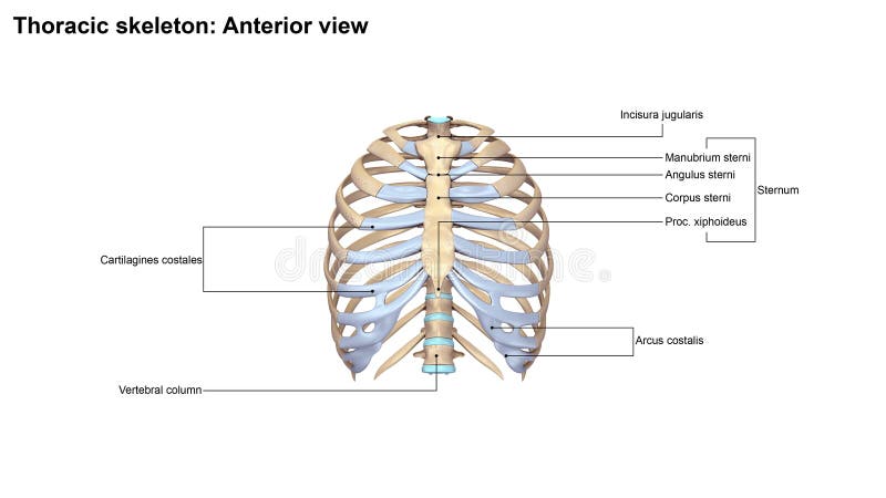
Free with trial In vertebrate anatomy, ribs are the long curved bones which form the rib cage. In most tetrapods, ribs surround the chest, enabling the lungs to expand and thus facilitate breathing by expanding the chest cavity. They serve to protect the lungs, heart, and other internal organs of the thorax. In some animals, especially snakes, ribs may provide support and protection for the entire body. View internal organs illustrations Thoracic Skeleton Anterior view. In vertebrate anatomy, ribs are the long curved bones which form the rib cage. In most tetrapods, ribs surround the chest, enabling the lungs to expand and thus facilitate breathing by expanding the chest cavity. They serve to protect the lungs, heart, and other internal organs of the thorax. In some animals, especially snakes, ribs may provide support and protection for the entire body.
Free with trial Realistic illustration of cirrhosis of human liver isolated on white. View internal organs illustrations Realistic illustration of cirrhosis of human liver
Free with trial Realistic illustration of cirrhosis of human liver isolated on white. View internal organs illustrations Realistic illustration of cirrhosis of human liver
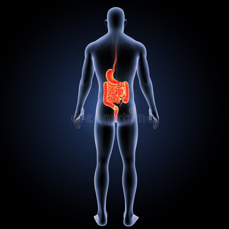
Free with trial The internal organ in which the major part of the digestion of food occurs, being in humans and many mammals a pear-shaped enlargement of the alimentary canal linking the oesophagus to the small intestine. The small intestine or small bowel is the part of the gastrointestinal tract between the stomach and the large intestine, and is where most of the end absorption of food takes place. The small intestine has three distinct regions – the duodenum, jejunum, and ileum. View internal organs illustrations Stomach with small intestine zoom with anatomy anterior view. The internal organ in which the major part of the digestion of food occurs, being in humans and many mammals a pear-shaped enlargement of the alimentary canal linking the oesophagus to the small intestine. The small intestine or small bowel is the part of the gastrointestinal tract between the stomach and the large intestine, and is where most of the end absorption of food takes place. The small intestine has three distinct regions – the duodenum, jejunum, and ileum.
Free with trial In human anatomy, the thoracic diaphragm, or simply the diaphragm , is a sheet of internal skeletal muscle that extends across the bottom of the rib cage. View internal organs illustrations Diaphragm with body lateral view. In human anatomy, the thoracic diaphragm, or simply the diaphragm , is a sheet of internal skeletal muscle that extends across the bottom of the rib cage.
Free with trial The stomach is a muscular, hollow, dilated part of the digestion system which functions as an important organ of the digestive tract in some animals, including vertebrates, echinoderms, insects mid-gut, and molluscs. It is involved in the second phase of digestion, following mastication chewing. View internal organs illustrations Stomach anterior view. The stomach is a muscular, hollow, dilated part of the digestion system which functions as an important organ of the digestive tract in some animals, including vertebrates, echinoderms, insects mid-gut, and molluscs. It is involved in the second phase of digestion, following mastication chewing.
Free with trial Microcopic view of micro objects with depth of field, Bacteria spheres 3d illustration. View internal organs illustrations Micro Bacteria spheres. Microcopic view of micro objects with depth of field, Bacteria spheres 3d illustration
Free with trial In human anatomy, the thoracic diaphragm, or simply the diaphragm , is a sheet of internal skeletal muscle that extends across the bottom of the rib cage. View internal organs illustrations Diaphragm lateral view. In human anatomy, the thoracic diaphragm, or simply the diaphragm , is a sheet of internal skeletal muscle that extends across the bottom of the rib cage.
Free with trial In human anatomy, the thoracic diaphragm, or simply the diaphragm , is a sheet of internal skeletal muscle that extends across the bottom of the rib cage. View internal organs illustrations Diaphragm lateral view. In human anatomy, the thoracic diaphragm, or simply the diaphragm , is a sheet of internal skeletal muscle that extends across the bottom of the rib cage.
Free with trial In human anatomy, the thoracic diaphragm, or simply the diaphragm , is a sheet of internal skeletal muscle that extends across the bottom of the rib cage. View internal organs illustrations Diaphragm anterior view. In human anatomy, the thoracic diaphragm, or simply the diaphragm , is a sheet of internal skeletal muscle that extends across the bottom of the rib cage.
Free with trial In human anatomy, the thoracic diaphragm, or simply the diaphragm , is a sheet of internal skeletal muscle that extends across the bottom of the rib cage. View internal organs illustrations Diaphragm posterior view. In human anatomy, the thoracic diaphragm, or simply the diaphragm , is a sheet of internal skeletal muscle that extends across the bottom of the rib cage.
Free with trial In human anatomy, the thoracic diaphragm, or simply the diaphragm , is a sheet of internal skeletal muscle that extends across the bottom of the rib cage. View internal organs illustrations Diaphragm posterior view. In human anatomy, the thoracic diaphragm, or simply the diaphragm , is a sheet of internal skeletal muscle that extends across the bottom of the rib cage.
Free with trial The human rib cage is made up of 12 paired rib bones; each are symmetrically paired on a right and left side. Of all 24 ribs, the first seven pairs are often labeled as `true. ` These bones are connected to the costal cartilage, while the five other `false` sets are not. The ribcage also encloses the thoracic cavity and helps protect the heart and lungs from damage. There are 24 ribs in the human body, divided into two sets of 12 curved, flat bones. Each one is attached by cartilage at the back to the thoracic vertebrae. MEN and women have 12 pairs of ribs a few individuals have 13 or 11 pairs. The idea that men have fewer ribs than women is widespread but wrong, perhaps deriving from the biblical story of Eve being made from one of Adam`s ribs. Both men and women have 24 ribs, twelve on each side. Floating rib: One of the last two ribs. A rib is said to be `floating` if it does not attach to the sternum the breast bone or to another rib. There are usually 12 pairs of ribs in all. Each pair of ribs is attached to the building blocks of the spine the vertebrae in the back. The ribs partially enclose and protect the chest cavity, where many vital organs including the heart and the lungs are located. The rib cage is collectively made up of long, curved individual bones with joint-connections to the spinal vertebrae. View internal organs illustrations Ribs with blood vessels and nerves lateral view. The human rib cage is made up of 12 paired rib bones; each are symmetrically paired on a right and left side. Of all 24 ribs, the first seven pairs are often labeled as `true.` These bones are connected to the costal cartilage, while the five other `false` sets are not. The ribcage also encloses the thoracic cavity and helps protect the heart and lungs from damage. There are 24 ribs in the human body, divided into two sets of 12 curved, flat bones. Each one is attached by cartilage at the back to the thoracic vertebrae. MEN and women have 12 pairs of ribs a few individuals have 13 or 11 pairs. The idea that men have fewer ribs than women is widespread but wrong, perhaps deriving from the biblical story of Eve being made from one of Adam`s ribs. Both men and women have 24 ribs, twelve on each side. Floating rib: One of the last two ribs. A rib is said to be `floating` if it does not attach to the sternum the breast bone or to another rib. There are usually 12 pairs of ribs in all. Each pair of ribs is attached to the building blocks of the spine the vertebrae in the back. The ribs partially enclose and protect the chest cavity, where many vital organs including the heart and the lungs are located. The rib cage is collectively made up of long, curved individual bones with joint-connections to the spinal vertebrae.
Free with trial In human anatomy, the thoracic diaphragm, or simply the diaphragm , is a sheet of internal skeletal muscle that extends across the bottom of the rib cage. View internal organs illustrations Diaphragm with skeleton anterior view. In human anatomy, the thoracic diaphragm, or simply the diaphragm , is a sheet of internal skeletal muscle that extends across the bottom of the rib cage.
Free with trial Microcopic view of micro objects with depth of field, Bacteria spheres 3d illustration. View internal organs illustrations Micro Bacteria spheres. Microcopic view of micro objects with depth of field, Bacteria spheres 3d illustration
Free with trial A detailed 3D anatomical illustration showcases the human torso, highlighting the respiratory system. The transparent rendering reveals the lungs, heart, and the crucial diaphragm muscle, prominently colored in reddish-brown. White arrows indicate the upward and downward movement of the diaphragm, illustrating its contraction and relaxation during the breathing process. This medical visualization is ideal for educating about human anatomy, physiology, and the mechanics of respiration, suitable for textbooks, health articles, and scientific presentations. View internal organs illustrations Human Diaphragm and Lungs Anatomy with Breathing Movement. A detailed 3D anatomical illustration showcases the human torso, highlighting the respiratory system. The transparent rendering reveals the lungs, heart, and the crucial diaphragm muscle, prominently colored in reddish-brown. White arrows indicate the upward and downward movement of the diaphragm, illustrating its contraction and relaxation during the breathing process. This medical visualization is ideal for educating about human anatomy, physiology, and the mechanics of respiration, suitable for textbooks, health articles, and scientific presentations.
Free with trial The human rib cage is made up of 12 paired rib bones; each are symmetrically paired on a right and left side. Of all 24 ribs, the first seven pairs are often labeled as `true. ` These bones are connected to the costal cartilage, while the five other `false` sets are not. The ribcage also encloses the thoracic cavity and helps protect the heart and lungs from damage. There are 24 ribs in the human body, divided into two sets of 12 curved, flat bones. Each one is attached by cartilage at the back to the thoracic vertebrae. MEN and women have 12 pairs of ribs a few individuals have 13 or 11 pairs. The idea that men have fewer ribs than women is widespread but wrong, perhaps deriving from the biblical story of Eve being made from one of Adam`s ribs. Both men and women have 24 ribs, twelve on each side. Floating rib: One of the last two ribs. A rib is said to be `floating` if it does not attach to the sternum the breast bone or to another rib. There are usually 12 pairs of ribs in all. Each pair of ribs is attached to the building blocks of the spine the vertebrae in the back. The ribs partially enclose and protect the chest cavity, where many vital organs including the heart and the lungs are located. The rib cage is collectively made up of long, curved individual bones with joint-connections to the spinal vertebrae. View internal organs illustrations Ribs with Ligments and arteries anterior view. The human rib cage is made up of 12 paired rib bones; each are symmetrically paired on a right and left side. Of all 24 ribs, the first seven pairs are often labeled as `true.` These bones are connected to the costal cartilage, while the five other `false` sets are not. The ribcage also encloses the thoracic cavity and helps protect the heart and lungs from damage. There are 24 ribs in the human body, divided into two sets of 12 curved, flat bones. Each one is attached by cartilage at the back to the thoracic vertebrae. MEN and women have 12 pairs of ribs a few individuals have 13 or 11 pairs. The idea that men have fewer ribs than women is widespread but wrong, perhaps deriving from the biblical story of Eve being made from one of Adam`s ribs. Both men and women have 24 ribs, twelve on each side. Floating rib: One of the last two ribs. A rib is said to be `floating` if it does not attach to the sternum the breast bone or to another rib. There are usually 12 pairs of ribs in all. Each pair of ribs is attached to the building blocks of the spine the vertebrae in the back. The ribs partially enclose and protect the chest cavity, where many vital organs including the heart and the lungs are located. The rib cage is collectively made up of long, curved individual bones with joint-connections to the spinal vertebrae.
Free with trial The human rib cage is made up of 12 paired rib bones; each are symmetrically paired on a right and left side. Of all 24 ribs, the first seven pairs are often labeled as `true. ` These bones are connected to the costal cartilage, while the five other `false` sets are not. The ribcage also encloses the thoracic cavity and helps protect the heart and lungs from damage. There are 24 ribs in the human body, divided into two sets of 12 curved, flat bones. Each one is attached by cartilage at the back to the thoracic vertebrae. MEN and women have 12 pairs of ribs a few individuals have 13 or 11 pairs. The idea that men have fewer ribs than women is widespread but wrong, perhaps deriving from the biblical story of Eve being made from one of Adam`s ribs. Both men and women have 24 ribs, twelve on each side. Floating rib: One of the last two ribs. A rib is said to be `floating` if it does not attach to the sternum the breast bone or to another rib. There are usually 12 pairs of ribs in all. Each pair of ribs is attached to the building blocks of the spine the vertebrae in the back. The ribs partially enclose and protect the chest cavity, where many vital organs including the heart and the lungs are located. The rib cage is collectively made up of long, curved individual bones with joint-connections to the spinal vertebrae. View internal organs illustrations Ribs with blood vessels and nerves anterior view. The human rib cage is made up of 12 paired rib bones; each are symmetrically paired on a right and left side. Of all 24 ribs, the first seven pairs are often labeled as `true.` These bones are connected to the costal cartilage, while the five other `false` sets are not. The ribcage also encloses the thoracic cavity and helps protect the heart and lungs from damage. There are 24 ribs in the human body, divided into two sets of 12 curved, flat bones. Each one is attached by cartilage at the back to the thoracic vertebrae. MEN and women have 12 pairs of ribs a few individuals have 13 or 11 pairs. The idea that men have fewer ribs than women is widespread but wrong, perhaps deriving from the biblical story of Eve being made from one of Adam`s ribs. Both men and women have 24 ribs, twelve on each side. Floating rib: One of the last two ribs. A rib is said to be `floating` if it does not attach to the sternum the breast bone or to another rib. There are usually 12 pairs of ribs in all. Each pair of ribs is attached to the building blocks of the spine the vertebrae in the back. The ribs partially enclose and protect the chest cavity, where many vital organs including the heart and the lungs are located. The rib cage is collectively made up of long, curved individual bones with joint-connections to the spinal vertebrae.
Free with trial The internal organ in which the major part of the digestion of food occurs, being in humans and many mammals a pear-shaped enlargement of the alimentary canal linking the oesophagus to the small intestine. The small intestine or small bowel is the part of the gastrointestinal tract between the stomach and the large intestine, and is where most of the end absorption of food takes place. The small intestine has three distinct regions – the duodenum, jejunum, and ileum. View internal organs illustrations Stomach and intestine with skeleton anterior view. The internal organ in which the major part of the digestion of food occurs, being in humans and many mammals a pear-shaped enlargement of the alimentary canal linking the oesophagus to the small intestine. The small intestine or small bowel is the part of the gastrointestinal tract between the stomach and the large intestine, and is where most of the end absorption of food takes place. The small intestine has three distinct regions – the duodenum, jejunum, and ileum.
Free with trial The internal organ in which the major part of the digestion of food occurs, being in humans and many mammals a pear-shaped enlargement of the alimentary canal linking the oesophagus to the small intestine. The small intestine or small bowel is the part of the gastrointestinal tract between the stomach and the large intestine, and is where most of the end absorption of food takes place. The small intestine has three distinct regions – the duodenum, jejunum, and ileum. View internal organs illustrations Stomach and intestine with body lateral view. The internal organ in which the major part of the digestion of food occurs, being in humans and many mammals a pear-shaped enlargement of the alimentary canal linking the oesophagus to the small intestine. The small intestine or small bowel is the part of the gastrointestinal tract between the stomach and the large intestine, and is where most of the end absorption of food takes place. The small intestine has three distinct regions – the duodenum, jejunum, and ileum.
Free with trial The internal organ in which the major part of the digestion of food occurs, being in humans and many mammals a pear-shaped enlargement of the alimentary canal linking the oesophagus to the small intestine. The small intestine or small bowel is the part of the gastrointestinal tract between the stomach and the large intestine, and is where most of the end absorption of food takes place. The small intestine has three distinct regions – the duodenum, jejunum, and ileum. View internal organs illustrations Stomach and small intestine zoom with anatomy lateral view. The internal organ in which the major part of the digestion of food occurs, being in humans and many mammals a pear-shaped enlargement of the alimentary canal linking the oesophagus to the small intestine. The small intestine or small bowel is the part of the gastrointestinal tract between the stomach and the large intestine, and is where most of the end absorption of food takes place. The small intestine has three distinct regions – the duodenum, jejunum, and ileum.
Free with trial Polycystic Kidney, artwork in high details. View internal organs illustrations Polycystic Kidney, artwork
Free with trial The internal organ in which the major part of the digestion of food occurs, being in humans and many mammals a pear-shaped enlargement of the alimentary canal linking the oesophagus to the small intestine. The small intestine or small bowel is the part of the gastrointestinal tract between the stomach and the large intestine, and is where most of the end absorption of food takes place. The small intestine has three distinct regions – the duodenum, jejunum, and ileum. View internal organs illustrations Stomach and small intestine with skeleton anterior view. The internal organ in which the major part of the digestion of food occurs, being in humans and many mammals a pear-shaped enlargement of the alimentary canal linking the oesophagus to the small intestine. The small intestine or small bowel is the part of the gastrointestinal tract between the stomach and the large intestine, and is where most of the end absorption of food takes place. The small intestine has three distinct regions – the duodenum, jejunum, and ileum.
Free with trial The internal organ in which the major part of the digestion of food occurs, being in humans and many mammals a pear-shaped enlargement of the alimentary canal linking the oesophagus to the small intestine. The small intestine or small bowel is the part of the gastrointestinal tract between the stomach and the large intestine, and is where most of the end absorption of food takes place. The small intestine has three distinct regions – the duodenum, jejunum, and ileum. View internal organs illustrations Stomach and small intestine with anatomy posterior view. The internal organ in which the major part of the digestion of food occurs, being in humans and many mammals a pear-shaped enlargement of the alimentary canal linking the oesophagus to the small intestine. The small intestine or small bowel is the part of the gastrointestinal tract between the stomach and the large intestine, and is where most of the end absorption of food takes place. The small intestine has three distinct regions – the duodenum, jejunum, and ileum.
Free with trial The internal organ in which the major part of the digestion of food occurs, being in humans and many mammals a pear-shaped enlargement of the alimentary canal linking the oesophagus to the small intestine. The small intestine or small bowel is the part of the gastrointestinal tract between the stomach and the large intestine, and is where most of the end absorption of food takes place. The small intestine has three distinct regions – the duodenum, jejunum, and ileum. View internal organs illustrations Stomach and intestine with circulatory system posterior view. The internal organ in which the major part of the digestion of food occurs, being in humans and many mammals a pear-shaped enlargement of the alimentary canal linking the oesophagus to the small intestine. The small intestine or small bowel is the part of the gastrointestinal tract between the stomach and the large intestine, and is where most of the end absorption of food takes place. The small intestine has three distinct regions – the duodenum, jejunum, and ileum.
Free with trial In human anatomy, the thoracic diaphragm, or simply the diaphragm , is a sheet of internal skeletal muscle that extends across the bottom of the rib cage. View internal organs illustrations Diaphragm with body anterior view. In human anatomy, the thoracic diaphragm, or simply the diaphragm , is a sheet of internal skeletal muscle that extends across the bottom of the rib cage.
Free with trial Bipolar neurons illustration in high details. View internal organs illustrations Bipolar neurons
Free with trial The human brain has the same general structure as the brains of other mammals, but has a more developed cortex than any other. The human brain has many properties that are common to all vertebrate brains, including a basic division into three parts called the forebrain, midbrain, and hindbrain, each with fluid-filled ventricles at their core, and a set of generic vertebrate brain structures including the medulla oblongata, pons, cerebellum, optic tectum, thalamus, hypothalamus, basal ganglia, olfactory bulb, and many others. View internal organs illustrations Human Brain with organs anterior view. The human brain has the same general structure as the brains of other mammals, but has a more developed cortex than any other. The human brain has many properties that are common to all vertebrate brains, including a basic division into three parts called the forebrain, midbrain, and hindbrain, each with fluid-filled ventricles at their core, and a set of generic vertebrate brain structures including the medulla oblongata, pons, cerebellum, optic tectum, thalamus, hypothalamus, basal ganglia, olfactory bulb, and many others.
Free with trial The human brain has the same general structure as the brains of other mammals, but has a more developed cortex than any other. The human brain has many properties that are common to all vertebrate brains, including a basic division into three parts called the forebrain, midbrain, and hindbrain, each with fluid-filled ventricles at their core, and a set of generic vertebrate brain structures including the medulla oblongata, pons, cerebellum, optic tectum, thalamus, hypothalamus, basal ganglia, olfactory bulb, and many others. View internal organs illustrations Human Brain with organs posterior view. The human brain has the same general structure as the brains of other mammals, but has a more developed cortex than any other. The human brain has many properties that are common to all vertebrate brains, including a basic division into three parts called the forebrain, midbrain, and hindbrain, each with fluid-filled ventricles at their core, and a set of generic vertebrate brain structures including the medulla oblongata, pons, cerebellum, optic tectum, thalamus, hypothalamus, basal ganglia, olfactory bulb, and many others.
Free with trial The human brain has the same general structure as the brains of other mammals, but has a more developed cortex than any other. The human brain has many properties that are common to all vertebrate brains, including a basic division into three parts called the forebrain, midbrain, and hindbrain, each with fluid-filled ventricles at their core, and a set of generic vertebrate brain structures including the medulla oblongata, pons, cerebellum, optic tectum, thalamus, hypothalamus, basal ganglia, olfactory bulb, and many others. View internal organs illustrations Human Brain with organs posterior view. The human brain has the same general structure as the brains of other mammals, but has a more developed cortex than any other. The human brain has many properties that are common to all vertebrate brains, including a basic division into three parts called the forebrain, midbrain, and hindbrain, each with fluid-filled ventricles at their core, and a set of generic vertebrate brain structures including the medulla oblongata, pons, cerebellum, optic tectum, thalamus, hypothalamus, basal ganglia, olfactory bulb, and many others.
Free with trial This highresolution image showcases a detailed crosssection of the human eye revealing its intricate anatomy with vivid clarity Perfect for medical education research and presentations it highlights key structures like the cornea iris lens retina and optic nerve The realistic depiction aids in understanding eye function and disorders Ideal for textbooks online courses and healthcare professionals. View internal organs illustrations Detailed CrossSection of Human Eye Anatomy for Educational Use. This highresolution image showcases a detailed crosssection of the human eye revealing its intricate anatomy with vivid clarity Perfect for medical education research and presentations it highlights key structures like the cornea iris lens retina and optic nerve The realistic depiction aids in understanding eye function and disorders Ideal for textbooks online courses and healthcare professionals
Free with trial The internal organ in which the major part of the digestion of food occurs, being in humans and many mammals a pear-shaped enlargement of the alimentary canal linking the oesophagus to the small intestine. The small intestine or small bowel is the part of the gastrointestinal tract between the stomach and the large intestine, and is where most of the end absorption of food takes place. The small intestine has three distinct regions – the duodenum, jejunum, and ileum. View internal organs illustrations Stomach and intestine with body posterior view. The internal organ in which the major part of the digestion of food occurs, being in humans and many mammals a pear-shaped enlargement of the alimentary canal linking the oesophagus to the small intestine. The small intestine or small bowel is the part of the gastrointestinal tract between the stomach and the large intestine, and is where most of the end absorption of food takes place. The small intestine has three distinct regions – the duodenum, jejunum, and ileum.
Free with trial A highly detailed and accurate medical illustration showcasing the intricate anatomy of the human liver including its vasculature This image highlights the livers lobes blood vessels and bile ducts making it an excellent resource for educational materials medical textbooks and healthcare presentations The clear labeling and precise rendering provide a comprehensive view of the livers structure. View internal organs illustrations Detailed Medical Illustration of Human Liver Anatomy with Vasculature. A highly detailed and accurate medical illustration showcasing the intricate anatomy of the human liver including its vasculature This image highlights the livers lobes blood vessels and bile ducts making it an excellent resource for educational materials medical textbooks and healthcare presentations The clear labeling and precise rendering provide a comprehensive view of the livers structure
Free with trial The human eye is a marvel of biological engineering, perfectly designed to capture and process visual information from the surrounding environment. Its intricate structure and precise functioning make it one of the most remarkable sensory organs in the human body. At the forefront of the eye lies the cornea, a transparent, dome-shaped tissue that covers the iris, pupil, and anterior chamber. Serving as the eye's outermost lens, the cornea plays a crucial role in refracting light rays onto the retina, the light-sensitive tissue located at the back of the eye. Surrounding the pupil, the iris adds a burst of color to the eye and regulates the amount of light entering through the pupil. This dynamic structure expands and contracts in response to changes in lighting conditions, adjusting the size of the pupil to optimize visual acuity. At the center of the iris lies the pupil, a black circular aperture through which light enters the eye. Its size is controlled by the iris muscles, which contract to constrict the pupil in bright light and dilate it in dim conditions, regulating the amount of light that reaches the retina. View internal organs vectors Human eye labeled vector. The human eye is a marvel of biological engineering, perfectly designed to capture and process visual information from the surrounding environment. Its intricate structure and precise functioning make it one of the most remarkable sensory organs in the human body. At the forefront of the eye lies the cornea, a transparent, dome-shaped tissue that covers the iris, pupil, and anterior chamber. Serving as the eye's outermost lens, the cornea plays a crucial role in refracting light rays onto the retina, the light-sensitive tissue located at the back of the eye. Surrounding the pupil, the iris adds a burst of color to the eye and regulates the amount of light entering through the pupil. This dynamic structure expands and contracts in response to changes in lighting conditions, adjusting the size of the pupil to optimize visual acuity. At the center of the iris lies the pupil, a black circular aperture through which light enters the eye. Its size is controlled by the iris muscles, which contract to constrict the pupil in bright light and dilate it in dim conditions, regulating the amount of light that reaches the retina.
Free with trial The internal organ in which the major part of the digestion of food occurs, being in humans and many mammals a pear-shaped enlargement of the alimentary canal linking the oesophagus to the small intestine. The small intestine or small bowel is the part of the gastrointestinal tract between the stomach and the large intestine, and is where most of the end absorption of food takes place. The small intestine has three distinct regions – the duodenum, jejunum, and ileum. View internal organs illustrations Stomach and small intestine with circulatory system anterior view. The internal organ in which the major part of the digestion of food occurs, being in humans and many mammals a pear-shaped enlargement of the alimentary canal linking the oesophagus to the small intestine. The small intestine or small bowel is the part of the gastrointestinal tract between the stomach and the large intestine, and is where most of the end absorption of food takes place. The small intestine has three distinct regions – the duodenum, jejunum, and ileum.
Free with trial Influenza complication, conceptual image showing brain infection encephalitis with close-up view of flu viruses, 3D illustration. View internal organs illustrations Influenza complication encephalitis. Influenza complication, conceptual image showing brain infection encephalitis with close-up view of flu viruses, 3D illustration
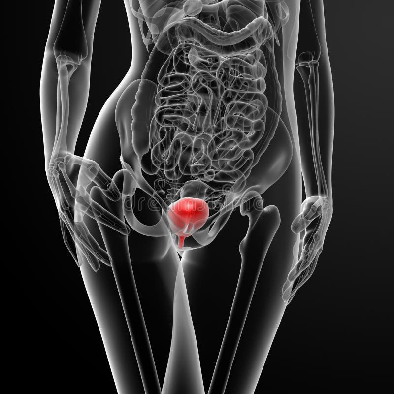
Free with trial 3d render female bladder anatomy x-ray - front view. View internal organs illustrations 3d render female bladder anatomy x-ray
Free with trial 3D illustration of semi-transparent female pelvis, with the uterus and ovaries rendered in soft pink hue. Two distinct, rounded fibroid nodules are prominently visible on the uterine wall. View internal organs illustrations 3D illustration of semi-transparent female pelvis, with the uterus and ovaries rendered in soft pink hue. Two distinct, rounded
Free with trial Realistic 3d illustration of cancer of human liver isolated on white. View internal organs illustrations Realistic illustration of cancer of human liver. Realistic 3d illustration of cancer of human liver isolated on white
Free with trial 3D anatomical diptych showing comparison of ovaries. Left ovary smooth and normal, right ovary enlarged with multiple peripheral cysts characteristic of PCOS. Clean clinical white background, Generative AI. View internal organs illustrations 3D anatomical diptych showing comparison of ovaries. Left ovary smooth and normal, right ovary enlarged with multiple peripheral
Free with trial 3d render female bladder anatomy x-ray - top view. View internal organs illustrations 3d render female bladder anatomy x-ray
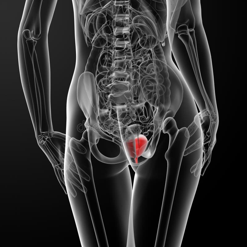
Free with trial 3d render female bladder anatomy x-ray - back view. View internal organs illustrations 3d render female bladder anatomy x-ray
Free with trial 3d render female bladder anatomy x-ray - side view. View internal organs illustrations 3d render female bladder anatomy x-ray
Free with trial 3d render female bladder anatomy x-ray - front view. View internal organs illustrations 3d render female bladder anatomy x-ray
Free with trial Flu viruses in human lungs, 3D illustration showing anatomy of human respiratory system and close-up view of influenza virus inside lungs. View internal organs illustrations Flu viruses in human lungs
Free with trial Medical abdominal ultrasonography concept. Sonogram of the stomach in flat style and set of abdomen organs. View internal organs illustrations Abdominal ultrasonography concept
Free with trial Influenza complication, conceptual image showing brain infection encephalitis with close-up view of flu viruses, 3D illustration. View internal organs illustrations Influenza complication encephalitis. Influenza complication, conceptual image showing brain infection encephalitis with close-up view of flu viruses, 3D illustration
Free with trial Human body organs Illustration of blood vessels: smooth blood muddy blood. View internal organs vectors Illustration of blood vessels: smooth blood muddy blood
Free with trial Human body infographic, man and organs highlighted in colors, lungs and brain, digestive system, vector illustration isolated on white background. View internal organs vectors Human Body Infographic Man Vector Illustration. Human body infographic, man and organs highlighted in colors, lungs and brain, digestive system, vector illustration isolated on white background
Free with trial Hand Drawn Coloured Human Brain on the Color Background with the Caption of Brain Areas or Lobes. Cerebral Cortex Areas. Free Hand Style Vector. View internal organs vectors Coloured Human Brain Areas. Lateral View. Hand Drawn Coloured Human Brain on the Color Background with the Caption of Brain Areas or Lobes. Cerebral Cortex Areas. Free Hand Style Vector.
Free with trial 3D illustration urinary system, kidneys, ureters and urinary bladder. - Ilustración. View internal organs illustrations 3D illustration urinary system, kidneys, ureters and urinary bladder. - Ilustración
Free with trial Medical illustration 3D human body kidneys, urethra, urinary bladder. View internal organs illustrations 3D illustration urinary system, kidneys, ureters and urinary bladder. - Ilustración. Medical illustration 3D human body kidneys, urethra, urinary bladder
Free with trial 3D illustration human body transparent prominent brain - Ilustración. View internal organs illustrations 3D illustration human body transparent prominent brain - Ilustración
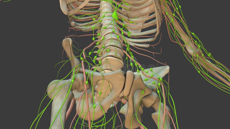
Free with trial Anatomy animation of the human lymphatic system is a visual representation or simulation that provides a detailed and dynamic view of the lymphatic system in the human body. The lymphatic system is a complex network of vessels, nodes, and organs that play a crucial role in maintaining the body's immune function, fluid balance, and the circulation of lymphatic fluid. View internal organs illustrations Human Lymphatic System 3d animation. Anatomy animation of the human lymphatic system is a visual representation or simulation that provides a detailed and dynamic view of the lymphatic system in the human body. The lymphatic system is a complex network of vessels, nodes, and organs that play a crucial role in maintaining the body's immune function, fluid balance, and the circulation of lymphatic fluid.
Free with trial Medical illustration 3D human body kidneys, urethra, urinary bladder. View internal organs illustrations 3D illustration urinary system, kidneys, ureters and urinary bladder. - Ilustración. Medical illustration 3D human body kidneys, urethra, urinary bladder
Free with trial A detailed image of a blood vessel in section, isolated on a white background. Ideal for medical, anatomical, or healthcare-related topics. View internal organs illustrations Blood Vessel In Section Isolated on White Background. A detailed image of a blood vessel in section, isolated on a white background. Ideal for medical, anatomical, or healthcare-related topics.
Free with trial Medical illustration of human stomach and lymphatic system. The image displays an anatomical view, highlighting the stomach, lymphatic vessels and lymph nodes. The colors are realistic and the image's style is detailed and educational. The background is a gradient of a muted red, complementing the detailed anatomy. This illustration could be used in educational materials, medical textbooks, or. View internal organs illustrations Stomach and lymphatic system anatomy illustration. Medical illustration of human stomach and lymphatic system. The image displays an anatomical view, highlighting the stomach, lymphatic vessels and lymph nodes. The colors are realistic and the image's style is detailed and educational. The background is a gradient of a muted red, complementing the detailed anatomy. This illustration could be used in educational materials, medical textbooks, or
Free with trial A high-resolution 3D rendering showcasing a detailed anatomical view of human lungs. The image clearly depicts the intricate bronchial tree branching throughout the lung tissue, set against a clean, neutral background. Ideal for medical education, publications, or presentations related to respiratory health, anatomy, and physiology. View internal organs illustrations Detailed 3D Render of Human Lungs with Bronchial Tree. A high-resolution 3D rendering showcasing a detailed anatomical view of human lungs. The image clearly depicts the intricate bronchial tree branching throughout the lung tissue, set against a clean, neutral background. Ideal for medical education, publications, or presentations related to respiratory health, anatomy, and physiology.
Free with trial High-quality 3D medical illustration of human lungs, showcasing the intricate bronchial tree structure. The image provides a clear and detailed view of the branching airways within the lungs, perfect for educational purposes, medical presentations, or anatomical studies. The realistic rendering and clear presentation make it suitable for use in textbooks, scientific articles, or healthcare websites. View internal organs illustrations Detailed 3D Anatomical Model of Human Lungs with Bronchial Tree. High-quality 3D medical illustration of human lungs, showcasing the intricate bronchial tree structure. The image provides a clear and detailed view of the branching airways within the lungs, perfect for educational purposes, medical presentations, or anatomical studies. The realistic rendering and clear presentation make it suitable for use in textbooks, scientific articles, or healthcare websites.
Free with trial Detailed anatomical representation of the human tongue and mouth. The image features a realistic, detailed view of the tongue and mouth structures. The colors are in a realistic, anatomical palette, with shades of pink and light peach for the tongue and surrounding tissues. The image has an overall detailed, medical aesthetic. The image presents a scientific and accurate view for medical or. View internal organs illustrations Tongue anatomy diagram illustration. Detailed anatomical representation of the human tongue and mouth. The image features a realistic, detailed view of the tongue and mouth structures. The colors are in a realistic, anatomical palette, with shades of pink and light peach for the tongue and surrounding tissues. The image has an overall detailed, medical aesthetic. The image presents a scientific and accurate view for medical or
Free with trial Detailed cutaway view of the human stomach. The stomach's inner lining and major blood vessels are visible. The image has a realistic, 3D style. The colors are light, desaturated pinks, reds, and light blues. The lighting is even, providing a clear anatomical study. The image is suitable for medical education, health presentations, and anatomical illustrations. View internal organs illustrations Human stomach cutaway medical illustration. Detailed cutaway view of the human stomach. The stomach's inner lining and major blood vessels are visible. The image has a realistic, 3D style. The colors are light, desaturated pinks, reds, and light blues. The lighting is even, providing a clear anatomical study. The image is suitable for medical education, health presentations, and anatomical illustrations
Free with trial This vivid 3D medical illustration provides a detailed view of the female reproductive system, showcasing the uterus and ovaries prominently. View internal organs illustrations 3D medical illustration of the female reproductive system highlighting the uterus and ovaries. This vivid 3D medical illustration provides a detailed view of the female reproductive system, showcasing the uterus and ovaries prominently.
Free with trial 3D illustration of the human heart in a close-up cross-section showing blocked arteries. This is a detailed closeup image portraying a human heart, set against a clean, white background that enhances its vivid features. View internal organs illustrations 3D illustration of the human heart in a close-up cross-section showing blocked arteries.A close up image of a human heart placed. 3D illustration of the human heart in a close-up cross-section showing blocked arteries.This is a detailed closeup image portraying a human heart, set against a clean, white background that enhances its vivid features
Free with trial An anatomical illustration depicts the muscles of the lower back. Key muscles include the erector spinae, located parallel to the spine, the multifidus, situated beneath the erector spinae, and the quadratus lumborum, positioned at the lower back's side. The spine is centrally aligned, with muscles highlighted in distinct colors for clarity. Labels indicate specific muscle names and locations, providing a clear visual guide to the musculature in this region. View internal organs illustrations Anatomical illustration showing the muscles of the lower back, including erector spinae, multifidus, and quadratus lumborum. An anatomical illustration depicts the muscles of the lower back. Key muscles include the erector spinae, located parallel to the spine, the multifidus, situated beneath the erector spinae, and the quadratus lumborum, positioned at the lower back's side. The spine is centrally aligned, with muscles highlighted in distinct colors for clarity. Labels indicate specific muscle names and locations, providing a clear visual guide to the musculature in this region.
Free with trial Clean 3D render of semi-transparent female pelvic model showing uterus and enlarged ovaries with multiple cystic follicles in ring pattern, characteristic of polycystic ovary syndrome, Generative AI. View internal organs illustrations Clean 3D render of semi-transparent female pelvic model showing uterus and enlarged ovaries with multiple cystic follicles in ring
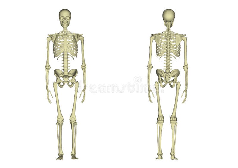
Free with trial Skeleton. Medical picture. The human skeleton. Internal organs. 3d rendering. View internal organs illustrations Skeleton on a white background. Front view. Back view. Anatomy. Part of the body. Medical examination. Human. 3d rendering. Skeleton. Medical picture. The human skeleton. Internal organs. 3d rendering.
Free with trial A digital illustration depicts a rabbit in side view facing left against a black background, with its body divided into three anatomical sections revealing internal organs highlighted in pink and purple tones, featuring an alert and curious expression with perked ears, elongated muscular body, prominently displayed heart centered in the chest flanked by smaller organs, lungs connected via white. View internal organs illustrations Digital illustration of a rabbit anatomy with visible internal organs in pink and purple on black background side view. A digital illustration depicts a rabbit in side view facing left against a black background, with its body divided into three anatomical sections revealing internal organs highlighted in pink and purple tones, featuring an alert and curious expression with perked ears, elongated muscular body, prominently displayed heart centered in the chest flanked by smaller organs, lungs connected via white
Free with trial Human body anatomy showing detailed internal organs like heart lungs and kidneys in a transparent blue light view. View internal organs illustrations Human body anatomy showing detailed internal organs like heart lungs and kidneys in a transparent blue light view
Free with trial Profile view of a futuristic female cyborg with transparent body showcasing internal organs and skeleton. View internal organs illustrations Futuristic transparent female cyborg showing internal organs and skeleton in profile view. Profile view of a futuristic female cyborg with transparent body showcasing internal organs and skeleton
Free with trial Detailed anatomical illustration of the human body displaying internal organs muscles and skeletal structure. The artwork provides a clear view of the abdomen and chest area in anatomical representation. View internal organs illustrations Human body anatomy illustration displaying the internal organs muscles and skeletal structure in a detailed anatomical view. Detailed anatomical illustration of. Detailed anatomical illustration of the human body displaying internal organs muscles and skeletal structure. The artwork provides a clear view of the abdomen and chest area in anatomical representation
Free with trial An x-ray view of a human body with internal organs. The skeleton and organs are visible. The image is isolated on a white background, showing the internal structure. View internal organs vectors Illustration of human body with internal organs isolated on transparent background, xray view. An x-ray view of a human body with internal organs. The skeleton and organs are visible. The image is isolated on a white background, showing the internal structure
Free with trial Frog inner anatomy scheme hand drawn side view detailed illustration. Amphibia internal organs anatomical structure on white background. Frog inner anatomy for zoology study, print table design. View internal organs illustrations Frog inner anatomy scheme hand drawn side view detailed illustration. Amphibia internal organs anatomical structure on
Free with trial 3d a detailed X-ray of a fish, revealing its internal skeletal structure. The spine, ribs, and other bones are clearly visible, along with the internal organs. The is in black and white, providing a stark contrast that highlights the anatomical details. The composition focuses on the side view of the fish, showcasing the intricate arrangement of its skeletal system. View internal organs illustrations Xray of fish skeleton with visible internal organs and spine 1. 3d a detailed X-ray of a fish, revealing its internal skeletal structure. The spine, ribs, and other bones are clearly visible, along with the internal organs. The is in black and white, providing a stark contrast that highlights the anatomical details. The composition focuses on the side view of the fish, showcasing the intricate arrangement of its skeletal system
Free with trial 3d a detailed x-ray of a fish skeleton, revealing its internal structure. The fish's bones, including the spine, ribs, and vertebrae, are clearly visible. The internal organs are also discernible, providing a comprehensive view of the fish's anatomy. The x-ray is set against a highlighting the blue hues of the skeletal structure. View internal organs illustrations Xray of fish skeleton with visible internal organs 3. 3d a detailed x-ray of a fish skeleton, revealing its internal structure. The fish's bones, including the spine, ribs, and vertebrae, are clearly visible. The internal organs are also discernible, providing a comprehensive view of the fish's anatomy. The x-ray is set against a highlighting the blue hues of the skeletal structure
Free with trial 3d This is an x-ray of a fish, revealing its internal skeleton and organs. The detailed structure of the fish's spine, ribs, fins, and other anatomical The black and white contrast highlights the intricate details of the fish's internal composition, providing a clear view of its skeletal system and internal organs. View internal organs illustrations X-ray of a fish showing internal skeleton and organs. 3d This is an x-ray of a fish, revealing its internal skeleton and organs. The detailed structure of the fish's spine, ribs, fins, and other anatomical The black and white contrast highlights the intricate details of the fish's internal composition, providing a clear view of its skeletal system and internal organs
Free with trial 3d This is an x-ray of a fish skeleton with visible internal organs. The detailed bone structure, including the spine, ribs, and vertebrae. The internal organs are also visible, providing a clear view of the fish's anatomy. The is in black and white, highlighting the contrast between the bones and the softer tissues. This type of imaging is commonly used in scientific and medical studies to. View internal organs illustrations Xray of a fish skeleton with visible internal organs 3. 3d This is an x-ray of a fish skeleton with visible internal organs. The detailed bone structure, including the spine, ribs, and vertebrae. The internal organs are also visible, providing a clear view of the fish's anatomy. The is in black and white, highlighting the contrast between the bones and the softer tissues. This type of imaging is commonly used in scientific and medical studies to
Free with trial 3d lungs pneumonia visualization - human body internal organs anatomy with red inflammation, front and back view on blue bg. Respiratory infection, disease treatment, medical concept. 3D illustration. View internal organs illustrations 3d lungs pneumonia visualization - human body internal organs anatomy with red inflammation, front and back view on blue bg
Free with trial 3d hepatitis and liver cirrhosis - human body internal organs anatomy model with acute kidney injury, front and back view on blue medical bg. Hepatorenal syndrome, disease treatment. 3D illustration. View internal organs illustrations 3d hepatitis and liver cirrhosis - human body internal organs anatomy model with acute kidney injury, front and back view on blue