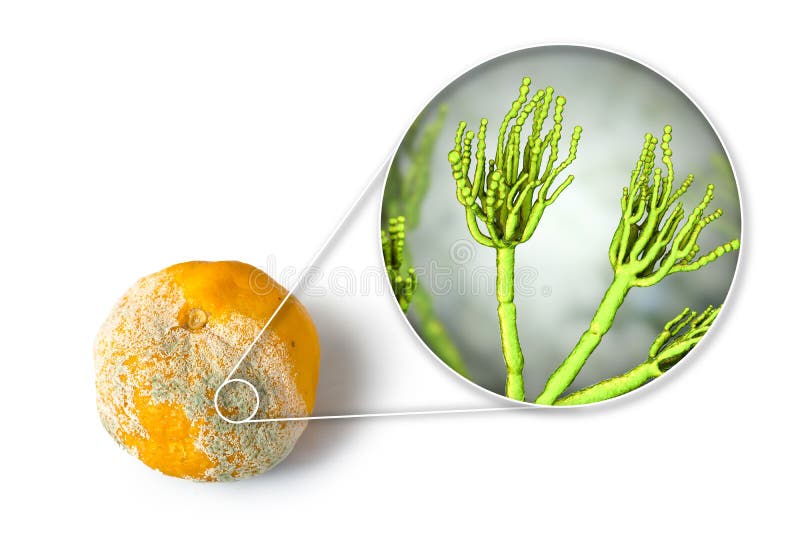
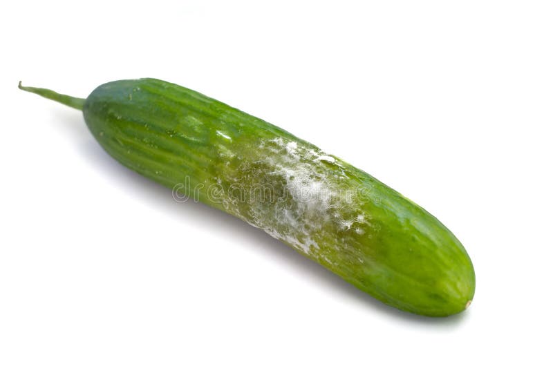
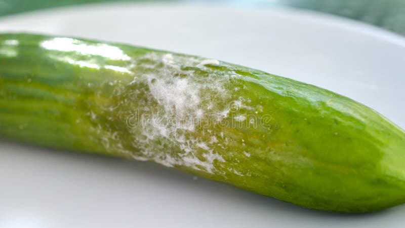
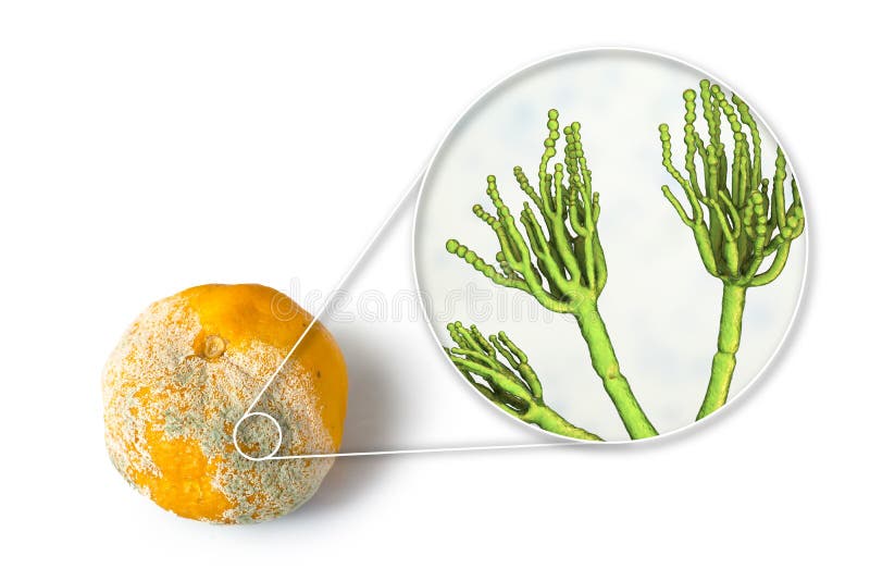
Search among 22 authentic chrysogenum stock photos, high-definition images, and pictures, or look at other food spoilage or fungi penicillium stock images to enhance your presentation with the perfect visual.

An ultrastructural view of Penicillium chrysogenum using color-enhanced DIC microscopy, capturing the release of spores from the sporangia. The image provides a detailed look at the mechanisms of spore dispersal in this medically significant mold. P. chrysogenum is the primary industrial source for the antibiotic penicillin. Chrysogenum stock images, royalty-free photos and pictures
Negative staining electron micrograph showing the brush-like conidiophores of Penicillium chrysogenum. The image provides high-surface detail of the chains of asexual conidia (spores). Chrysogenum stock images, royalty-free photos and pictures
Differential interference contrast micrograph of Penicillium notatum showing the internal ultrastructure of the ascus. Multiple ascospores are visible contained within the reproductive sac. Chrysogenum stock images, royalty-free photos and pictures
This phase contrast micrograph shows the dense, hardened resting bodies known as sclerotia of the fungus Penicillium notatum. The image reveals the compact cellular organization and protective outer layers of the fungal tissue. These structures are vital for the long-term survival of the organism in adverse environmental conditions. Chrysogenum stock images, royalty-free photos and pictures