Free with trial Nissl bodies stained with cresyl violet. Motor neuron located in the ventral horn of the spinal cord. Cresyl stock images, royalty-free photos and pictures Neuron. Nissl bodies. Nissl bodies stained with cresyl violet. Motor neuron located in the ventral horn of the spinal cord.
Free with trial Motor neurons of the anterior horn of the spinal cord stained with cresyl violet. They contain in their cytoplasm coarse Nissl bodies and a large nuclei with a striking nucleolus. Cresyl stock images, royalty-free photos and pictures Motor neurons. Nissl bodies. Motor neurons of the anterior horn of the spinal cord stained with cresyl violet. They contain in their cytoplasm coarse Nissl bodies and a large nuclei with a striking nucleolus.
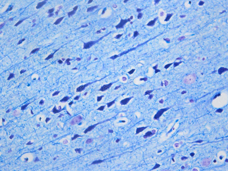
Free with trial Ever wonder what your brain looks like under the microscope? This is a 400x picture of the brain after it has been stained with luxol fast blue + cresyl violet. This stain reveals the nerve bodies (dark blue), their axons (blue lines) and the nuclei (light purple). Cresyl stock images, royalty-free photos and pictures Histology of the normal brain using LFB/CV. Ever wonder what your brain looks like under the microscope? This is a 400x picture of the brain after it has been stained with luxol fast blue + cresyl violet. This stain reveals the nerve bodies (dark blue), their axons (blue lines) and the nuclei (light purple).
Free with trial Neuronal cell bodies stained with cresyl violet to show the Nissl bodies. In the cerebellar cortex, the staining of the cell bodies of Purkinje neurons and the granular layer stand out. Cresyl stock images, royalty-free photos and pictures Cerebellar cortex. Cresyl violet. Neuronal cell bodies stained with cresyl violet to show the Nissl bodies. In the cerebellar cortex, the staining of the cell bodies of Purkinje neurons and the granular layer stand out
Free with trial Microscope and tissue staining kit for making biopsy. Cresyl stock images, royalty-free photos and pictures Microscope and tissue staining kit
Free with trial Light micrograph of cerebellar folium stained with cresyl violet showing from left to right: a thin meningeal layer (pia mater), molecular layer, Purkinje cell layer with large cell bodies, granular layer with many small granule cells and central axis of white matter. Cresyl stock images, royalty-free photos and pictures Cerebellar folium. Light micrograph of cerebellar folium stained with cresyl violet showing from left to right: a thin meningeal layer (pia mater), molecular. Light micrograph of cerebellar folium stained with cresyl violet showing from left to right: a thin meningeal layer (pia mater), molecular layer, Purkinje cell layer with large cell bodies, granular layer with many small granule cells and central axis of white matter
Free with trial Motor neurons of the anterior horn of the spinal cord stained with a basic dye, the cresyl violet. They contain in their cytoplasm coarse Nissl bodies and a large nucleus with a striking nucleolus. Cresyl stock images, royalty-free photos and pictures Spinal cord. Motor neurons of anterior horn. Motor neurons of the anterior horn of the spinal cord stained with a basic dye, the cresyl violet. They contain in their cytoplasm coarse Nissl bodies and a large nucleus with a striking nucleolus
Free with trial Motor neurons of the anterior horn of the spinal cord stained with a basic dye, the cresyl violet. They contain in their cytoplasm coarse Nissl bodies and a large nucleus with a striking nucleolus. Cresyl stock images, royalty-free photos and pictures Spinal cord. Motor neurons of anterior horn. Motor neurons of the anterior horn of the spinal cord stained with a basic dye, the cresyl violet. They contain in their cytoplasm coarse Nissl bodies and a large nucleus with a striking nucleolus
Free with trial Motor neurons of the anterior horn of the spinal cord stained with a basic dye, the cresyl violet. They contain in their cytoplasm coarse Nissl bodies and a large nucleus with a striking nucleolus. Cresyl stock images, royalty-free photos and pictures Spinal cord. Motor neurons of anterior horn. Motor neurons of the anterior horn of the spinal cord stained with a basic dye, the cresyl violet. They contain in their cytoplasm coarse Nissl bodies and a large nucleus with a striking nucleolus
Free with trial Motor neurons of the anterior horn of the spinal cord stained with a basic dye, the cresyl violet. They contain in their cytoplasm coarse Nissl bodies and a large nucleus with a striking nucleolus. Cresyl stock images, royalty-free photos and pictures Spinal cord. Motor neurons of anterior horn. Motor neurons of the anterior horn of the spinal cord stained with a basic dye, the cresyl violet. They contain in their cytoplasm coarse Nissl bodies and a large nucleus with a striking nucleolus
Free with trial Motor neurons of the anterior horn of the spinal cord stained with a basic dye, the cresyl violet. They contain in their cytoplasm coarse Nissl bodies and a large nucleus with a striking nucleolus. Cresyl stock images, royalty-free photos and pictures Spinal cord. Motor neurons of anterior horn. Motor neurons of the anterior horn of the spinal cord stained with a basic dye, the cresyl violet. They contain in their cytoplasm coarse Nissl bodies and a large nucleus with a striking nucleolus
Free with trial Motor neurons of the anterior horn of the spinal cord stained with a basic dye, the cresyl violet. They contain in their cytoplasm coarse Nissl bodies and a large nucleus with a striking nucleolus. Cresyl stock images, royalty-free photos and pictures Spinal cord. Motor neurons of anterior horn. Motor neurons of the anterior horn of the spinal cord stained with a basic dye, the cresyl violet. They contain in their cytoplasm coarse Nissl bodies and a large nucleus with a striking nucleolus
Free with trial Neuronal cell bodies stained with cresyl violet to show the Nissl bodies. In the cerebellar cortex, the staining of the cell bodies of Purkinje neurons and the granular layer stand out. Cresyl stock images, royalty-free photos and pictures Cerebellar cortex. Cresyl violet. Neuronal cell bodies stained with cresyl violet to show the Nissl bodies. In the cerebellar cortex, the staining of the cell bodies of Purkinje neurons and the granular layer stand out
Free with trial Motor neurons of the anterior horn of the spinal cord stained with a basic dye, the cresyl violet. They contain in their cytoplasm coarse Nissl bodies and a large nucleus with a striking nucleolus. Cresyl stock images, royalty-free photos and pictures Spinal cord. Motor neurons of anterior horn. Motor neurons of the anterior horn of the spinal cord stained with a basic dye, the cresyl violet. They contain in their cytoplasm coarse Nissl bodies and a large nucleus with a striking nucleolus
Free with trial Motor neurons of the anterior horn of the spinal cord stained with a basic dye, the cresyl violet. They contain in their cytoplasm coarse Nissl bodies and a large nucleus with a striking nucleolus. Cresyl stock images, royalty-free photos and pictures Spinal cord. Motor neurons of anterior horn. Motor neurons of the anterior horn of the spinal cord stained with a basic dye, the cresyl violet. They contain in their cytoplasm coarse Nissl bodies and a large nucleus with a striking nucleolus
Free with trial Motor neurons of the anterior horn of the spinal cord stained with a basic dye, the cresyl violet. They contain in their cytoplasm coarse Nissl bodies and a large nucleus with a striking nucleolus. Cresyl stock images, royalty-free photos and pictures Spinal cord. Motor neurons of anterior horn. Motor neurons of the anterior horn of the spinal cord stained with a basic dye, the cresyl violet. They contain in their cytoplasm coarse Nissl bodies and a large nucleus with a striking nucleolus
Free with trial Cerebellar cortex showing from top to bottom: molecular layer, Purkinje cell layer, and granular layer. A Golgi cell can be seen in the granular layer. Cresyl violet stain. Cresyl stock images, royalty-free photos and pictures Cerebellar cortex. Golgi cell. Cerebellar cortex showing from top to bottom: molecular layer, Purkinje cell layer, and granular layer. A Golgi cell can be seen in the granular layer. Cresyl violet stain
Free with trial Hippocampus or Ammon horn showing, from top, stratum oriens, cell bodies of neurons of pyramidal layer with stained Nissl bodies and stratum radiatum. Cresyl violet. Cresyl stock images, royalty-free photos and pictures Ammon horn. Hippocampus. Hippocampus or Ammon horn showing, from top, stratum oriens, cell bodies of neurons of pyramidal layer with stained Nissl bodies and stratum radiatum. Cresyl violet
Free with trial Hippocampus or Ammon horn showing, from top, stratum oriens, cell bodies of neurons of pyramidal layer with stained Nissl bodies and stratum radiatum. Cresyl violet. Cresyl stock images, royalty-free photos and pictures Ammon horn. Hippocampus. Hippocampus or Ammon horn showing, from top, stratum oriens, cell bodies of neurons of pyramidal layer with stained Nissl bodies and stratum radiatum. Cresyl violet
Free with trial Dorsal root ganglion neurons. The micrograph shows small, thin Nissl bodies characteristic of sensitive neurons and a large nucleus with a prominent nucleolus. The pale region in the cytoplasm is the axon hillock. The small dense nuclei correspond to? of? satellite glial cells. Cresyl violet. Cresyl stock images, royalty-free photos and pictures Dorsal root ganglion. Nissl bodies. Dorsal root ganglion neurons. The micrograph shows small, thin Nissl bodies characteristic of sensitive neurons and a large nucleus with a prominent nucleolus. The pale region in the cytoplasm is the axon hillock. The small dense nuclei correspond to? of? satellite glial cells. Cresyl violet
Free with trial Cross section of a spinal cord stained with cresyl violet. The central gray matter shows many neuronal somata. In the large anterior horns, big motor neurons bottom are observed. Cresyl stock images, royalty-free photos and pictures Spinal cord. Cresyl violet stain. Cross section of a spinal cord stained with cresyl violet. The central gray matter shows many neuronal somata. In the large anterior horns, big motor neurons bottom are observed.
Free with trial Motor neuron of the anterior horn of the spinal cord stained with cresyl violet. They contain in their cytoplasm coarse Nissl bodies and a large nuclei with a striking nucleolus. Cresyl stock images, royalty-free photos and pictures Motor neuron. Nissl bodies. Motor neuron of the anterior horn of the spinal cord stained with cresyl violet. They contain in their cytoplasm coarse Nissl bodies and a large nuclei with a striking nucleolus
Free with trial Four cerebellum sections stained with H&E top right, cresyl violet top right, silver nitrate method for nerve fibers bottom left and Kluver-Barrera technique for myelinated fibers bottom right. Cresyl stock images, royalty-free photos and pictures Cerebellum. Comparison of four stain methods. Four cerebellum sections stained with H&E top right, cresyl violet top right, silver nitrate method for nerve fibers bottom left and Kluver-Barrera technique for myelinated fibers bottom right
Free with trial Light micrograph of motor neurons from the spinal cord stained with cresyl violet. The soma of the neurons shows coarse Nissl bodies in their cytoplasm and a large nuclei with a striking nucleolus. Cresyl stock images, royalty-free photos and pictures Spinal cord. Nissl bodies. Light micrograph of motor neurons from the spinal cord stained with cresyl violet. The soma of the neurons shows coarse Nissl bodies in their cytoplasm and a large nuclei with a striking nucleolus
Free with trial Light micrograph of the hippocampus (allocortex), example of the archicortex, showing the Ammon horn (curve open to the right), the dentate gyrus (curve open to the left), and, on bottom, the fimbria of hippocampus. Cresyl violet stain. Cresyl stock images, royalty-free photos and pictures Hippocampus. Ammon’s horn. Light micrograph of the hippocampus (allocortex), example of the archicortex, showing the Ammon horn (curve open to the right), the dentate gyrus (curve open to the left), and, on bottom, the fimbria of hippocampus. Cresyl violet stain
Free with trial Light microscope micrograph showing neurons in a dorsal root ganglion stained with cresyl violet. They are pseudounipolar neurons of rounded soma showing small, thin Nissl bodies characteristic of sensitive neurons and a large nucleus with a prominent nucleolus. Each cell body is surrounded by small dense nuclei corresponding to glia cells called satellite cells. Cresyl stock images, royalty-free photos and pictures Neurons. Dorsal root ganglion. Light microscope micrograph showing neurons in a dorsal root ganglion stained with cresyl violet. They are pseudounipolar neurons of rounded soma showing small, thin Nissl bodies characteristic of sensitive neurons and a large nucleus with a prominent nucleolus. Each cell body is surrounded by small dense nuclei corresponding to glia cells called satellite cells
Free with trial Light microscope micrograph showing neurons in a dorsal root ganglion stained with cresyl violet. They are pseudounipolar neurons of rounded soma showing small, thin Nissl bodies characteristic of sensitive neurons and a large nucleus with a prominent nucleolus. Each cell body is surrounded by small dense nuclei corresponding to glia cells called satellite cells. Cresyl stock images, royalty-free photos and pictures Neurons. Dorsal root ganglion. Light microscope micrograph showing neurons in a dorsal root ganglion stained with cresyl violet. They are pseudounipolar neurons of rounded soma showing small, thin Nissl bodies characteristic of sensitive neurons and a large nucleus with a prominent nucleolus. Each cell body is surrounded by small dense nuclei corresponding to glia cells called satellite cells
Free with trial Light microscope micrograph showing neurons in a dorsal root ganglion stained with cresyl violet. They are pseudounipolar neurons of rounded soma showing small, thin Nissl bodies characteristic of sensitive neurons and a large nucleus with a prominent nucleolus. Each cell body is surrounded by small dense nuclei corresponding to glia cells called satellite cells. The large neuron located on right. Cresyl stock images, royalty-free photos and pictures Neurons. Dorsal root ganglion. Light microscope micrograph showing neurons in a dorsal root ganglion stained with cresyl violet. They are pseudounipolar neurons of rounded soma showing small, thin Nissl bodies characteristic of sensitive neurons and a large nucleus with a prominent nucleolus. Each cell body is surrounded by small dense nuclei corresponding to glia cells called satellite cells. The large neuron located on right
Free with trial Light microscope micrograph showing neurons in a dorsal root ganglion stained with cresyl violet. They are pseudounipolar neurons of rounded soma showing small, thin Nissl bodies characteristic of sensitive neurons and a large nucleus with a prominent nucleolus. Each cell body is surrounded by small dense nuclei corresponding to glia cells called satellite cells. The large neuron located on the. Cresyl stock images, royalty-free photos and pictures Neurons. Dorsal root ganglion. Light microscope micrograph showing neurons in a dorsal root ganglion stained with cresyl violet. They are pseudounipolar neurons of rounded soma showing small, thin Nissl bodies characteristic of sensitive neurons and a large nucleus with a prominent nucleolus. Each cell body is surrounded by small dense nuclei corresponding to glia cells called satellite cells. The large neuron located on the
Free with trial Light microscope micrograph showing neurons in a dorsal root ganglion stained with cresyl violet. They are pseudounipolar neurons of rounded soma showing small, thin Nissl bodies characteristic of sensitive neurons and a large nucleus with a prominent nucleolus. Each cell body is surrounded by small dense nuclei corresponding to glia cells called satellite cells. Cresyl stock images, royalty-free photos and pictures Neurons. Dorsal root ganglion. Light microscope micrograph showing neurons in a dorsal root ganglion stained with cresyl violet. They are pseudounipolar neurons of rounded soma showing small, thin Nissl bodies characteristic of sensitive neurons and a large nucleus with a prominent nucleolus. Each cell body is surrounded by small dense nuclei corresponding to glia cells called satellite cells
Free with trial Light microscope micrograph showing neurons in a dorsal root ganglion stained with cresyl violet. They are pseudounipolar neurons of rounded soma showing small, thin Nissl bodies characteristic of sensitive neurons and a large nucleus with a prominent nucleolus. Each cell body is surrounded by small dense nuclei corresponding to glia cells called satellite cells. Cresyl stock images, royalty-free photos and pictures Neurons. Dorsal root ganglion. Light microscope micrograph showing neurons in a dorsal root ganglion stained with cresyl violet. They are pseudounipolar neurons of rounded soma showing small, thin Nissl bodies characteristic of sensitive neurons and a large nucleus with a prominent nucleolus. Each cell body is surrounded by small dense nuclei corresponding to glia cells called satellite cells
Free with trial Very low magnification micrograph of a sagittal section of a rat cerebellum stained with cresyl violet, showing the ramified cerebellar folia. Cresyl stock images, royalty-free photos and pictures Cerebellum. Cerebellar folia. Very low magnification micrograph of a sagittal section of a rat cerebellum stained with cresyl violet, showing the ramified cerebellar folia
Free with trial A large teak tree that was cut from the trunk and placed on the ground. Is a tree that has a very high economic value With a special characteristic that is softwood that is more durable than hardwood. Cresyl stock images, royalty-free photos and pictures A large teak tree that was cut from the trunk and placed on the ground