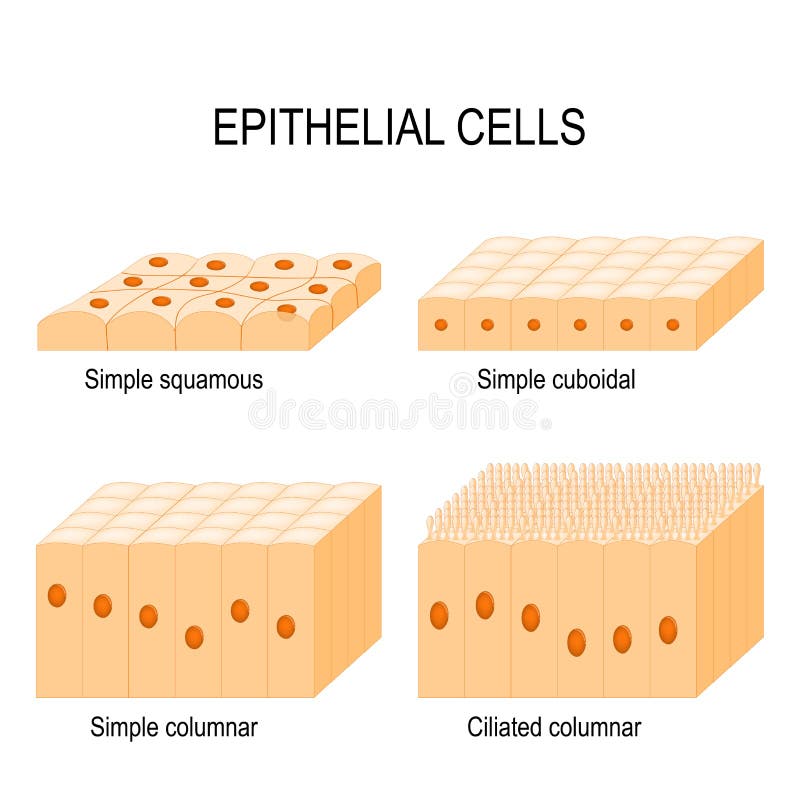
Free with trial Types of epithelial cells: cilliated columnar, simple columnar, simple cuboidal, and simple squamous cells. Ciliated vectors Types of epithelial cells. Cilliated columnar, simple columnar, simple cuboidal, and simple squamous cells
Free with trial Nasal mucosa cells. Nasal secretions. Ciliated, basal and goblet cells. Olfactory epithelium. Cells act as a low resistance filter. Vector illustration. Ciliated vectors Nasal mucosa cells. Nasal secretions. Ciliated, basal and goblet cells. Olfactory epithelium. Cells act as a low resistance filter. Vector illustration
Free with trial Types of epithelial cells: cilliated columnar, simple columnar, simple cuboidal, and simple squamous cells. Ciliated vectors Types of epithelial cells. Cilliated columnar, simple columnar, simple cuboidal, and simple squamous cells
Free with trial Nasal mucosa anatomy. Nasal mucous membrane lining the respiratory tract. Ciliated, basal and goblet cells. Flat vector illustration. Ciliated vectors Nasal mucosa anatomy. Nasal mucous membrane lining the respiratory tract. Ciliated, basal and goblet cells. Flat vector illustration
Free with trial Skin Cells and Structure Layers of epidermis. cornified stratum corneum, clear or translucent layer lucidum, granular stratum granulosum, spinous spinosum, basal or germinal layer stratum basale or germinativum. Ciliated vectors Skin Cells and Structure Layers of epidermis. Cornified stratum corneum, clear or translucent layer lucidum, granular stratum granulosum, spinous spinosum
Free with trial Simple columnar epithelium / Epithelial tissue. On white. Ciliated vectors Simple columnar epithelium / Epithelial tissue. On white
Free with trial Vaginal atrophy. Thinning, drying and inflammation of the vaginal walls that may occur when your body has less estrogen. Epithelial cells destruction. Flat vector illustration. Ciliated vectors Vaginal atrophy. Thinning, drying and inflammation of the vaginal walls. That may occur when your body has less estrogen. Epithelial cells destruction. Flat
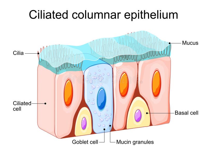
Free with trial Ciliated columnar epithelium. Epithelial cells forms the lining of the stomach and intestines, duodenum, fallopian tubes, uterus, central canal of the spinal cord, nose, ears and the taste buds. Nasal epithelium. Ciliated cells. Respiratory defense mechanisms. Vector illustration. Medical poster. Schematic diagram. Ciliated vectors Ciliated columnar epithelium. Nasal epithelium. Ciliated columnar epithelium. Epithelial cells forms the lining of the stomach and intestines, duodenum. Ciliated columnar epithelium. Epithelial cells forms the lining of the stomach and intestines, duodenum, fallopian tubes, uterus, central canal of the spinal cord, nose, ears and the taste buds. Nasal epithelium. Ciliated cells. Respiratory defense mechanisms. Vector illustration. Medical poster. Schematic diagram
Free with trial Respiratory Epithelium Cell Structure Illustration. Ciliated, Basal, and Goblet Cells Diagram. Mucociliary Epithelium. Cross Section Illustration. Ciliated vectors Respiratory Epithelium Cell Structure Illustration. Ciliated, Basal, and Goblet Cells Diagram. Mucociliary Epithelium. Cross Section Illustration
Free with trial Ciliated columnar epithelium. epithelial cells forms the lining of the stomach and intestines, duodenum, fallopian tubes, uterus, central canal of the spinal cord, nose, ears and the taste buds. Ciliated vectors Ciliated columnar epithelium. Epithelial cells forms the lining of the stomach and intestines, duodenum, fallopian tubes, uterus, central canal of the spinal
Free with trial Nasal epithelium. Ciliated columnar epithelium. epithelial cells forms the lining of the stomach and intestines, duodenum, fallopian tubes, uterus, central canal of the spinal cord, nose, ears and the taste buds. Ciliated cells. Respiratory defense mechanisms. Vector poster. Isometric Flat illustration. Ciliated vectors Nasal epithelium. Ciliated columnar epithelium. Epithelial cells forms the lining of the stomach and intestines, duodenum, fallopian tubes, uterus, central
Free with trial Ciliated epithelium medical background. Stock vector iluustration of typical human cell. Medicine and biology collection. Ciliated vectors Ciliated epithelium medical background. Stock vector iluustration of typical human cell. Medicine and biology collection
Free with trial Ciliated epithelium duct medical background. Stock vector iluustration of typical human cell. Medicine and biology collection. Ciliated vectors Ciliated epithelium medical background. Ciliated epithelium duct medical background. Stock vector iluustration of typical human cell. Medicine and biology. Ciliated epithelium duct medical background. Stock vector iluustration of typical human cell. Medicine and biology collection.
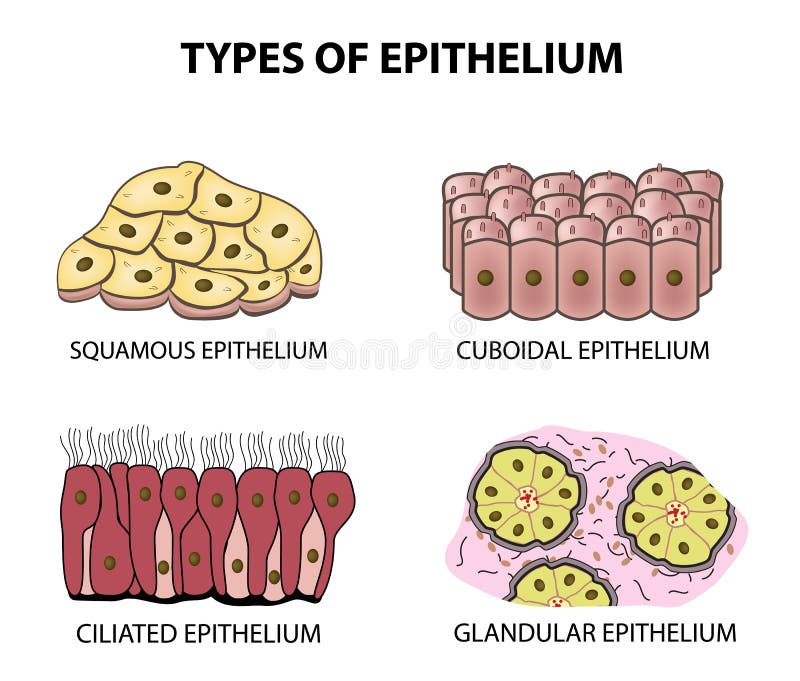
Free with trial Types of epithelium. squamous, cubic, ciliated, glandular. Set Infographics Vector illustration on background. Ciliated vectors Types of epithelium. squamous, cubic, ciliated, glandular. Set. Infographics. Vector illustration on background. Types of epithelium. squamous, cubic, ciliated. Types of epithelium. squamous, cubic, ciliated, glandular. Set Infographics Vector illustration on background
Free with trial Medicine Respiratory prismatic ciliated pseudostratified epithelium. The apical border of the epithelium has a layer of cilia supported in their basal bodies. Generative ai. Ciliated vectors Medicine Respiratory prismatic ciliated pseudostratified epithelium. The apical border of the epithelium has a layer of cilia. Supported in their basal bodies
Free with trial Medicine Respiratory prismatic ciliated pseudostratified epithelium. The apical border of the epithelium has a layer of cilia supported in their basal bodies. Generative ai. Ciliated vectors Medicine Respiratory prismatic ciliated pseudostratified epithelium. The apical border of the epithelium has a layer of cilia. Supported in their basal bodies
Free with trial Medicine Respiratory prismatic ciliated pseudostratified epithelium. The apical border of the epithelium has a layer of cilia supported in their basal bodies. Generative ai. Ciliated vectors Medicine Respiratory prismatic ciliated pseudostratified epithelium. The apical border of the epithelium has a layer of cilia. Supported in their basal bodies
Free with trial Medical illustration shows a comparison between normal ciliated respiratory epithelium and damaged epithelium with impaired mucociliary clearance. Healthy Versus Impaired Ciliated Epithelium. Ciliated vectors Medical illustration shows a comparison between normal ciliated respiratory epithelium and damaged epithelium with impaired. Mucociliary clearance. Healthy. Medical illustration shows a comparison between normal ciliated respiratory epithelium and damaged epithelium with impaired mucociliary clearance. Healthy Versus Impaired Ciliated Epithelium
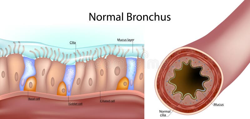
Free with trial Structure of a normal human bronchus. Cross-section of the airway and a close-up of the pseudostratified ciliated epithelium with ciliated cells, goblet cells, basal cells, and the mucus layer. Ciliated vectors Structure of a normal human bronchus. Cross-section of the airway and a close-up of the pseudostratified ciliated epithelium with. Ciliated cells, goblet cells. Structure of a normal human bronchus. Cross-section of the airway and a close-up of the pseudostratified ciliated epithelium with ciliated cells, goblet cells, basal cells, and the mucus layer.
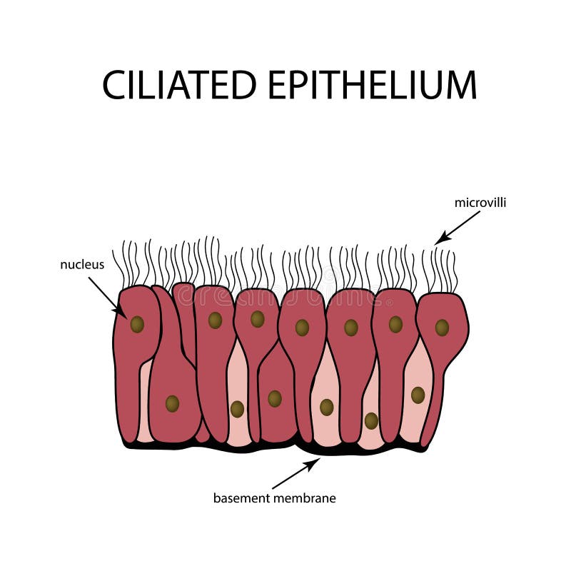
Free with trial The structure of the ciliated epithelium. Infographics. Vector illustration on background. Ciliated vectors The structure of the ciliated epithelium. Infographics. Vector illustration on background.
Free with trial Bronchus. Medical illustration depicting Respiratory cilia and mucus. Pseudostratified columnar epithelium. Structure. Ciliated Epithelium Infographics. Ciliated vectors Bronchus. Medical illustration depicting Respiratory cilia and mucus. Pseudostratified columnar epithelium. Structure. Ciliated Epithelium Infographics
Free with trial Nasal mucosa flat vector anatomical icon. Epithelium in the nose, gut, stomach or colon. Human olfactory system concept. Mucus, ciliated cells, villi and cilia poster for clinic or education. Ciliated vectors Epithelium of nasal mucosa. Nasal mucosa flat vector anatomical icon. Epithelium in the nose, gut, stomach or colon. Human olfactory system concept. Mucus. Nasal mucosa flat vector anatomical icon. Epithelium in the nose, gut, stomach or colon. Human olfactory system concept. Mucus, ciliated cells, villi and cilia poster for clinic or education.
Free with trial Nasal epithelium anatomical poster. Human respiratory system concept. Mucus, goblet, ciliated and basal cells. Smell organs respiratory system. Medical flat vector illustration for clinic or education. Ciliated vectors Nasal epithelium concept. Nasal epithelium anatomical poster. Human respiratory system concept. Mucus, goblet, ciliated and basal cells. Smell organs respiratory. Nasal epithelium anatomical poster. Human respiratory system concept. Mucus, goblet, ciliated and basal cells. Smell organs respiratory system. Medical flat vector illustration for clinic or education
Free with trial Nasal mucosa flat vector anatomical icon. Epithelium in the nose, gut, stomach or colon. Human olfactory system concept. Mucus, ciliated cells, villi and cilia poster for clinic or education. Ciliated vectors Epithelium of nasal mucosa. Nasal mucosa flat vector anatomical icon. Epithelium in the nose, gut, stomach or colon. Human olfactory system concept. Mucus. Nasal mucosa flat vector anatomical icon. Epithelium in the nose, gut, stomach or colon. Human olfactory system concept. Mucus, ciliated cells, villi and cilia poster for clinic or education.
Free with trial Nasal mucosa flat vector anatomical icon. Epithelium in the nose, gut, stomach or colon. Human olfactory system concept. Mucus, ciliated cells, villi and cilia poster for clinic or education. Ciliated vectors Epithelium of nasal mucosa. Nasal mucosa flat vector anatomical icon. Epithelium in the nose, gut, stomach or colon. Human olfactory system concept. Mucus. Nasal mucosa flat vector anatomical icon. Epithelium in the nose, gut, stomach or colon. Human olfactory system concept. Mucus, ciliated cells, villi and cilia poster for clinic or education.
Free with trial Nasal mucosa flat vector anatomical icon. Epithelium in the nose, gut, stomach or colon. Human olfactory system concept. Mucus, ciliated cells, villi and cilia poster for clinic or education. Ciliated vectors Epithelium of nasal mucosa. Nasal mucosa flat vector anatomical icon. Epithelium in the nose, gut, stomach or colon. Human olfactory system concept. Mucus. Nasal mucosa flat vector anatomical icon. Epithelium in the nose, gut, stomach or colon. Human olfactory system concept. Mucus, ciliated cells, villi and cilia poster for clinic or education.
Free with trial Nasal epithelium anatomical poster. Human respiratory system concept. Mucus, goblet, ciliated and basal cells. Smell organs respiratory system. Medical flat vector illustration for clinic or education. Ciliated vectors Nasal epithelium concept. Nasal epithelium anatomical poster. Human respiratory system concept. Mucus, goblet, ciliated and basal cells. Smell organs respiratory. Nasal epithelium anatomical poster. Human respiratory system concept. Mucus, goblet, ciliated and basal cells. Smell organs respiratory system. Medical flat vector illustration for clinic or education
Free with trial Nasal epithelium anatomical poster. Human respiratory system concept. Mucus, goblet, ciliated and basal cells. Smell organs respiratory system. Medical flat vector illustration for clinic or education. Ciliated vectors Nasal epithelium concept. Nasal epithelium anatomical poster. Human respiratory system concept. Mucus, goblet, ciliated and basal cells. Smell organs respiratory. Nasal epithelium anatomical poster. Human respiratory system concept. Mucus, goblet, ciliated and basal cells. Smell organs respiratory system. Medical flat vector illustration for clinic or education
Free with trial Olfactory Bulb Anatomy. Epithelium receptor cells. Nasal mucosa cells. Nasal mucosa. Ciliated vectors Olfactory Bulb Anatomy. Epithelium receptor cells. Nasal mucosa cells. Olfactory Bulb Anatomy. Epithelium receptor cells. Nasal mucosa
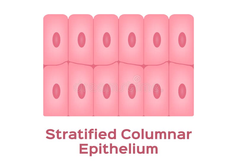
Free with trial Stratified columnar epithelium / Epithelial tissue. On white. Ciliated vectors Stratified columnar epithelium / Epithelial tissue. On white
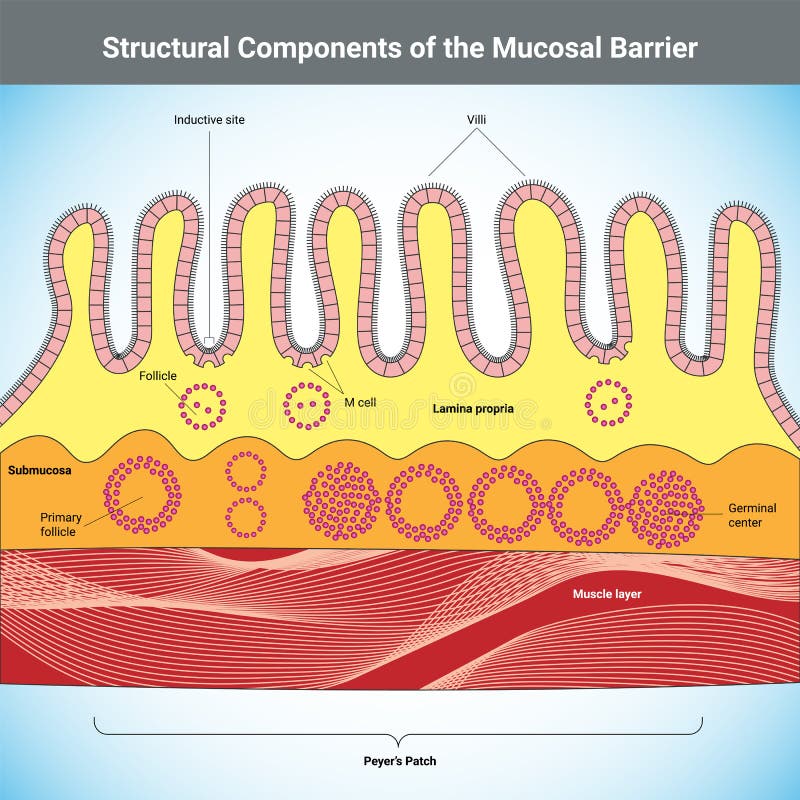
Free with trial Structural Components of the Mucosal Barrier medical illustration. Ciliated vectors Structural Components of the Mucosal Barrier medical illustration. Structural Components of the Mucosal Barrier vector medical illustration
Free with trial Goblet cells are specialized epithelial cells found in the lining of various organs, particularly in the respiratory and digestive tracts. They are named for their goblet-like shape, with a wide base and a narrower opening at the top. Goblet cells are responsible for secreting mucins, which are glycoproteins that form mucous. Ciliated vectors Goblet cell. S are specialized epithelial cells found in the lining of various organs, particularly in the respiratory and digestive tracts. They are named for. Goblet cells are specialized epithelial cells found in the lining of various organs, particularly in the respiratory and digestive tracts. They are named for their goblet-like shape, with a wide base and a narrower opening at the top. Goblet cells are responsible for secreting mucins, which are glycoproteins that form mucous.
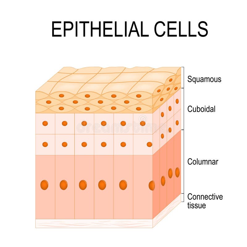
Free with trial Types of EpitheliumTypes of epithelium. Epithelial cells in a variety of configurations. Flat illustration. Ciliated vectors Types of epithelium. Epithelial cells in a variety of configurations. Types of EpitheliumTypes of epithelium. Epithelial cells in a variety of configurations. Types of EpitheliumTypes of epithelium. Epithelial cells in a variety of configurations. Flat illustration.
Free with trial Tall and slender cells with oval-shaped nuclei. Lines most organs of the digestive tract like stomach, intestines. Medical illustration. Vector. Ciliated vectors Simple columnar epithelium. Epithelial tissue types. Tall and slender cells with oval-shaped nuclei. Lines most organs of the digestive tract like stomach. Tall and slender cells with oval-shaped nuclei. Lines most organs of the digestive tract like stomach, intestines. Medical illustration. Vector.
Free with trial Single layer of cells. Each cell is in contact with the basement membrane. Found in respiratory tract, prostate, endometrium. Vector illustration. Ciliated vectors Pseudostratified epithelium. Epithelial tissue types. Single layer of cells. Each cell is in contact with the basement membrane. Found in respiratory tract. Single layer of cells. Each cell is in contact with the basement membrane. Found in respiratory tract, prostate, endometrium. Vector illustration
Free with trial Human Intestinal Cells seamless border. Unhelthy guts epithelium cells pattern flat vector illustration. Unhelthy guts. Ciliated vectors Human Intestinal Cells seamless border. Unhelthy guts epithelium cells pattern flat vector illustration. Human Intestinal Cells seamless border. Unhelthy guts
Free with trial Cellular metaplasia illustrated, airway epithelium shifts from ciliated columnar to stratified squamous under stress, lungs, cilia, squamous cells highlighted. Outline diagram. Ciliated vectors Cellular metaplasia illustrated, airway epithelium shifts from ciliated columnar to ... Cellular metaplasia illustrated, airway epithelium shifts from ciliated. Cellular metaplasia illustrated, airway epithelium shifts from ciliated columnar to stratified squamous under stress, lungs, cilia, squamous cells highlighted. Outline diagram
Free with trial Nasal Epithelium structure anatomy, labeled with cilia, mucus, ciliated cell, goblet cell, basal cell. Human tissue nasal cavity, immune response to nasal diseases, diagram biology education. Ciliated vectors Nasal Epithelium structure anatomy, labeled with cilia, mucus, ciliated cell, goblet cell, basal cell. Human tissue nasal cavity,. Immune response to nasal. Nasal Epithelium structure anatomy, labeled with cilia, mucus, ciliated cell, goblet cell, basal cell. Human tissue nasal cavity, immune response to nasal diseases, diagram biology education
Free with trial Nasal Epithelium structure anatomy, labeled with cilia, mucus, ciliated cell, goblet cell, basal cell. Human tissue nasal cavity, immune response to nasal diseases, diagram biology education. Ciliated vectors Nasal Epithelium structure anatomy, labeled with cilia, mucus, ciliated cell, goblet cell, basal cell. Human tissue nasal cavity,. Immune response to nasal. Nasal Epithelium structure anatomy, labeled with cilia, mucus, ciliated cell, goblet cell, basal cell. Human tissue nasal cavity, immune response to nasal diseases, diagram biology education
Free with trial Medical infographic diagram illustrating the diverse cell types of the respiratory or airway epithelium. This cross-section diagram displays various specialized cells aligned on the basement membrane, including ciliated cells, deuterosomal cells, club cells, tuft cells, goblet cells, basal cells, basal progenitor cells, suprabasal cells, ionocytes, and pulmonary neuroendocrine cells. Excellent for pulmonology, histology, anatomy textbooks, respiratory system studies, and medical education. Ciliated vectors Respiratory Airway Epithelium Cell Types Diagram Vector Illustration. Medical infographic diagram illustrating the diverse cell types of the respiratory or. Medical infographic diagram illustrating the diverse cell types of the respiratory or airway epithelium. This cross-section diagram displays various specialized cells aligned on the basement membrane, including ciliated cells, deuterosomal cells, club cells, tuft cells, goblet cells, basal cells, basal progenitor cells, suprabasal cells, ionocytes, and pulmonary neuroendocrine cells. Excellent for pulmonology, histology, anatomy textbooks, respiratory system studies, and medical education.
Free with trial A ciliated rod bacterium microbe germ silhouette for healthcare research, detailed graphic. Ciliated vectors Ciliated rod bacterium microbe germ silhouette for healthcare research. A ciliated rod bacterium microbe germ silhouette for healthcare research, ai generated. A ciliated rod bacterium microbe germ silhouette for healthcare research, detailed graphic
Free with trial Medical illustration showing damage to the respiratory epithelium, impaired cilia, increased mucus production, airway inflammation, and bacterial infection. Cross-section of the bronchial lumen. Ciliated vectors Medical illustration showing damage to the respiratory epithelium, impaired cilia, increased mucus production, airway inflammation. And bacterial infection. Medical illustration showing damage to the respiratory epithelium, impaired cilia, increased mucus production, airway inflammation, and bacterial infection. Cross-section of the bronchial lumen
Free with trial Science and laboratory elements collection in flat style, featuring scientific equipment, research tools, chemistry, biology, and educational science icons isolated on a white background. Ciliated vectors Airway Epithelium Cell Types Scientific Illustration. Science and laboratory elements collection in flat style, featuring scientific equipment, research tools. Science and laboratory elements collection in flat style, featuring scientific equipment, research tools, chemistry, biology, and educational science icons isolated on a white background.
Free with trial Science and laboratory elements collection in flat style, featuring scientific equipment, research tools, chemistry, biology, and educational science icons isolated on a white background. Ciliated vectors Airway Epithelium Cell Types Scientific Illustration. Science and laboratory elements collection in flat style, featuring scientific equipment, research tools. Science and laboratory elements collection in flat style, featuring scientific equipment, research tools, chemistry, biology, and educational science icons isolated on a white background.
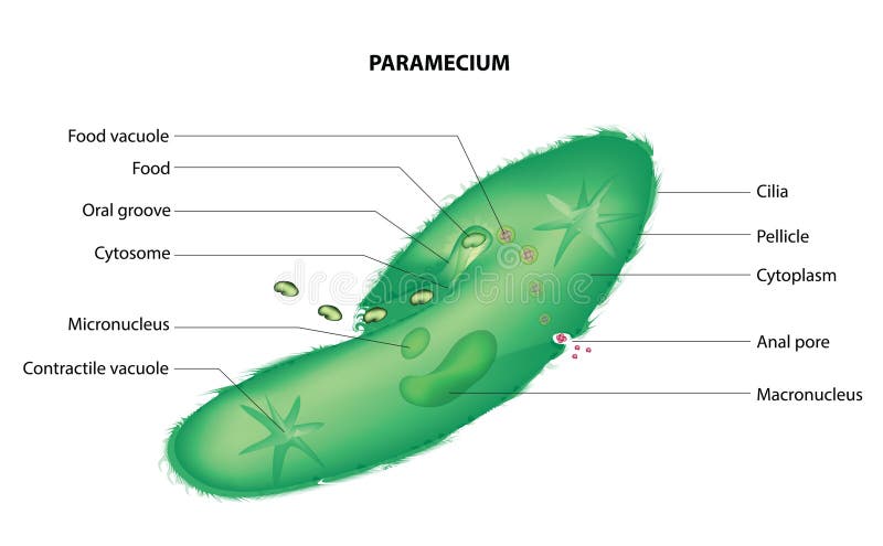
Free with trial Paramecium is a genus of single-celled, ciliate protists belonging to the phylum Ciliophora. These organisms are commonly found in freshwater environments, such as ponds, lakes, and slow-moving streams, where they feed on bacteria, algae, and other small microorganisms. Paramecium has a distinctive shape and is well-known for its ciliated surface, which it uses for movement and feeding. Ciliated vectors Paramecium. Is a genus of single-celled, ciliate protists belonging to the phylum Ciliophora. These organisms are commonly found in freshwater environments. Paramecium is a genus of single-celled, ciliate protists belonging to the phylum Ciliophora. These organisms are commonly found in freshwater environments, such as ponds, lakes, and slow-moving streams, where they feed on bacteria, algae, and other small microorganisms. Paramecium has a distinctive shape and is well-known for its ciliated surface, which it uses for movement and feeding.
Free with trial Stratified columnar epithelium is a type of epithelial tissue characterized by multiple layers of cells, with the superficial layer consisting of columnar cells. This type of tissue is less common compared to other epithelial types like simple squamous or simple cuboidal epithelium. Ciliated vectors Stratified Columnar Epithelium. Is a type of epithelial tissue characterized by multiple layers of cells, with the superficial layer consisting of columnar. Stratified columnar epithelium is a type of epithelial tissue characterized by multiple layers of cells, with the superficial layer consisting of columnar cells. This type of tissue is less common compared to other epithelial types like simple squamous or simple cuboidal epithelium.
Free with trial Stratified cuboidal epithelium is a type of epithelial tissue characterized by multiple layers of cube-shaped cells. This type of tissue is less common compared to other epithelial types like simple squamous or simple cuboidal epithelium. It is typically found lining ducts and tubules in various glands and organs where protection and secretion are required. Ciliated vectors Stratified cuboidal epithelium. Is a type of epithelial tissue characterized by multiple layers of cube-shaped cells. This type of tissue is less common. Stratified cuboidal epithelium is a type of epithelial tissue characterized by multiple layers of cube-shaped cells. This type of tissue is less common compared to other epithelial types like simple squamous or simple cuboidal epithelium. It is typically found lining ducts and tubules in various glands and organs where protection and secretion are required.
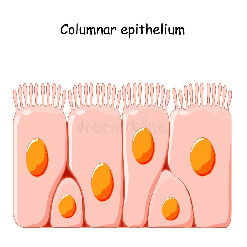
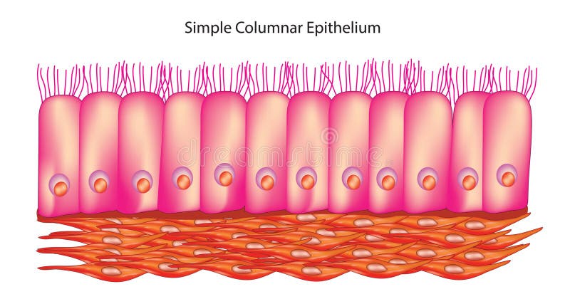
Free with trial Simple columnar epithelium is a type of epithelial tissue characterized by a single layer of tall, column-shaped cells. These cells are closely packed together and have elongated nuclei located near the basal surface. Simple columnar epithelium lines various structures in the body and performs specialized functions. Ciliated vectors Simple columnar epithelium. Is a type of epithelial tissue characterized by a single layer of tall, column-shaped cells. These cells are closely packed together. Simple columnar epithelium is a type of epithelial tissue characterized by a single layer of tall, column-shaped cells. These cells are closely packed together and have elongated nuclei located near the basal surface. Simple columnar epithelium lines various structures in the body and performs specialized functions.
Free with trial Pseudostratified columnar epithelium is a type of epithelial tissue that appears to be stratified but is actually a single layer of cells. Each cell is attached to the basement membrane, but not all cells reach the apical surface, giving the appearance of multiple layers. Ciliated vectors Pseudostratified columnar epithelium. Is a type of epithelial tissue that appears to be stratified but is actually a single layer of cells. Each cell is. Pseudostratified columnar epithelium is a type of epithelial tissue that appears to be stratified but is actually a single layer of cells. Each cell is attached to the basement membrane, but not all cells reach the apical surface, giving the appearance of multiple layers.
Free with trial Epithelium. squamous, cubic, ciliated glandular Set Infographics Vector illustration. Ciliated vectors Epithelium. squamous, cubic, ciliated, glandular. Set. Infographics. Vector illustration. Epithelium. squamous, cubic, ciliated glandular Set Infographics. Epithelium. squamous, cubic, ciliated glandular Set Infographics Vector illustration
Free with trial Epithelium. squamous, cubic, ciliated glandular Set Infographics Vector illustration. Ciliated vectors Epithelium. squamous, cubic, ciliated, glandular. Set. Infographics. Vector illustration. Epithelium. squamous, cubic, ciliated glandular Set Infographics. Epithelium. squamous, cubic, ciliated glandular Set Infographics Vector illustration
Free with trial Ciliated Epithelium, vintage illustration. Ciliated epithelium from the human trachea, vintage line drawing or engraving illustration. Ciliated vectors Ciliated Epithelium, vintage illustration. Ciliated epithelium from the human trachea, vintage line drawing or engraving illustration
Free with trial The spheroidal ciliated cells from the mouth of a frog, vintage line drawing or engraving illustration. Ciliated vectors Spheroidal Ciliated Cells from a Frog, vintage illustration. The spheroidal ciliated cells from the mouth of a frog, vintage line drawing or engraving. The spheroidal ciliated cells from the mouth of a frog, vintage line drawing or engraving illustration
Free with trial Ciliated epithelium cells from the tracheawindpipe, vintage engraving. Ciliated vectors Ciliated epithelium cells from the tracheawindpipe, vintage engraving. Ciliated epithelium cells from the tracheawindpipe, vintage engraved illustration
Free with trial Ciliated Antenna, vintage illustration. Ciliated Antenna which is covered in microscopic projections, vintage line drawing or engraving illustration. Ciliated vectors Ciliated Antenna, vintage illustration. Ciliated Antenna which is covered in microscopic projections, vintage line drawing or engraving illustration
Free with trial Vector illustration background of pseudostratified ciliated epithelial cells, tissue found along the respiratory tract, pseudostratified ciliated columnar epithelial cells help trap and transport particles brought in through the nasal passages and lungs. Ciliated vectors Pseudostratified Ciliated Epithelium science background vector. Vector illustration background of pseudostratified ciliated epithelial cells, tissue found along. Vector illustration background of pseudostratified ciliated epithelial cells, tissue found along the respiratory tract, pseudostratified ciliated columnar epithelial cells help trap and transport particles brought in through the nasal passages and lungs.
Free with trial Ciliated Cells, vintage illustration. Ciliated cells such as those lining the windpipe, vintage line drawing or engraving illustration. Ciliated vectors Ciliated Cells, vintage illustration. Ciliated cells such as those lining the windpipe, vintage line drawing or engraving illustration
Free with trial Ciliated infusoria origin and development modes, vector illustration. Hand drawn euglena transforming, chlorococus vesicle cell. Process in microorganism structure, isolated on white. Ciliated vectors Ciliated infusoria origin and development modes, vector illustration. Hand drawn euglena transforming, chlorococus vesicle cell. Process in microorganism
Free with trial Medical vector illustration of phospholipids and respiratory mucus cough clearance. Ciliated and mucous cell structure. Ciliated vectors Medical vector illustration of phospholipids and respiratory mucus cough clearance. Ciliated and mucous cell structure
Free with trial It performs the function of moving particles or fluid over the epithelial surface. Found in trachea, bronchial tubes, nasal cavities. Vector illustration. Ciliated vectors Ciliated epithelium. Epithelial tissue types. It performs the function of moving particles or fluid over the epithelial surface. Found in trachea, bronchial. It performs the function of moving particles or fluid over the epithelial surface. Found in trachea, bronchial tubes, nasal cavities. Vector illustration
Free with trial The lancelet, also known as amphioxus, is a small marine animal belonging to the chordate phylum. It has a slender, fish-like body and is considered one of the closest living relatives to vertebrates. Lancelets are filter feeders, using their ciliated gill slits to capture plankton and small organisms for food. Ciliated vectors Lancelet or Amphioxus (Branchiostoma). The lancelet is a small, translucent, fish-like animal. The lancelet, also known as amphioxus, is a small marine animal. The lancelet, also known as amphioxus, is a small marine animal belonging to the chordate phylum. It has a slender, fish-like body and is considered one of the closest living relatives to vertebrates. Lancelets are filter feeders, using their ciliated gill slits to capture plankton and small organisms for food.
Free with trial Nasal mucosa flat vector anatomical icon. Epithelium in the nose, gut, stomach or colon. Human olfactory system concept. Mucus, ciliated cells, villi and cilia poster for clinic or education. Ciliated vectors Epithelium of nasal mucosa. Nasal mucosa flat vector anatomical icon. Epithelium in the nose, gut, stomach or colon. Human olfactory system concept. Mucus. Nasal mucosa flat vector anatomical icon. Epithelium in the nose, gut, stomach or colon. Human olfactory system concept. Mucus, ciliated cells, villi and cilia poster for clinic or education.
Free with trial Nasal mucosa flat vector anatomical icon. Epithelium in the nose, gut, stomach or colon. Human olfactory system concept. Mucus, ciliated cells, villi and cilia poster for clinic or education. Ciliated vectors Epithelium of nasal mucosa. Nasal mucosa flat vector anatomical icon. Epithelium in the nose, gut, stomach or colon. Human olfactory system concept. Mucus. Nasal mucosa flat vector anatomical icon. Epithelium in the nose, gut, stomach or colon. Human olfactory system concept. Mucus, ciliated cells, villi and cilia poster for clinic or education.
Free with trial Nasal epithelium anatomical poster. Human respiratory system concept. Mucus, goblet, ciliated and basal cells. Smell organs respiratory system. Medical flat vector illustration for clinic or education. Ciliated vectors Nasal epithelium concept. Nasal epithelium anatomical poster. Human respiratory system concept. Mucus, goblet, ciliated and basal cells. Smell organs respiratory. Nasal epithelium anatomical poster. Human respiratory system concept. Mucus, goblet, ciliated and basal cells. Smell organs respiratory system. Medical flat vector illustration for clinic or education
Free with trial Nasal epithelium anatomical poster. Human respiratory system concept. Mucus, goblet, ciliated and basal cells. Smell organs respiratory system. Medical flat vector illustration for clinic or education. Ciliated vectors Nasal epithelium concept. Nasal epithelium anatomical poster. Human respiratory system concept. Mucus, goblet, ciliated and basal cells. Smell organs respiratory. Nasal epithelium anatomical poster. Human respiratory system concept. Mucus, goblet, ciliated and basal cells. Smell organs respiratory system. Medical flat vector illustration for clinic or education
Free with trial Nasal mucosa flat vector anatomical icon. Epithelium in the nose, gut, stomach or colon. Human olfactory system concept. Mucus, ciliated cells, villi and cilia poster for clinic or education. Ciliated vectors Epithelium of nasal mucosa. Nasal mucosa flat vector anatomical icon. Epithelium in the nose, gut, stomach or colon. Human olfactory system concept. Mucus. Nasal mucosa flat vector anatomical icon. Epithelium in the nose, gut, stomach or colon. Human olfactory system concept. Mucus, ciliated cells, villi and cilia poster for clinic or education.
Free with trial Nasal mucosa flat vector anatomical icon. Epithelium in the nose, gut, stomach or colon. Human olfactory system concept. Mucus, ciliated cells, villi and cilia poster for clinic or education. Ciliated vectors Epithelium of nasal mucosa. Nasal mucosa flat vector anatomical icon. Epithelium in the nose, gut, stomach or colon. Human olfactory system concept. Mucus. Nasal mucosa flat vector anatomical icon. Epithelium in the nose, gut, stomach or colon. Human olfactory system concept. Mucus, ciliated cells, villi and cilia poster for clinic or education.
Free with trial Nasal mucosa flat vector anatomical icon. Epithelium in the nose, gut, stomach or colon. Human olfactory system concept. Mucus, ciliated cells, villi and cilia poster for clinic or education. Ciliated vectors Epithelium of nasal mucosa. Nasal mucosa flat vector anatomical icon. Epithelium in the nose, gut, stomach or colon. Human olfactory system concept. Mucus. Nasal mucosa flat vector anatomical icon. Epithelium in the nose, gut, stomach or colon. Human olfactory system concept. Mucus, ciliated cells, villi and cilia poster for clinic or education.
Free with trial Nasal mucosa flat vector anatomical icon. Epithelium in the nose, gut, stomach or colon. Human olfactory system concept. Mucus, ciliated cells, villi and cilia poster for clinic or education. Ciliated vectors Epithelium of nasal mucosa. Nasal mucosa flat vector anatomical icon. Epithelium in the nose, gut, stomach or colon. Human olfactory system concept. Mucus. Nasal mucosa flat vector anatomical icon. Epithelium in the nose, gut, stomach or colon. Human olfactory system concept. Mucus, ciliated cells, villi and cilia poster for clinic or education.
Free with trial Nasal mucosa flat vector anatomical icon. Epithelium in the nose, gut, stomach or colon. Human olfactory system concept. Mucus, ciliated cells, villi and cilia poster for clinic or education. Ciliated vectors Epithelium of nasal mucosa. Nasal mucosa flat vector anatomical icon. Epithelium in the nose, gut, stomach or colon. Human olfactory system concept. Mucus. Nasal mucosa flat vector anatomical icon. Epithelium in the nose, gut, stomach or colon. Human olfactory system concept. Mucus, ciliated cells, villi and cilia poster for clinic or education.
Free with trial Nasal mucosa flat vector anatomical icon. Epithelium in the nose, gut, stomach or colon. Human olfactory system concept. Mucus, ciliated cells, villi and cilia poster for clinic or education. Ciliated vectors Epithelium of nasal mucosa. Nasal mucosa flat vector anatomical icon. Epithelium in the nose, gut, stomach or colon. Human olfactory system concept. Mucus. Nasal mucosa flat vector anatomical icon. Epithelium in the nose, gut, stomach or colon. Human olfactory system concept. Mucus, ciliated cells, villi and cilia poster for clinic or education.
Free with trial Nasal mucosa flat vector anatomical icon. Epithelium in the nose, gut, stomach or colon. Human olfactory system concept. Mucus, ciliated cells, villi and cilia poster for clinic or education. Ciliated vectors Epithelium of nasal mucosa. Nasal mucosa flat vector anatomical icon. Epithelium in the nose, gut, stomach or colon. Human olfactory system concept. Mucus. Nasal mucosa flat vector anatomical icon. Epithelium in the nose, gut, stomach or colon. Human olfactory system concept. Mucus, ciliated cells, villi and cilia poster for clinic or education.
Free with trial Protonephridium of a Shark vintage illustration. Protonephridium of a Shark in which ciliated funnels is present vintage line drawing or engraving illustration. Ciliated vectors Protonephridium of a Shark vintage illustration. Protonephridium of a Shark in which ciliated funnels is present vintage line drawing or engraving illustration
Free with trial Intestinal epithelial cells, also known as enterocytes, are the cells that line the inner surface of the intestine, forming the intestinal epithelium. They play crucial roles in nutrient absorption, secretion of mucus, and protection against pathogens and toxins. Ciliated vectors Intestinal epithelial cell. S, also known as enterocytes, are the cells that line the inner surface of the intestine, forming the intestinal epithelium. They. Intestinal epithelial cells, also known as enterocytes, are the cells that line the inner surface of the intestine, forming the intestinal epithelium. They play crucial roles in nutrient absorption, secretion of mucus, and protection against pathogens and toxins.
Free with trial Simple cuboidal epithelium is a specialized type of tissue that lines small ducts and tubules in various organs, where it facilitates absorption, secretion, and transport functions. Its cube-shaped cells provide a balance between surface area and protection, making it well-suited for its physiological roles. Ciliated vectors Simple cuboidal epithelium. Is a specialized type of tissue that lines small ducts and tubules in various organs, where it facilitates absorption, secretion. Simple cuboidal epithelium is a specialized type of tissue that lines small ducts and tubules in various organs, where it facilitates absorption, secretion, and transport functions. Its cube-shaped cells provide a balance between surface area and protection, making it well-suited for its physiological roles.
Free with trial Nomenclature of schizomycetes, vintage engraving. Nomenclature of schizomycetes, vintage engraved illustration. Ciliated vectors Nomenclature of schizomycetes, vintage engraving. Nomenclature of schizomycetes, vintage engraved illustration
Free with trial A vertical section of an intestinal villus of a cat have a striated basilar border of the epithelium, vintage line drawing or engraving illustration. Ciliated vectors Intestinal Villus of a Cat, vintage illustration. A vertical section of an intestinal villus of a cat have a striated basilar border of the epithelium, vintage. A vertical section of an intestinal villus of a cat have a striated basilar border of the epithelium, vintage line drawing or engraving illustration
Free with trial Tall and slender cells with oval-shaped nuclei. It is found in the conjunctiva, pharynx, anus, and male urethra. Medical illustration. Vector. Ciliated vectors Stratified columnar epithelium. Epithelial tissue types. Tall and slender cells with oval-shaped nuclei. It is found in the conjunctiva, pharynx, anus, and male. Tall and slender cells with oval-shaped nuclei. It is found in the conjunctiva, pharynx, anus, and male urethra. Medical illustration. Vector.
Free with trial 3D Isometric Flat Vector Conceptual Illustration of Nasal Mucosa Cells, Medical Educational Diagram. Ciliated vectors 3D Isometric Flat Vector Conceptual Illustration of Nasal Mucosa Cells. Medical Educational Diagram
Free with trial Tall and slender cells with oval-shaped nuclei. Lines most organs of the digestive tract like stomach, intestines. Medical illustration. Vector. Ciliated vectors Simple columnar epithelium. Epithelial tissue types. Tall and slender cells with oval-shaped nuclei. Lines most organs of the digestive tract like stomach. Tall and slender cells with oval-shaped nuclei. Lines most organs of the digestive tract like stomach, intestines. Medical illustration. Vector.
 Sign in