Free with trial Photoreceptor cells in the retina of the eye. Structure and function rod cells and cone cells. Vector scheme. Conjunctiva vectors Photoreceptor cells in the retina of the eye. Structure and function rod cells and cone cells. Vector scheme
Free with trial Structure of the human eye and organization of the retina. Optic part of retina. Conjunctiva vectors Optic part of retina. Structure of the human eye and organization of the retina. Optic part of retina. Structure of the human eye and organization of the retina. Optic part of retina.
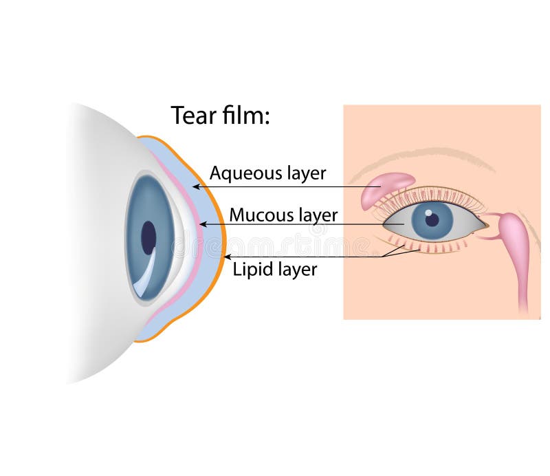
Free with trial Tear film formation. The lacrimal gland, meibomian glands and conjunctival goblet cells secretion. Conjunctiva vectors Tear film formation. The lacrimal gland, meibomian glands and conjunctival goblet cells secretion
Free with trial Tears chemical composition. The lacrimal gland, meibomian glands and conjunctival goblet cells secretion, anterior and lateral view. Conjunctiva vectors Tears chemical composition. The lacrimal gland, meibomian glands and conjunctival goblet cells secretion, anterior and lateral view
Free with trial Eye anatomy. Rod cells and cone cells. The arrangement of retinal cells is shown in a cross section. Vector diagram for your design, educational, biological, science and medical use. Conjunctiva vectors Eye anatomy. Rod cells and cone cells. The arrangement of retinal cells is shown in a cross section. Vector diagram for your design, educational, biological
Free with trial Photoreceptor cells in the retina of the eye. retinal cells. rod cell and cone cell. The arrangement of retinal cells is shown in a cross section. Conjunctiva vectors Photoreceptor cells in the retina of the eye. Retinal cells. rod cell and cone cell. The arrangement of retinal cells is shown in a cross section
Free with trial Eye oblique. Oblique view of a section through a human eye showing all major structures. Conjunctiva vectors Eye oblique. Oblique view of a section through a human eye showing all major structures
Free with trial Conjunctivitis. Medical illustration of the effects of conjunctivitis. Conjunctiva vectors Conjunctivitis. Medical illustration of the effects of conjunctivitis
Free with trial Cataract Surgery. Steps laser cataract surgery. Conjunctiva vectors Cataract Surgery. Steps laser
Free with trial The lacrimal apparatus. The lacrimal gland and other structure of tear producing organ, eps10. Conjunctiva vectors The lacrimal apparatus. The lacrimal gland and other structure of tear producing organ, eps10
Free with trial Eye conjunctiva. And other accessory structures of the orbit, eps10. Conjunctiva vectors Eye conjunctiva. And other accessory structures of the orbit, eps10
Free with trial Conjunctiva of the eye. Diagram to show the conjuntiva of the eye, including the eyelids and lashes. Conjunctiva vectors Conjunctiva of the eye. Diagram to show the conjuntiva of the eye, including the eyelids and lashes
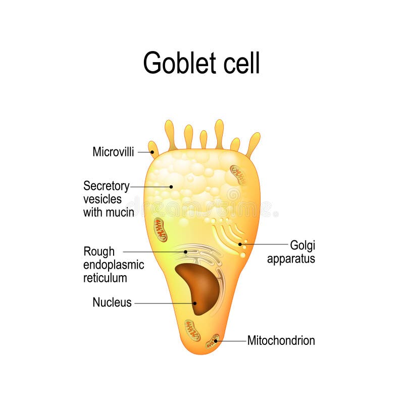
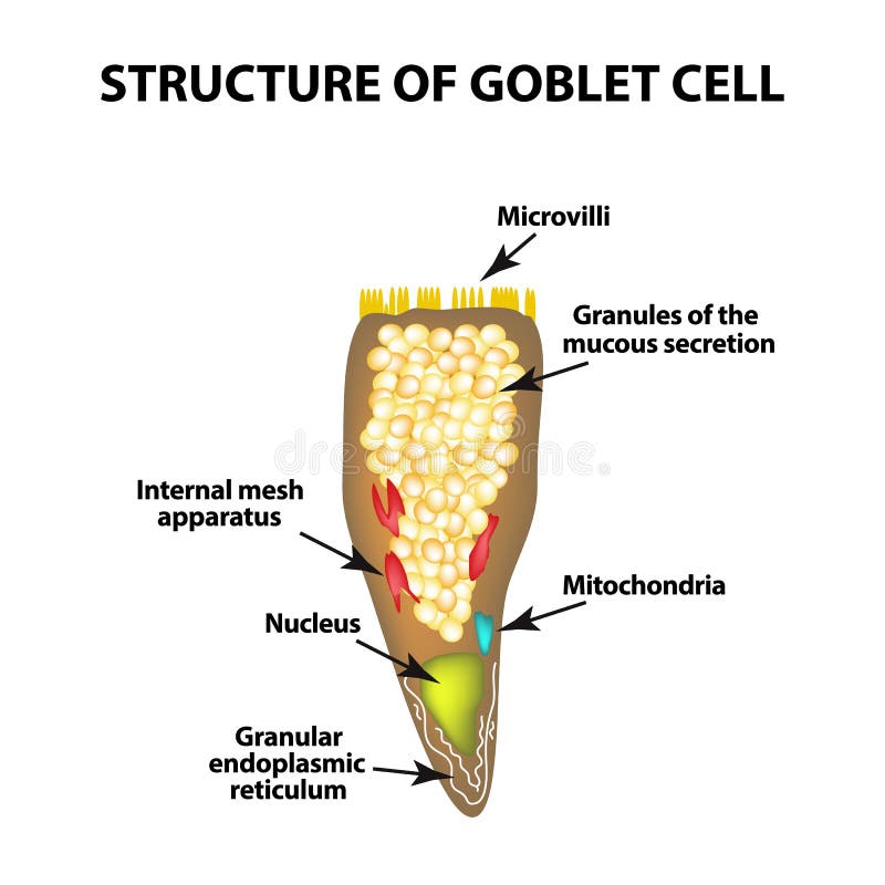
Free with trial Goblet cell. simple columnar epithelial cell for secrete mucus. They are found inside the trachea, bronchi, small and large intestine, and conjunctiva in the eyes. Structure cell nucleus and other organelles. Conjunctiva vectors Goblet cell. Structure cell nucleus and other organelles. Goblet cell. simple columnar epithelial cell for secrete mucus. They are found inside the trachea. Goblet cell. simple columnar epithelial cell for secrete mucus. They are found inside the trachea, bronchi, small and large intestine, and conjunctiva in the eyes. Structure cell nucleus and other organelles.
Free with trial Chalazion & sty. Medical illustration of symptoms of chalazion and sty. Conjunctiva vectors Chalazion & sty. Medical illustration of symptoms of chalazion and sty
Free with trial Cone cells. Vector. Cone cells, or cones, are photoreceptor cells in the retina of the eye that are responsible for color vision. Conjunctiva vectors Cone cells. Vector. Cone cells, or cones, are photoreceptor cells in the retina of the eye that are responsible for color vision. Vector
Free with trial Conjunctivitis. An eye cut saggital labeled diagram with conjunctivitis. Conjunctiva vectors Conjunctivitis. An eye cut saggital labeled diagram with conjunctivitis
Free with trial Schematic structure of the rod cell. Peripheral vision. Rod cells, or rods, are photoreceptor cells in the retina of the eye that can function in less intense light. Vector scheme. Conjunctiva vectors The Rod cell. Model Photoreceptor. Vector. Schematic structure of the rod cell. Peripheral vision. Rod cells, or rods, are photoreceptor cells in the retina of. Schematic structure of the rod cell. Peripheral vision. Rod cells, or rods, are photoreceptor cells in the retina of the eye that can function in less intense light. Vector scheme
Free with trial Anatomy of the cat`s or dog`s eye. Vertical section of the eye and eyelids. Third eyelid and Tapetum lucidum. Schematic diagram. detailed illustration. Conjunctiva vectors Anatomy of the cat`s or dog`s eye. Vertical section of the eye and eyelids. Third eyelid and Tapetum lucidum. Schematic diagram. detailed illustration
Free with trial Pterygium. Medical illustration of the symptoms of pterygium. Conjunctiva vectors Pterygium. Medical illustration of the symptoms of pterygium
Free with trial Conjunctivitis. Healthy eye and pink eye with inflammation. Conjunctiva vectors Conjunctivitis. Healthy eye and pink eye with inflammation
Free with trial Viral conjunctivitis. Adenoviruses is the cause of viral conjunctivitis. Conjunctiva vectors Viral conjunctivitis. Adenoviruses is the cause of
Free with trial Human eye anatomy in front view. Illustration about physical. Conjunctiva vectors Human eye anatomy in front view. Illustration about physical
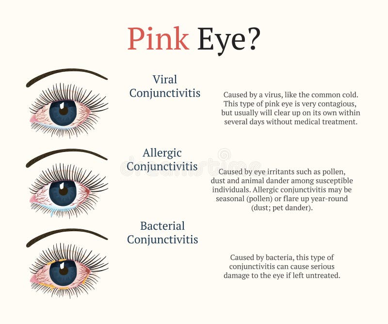
Free with trial Conjunctivitis types. Inflammatory diseases of eyes. Conjunctiva vectors Conjunctivitis types. Inflammatory diseases of eyes.
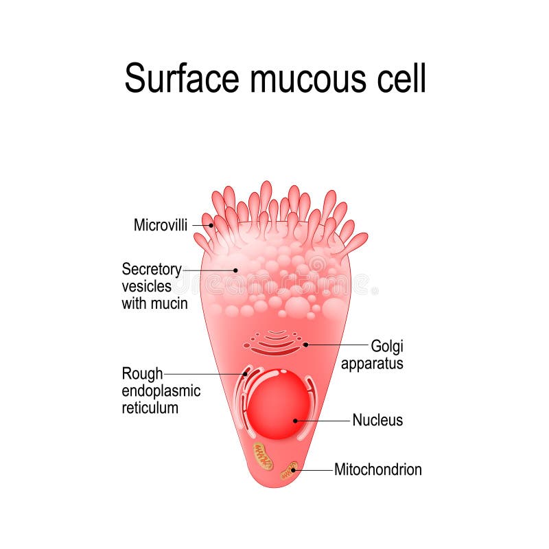
Free with trial Surface mucous cell is a foveolar mucus-producing cell that covering the inside of the stomach. Structure cell: golgi apparatus, secretory vesicle, mucin, nucleus, mitochondrion, microvilli. Conjunctiva vectors Surface mucous cell. Is a foveolar mucus-producing cell that covering the inside of the stomach. Structure cell: golgi apparatus, secretory vesicle, mucin. Surface mucous cell is a foveolar mucus-producing cell that covering the inside of the stomach. Structure cell: golgi apparatus, secretory vesicle, mucin, nucleus, mitochondrion, microvilli
Free with trial Bacterial conjunctivitis. Eye with conjunctivitis and bacteria that cause it. Staphylococci. Conjunctiva vectors Bacterial conjunctivitis. Eye with conjunctivitis and bacteria that cause it. Staphylococci
Free with trial Photoreceptor cells. retinal cells: rod and cone cells, amacrine, ganglion, bipolar cells and RPE. The arrangement of retinal cells is shown in a cross section. infographic of human eye. Conjunctiva vectors Retinal cells: rod and cone cells. Photoreceptor cells. retinal cells: rod and cone cells, amacrine, ganglion, bipolar cells and RPE. The arrangement of retinal. Photoreceptor cells. retinal cells: rod and cone cells, amacrine, ganglion, bipolar cells and RPE. The arrangement of retinal cells is shown in a cross section. infographic of human eye
Free with trial Cross section through the eye showing the major structures, chambers and muscle attachments. Created in Adobe Illustrator. Contains transparent objects and gradient fills. EPS 10. Conjunctiva vectors Cross section through the eye. Showing the major structures, chambers and muscle attachments. Created in Adobe Illustrator. Contains transparent objects and. Cross section through the eye showing the major structures, chambers and muscle attachments. Created in Adobe Illustrator. Contains transparent objects and gradient fills. EPS 10.
Free with trial Cataract operation, showing a normal eye and a lens with a cataract. The procedure for surgical removal is shown. Created in Adobe Illustrator. Contains transparencies and gradient meshes. EPS 10. Conjunctiva vectors Cataract removal. Cataract operation, showing a normal eye and a lens with a cataract. The procedure for surgical removal is shown. Created in Adobe Illustrator. Cataract operation, showing a normal eye and a lens with a cataract. The procedure for surgical removal is shown. Created in Adobe Illustrator. Contains transparencies and gradient meshes. EPS 10.
Free with trial The Comparison between normal skin people and yellowing from Jaundice. Illustration about health problems condition. Conjunctiva vectors The Comparison between normal skin people and yellowing from Jaundice. Illustration about health problems condition
Free with trial Conjunctivitis. pink eye with inflammation. External View and Vertical section of the human eyes and eyelids. Schematic diagram. detailed illustration. Conjunctiva vectors Conjunctivitis. External View and Vertical section of the human. Conjunctivitis. pink eye with inflammation. External View and Vertical section of the human eyes. Conjunctivitis. pink eye with inflammation. External View and Vertical section of the human eyes and eyelids. Schematic diagram. detailed illustration
Free with trial Anatomy of the eyeball, functions and structure of the retina, projection of the image in the eye, the mechanism of visual perception. Conjunctiva vectors Eyeball in section, structure of the retina, close-up. Anatomy of the eyeball, functions and structure of the retina, projection of the image in the eye, the. anatomy of the eyeball, functions and structure of the retina, projection of the image in the eye, the mechanism of visual perception
Free with trial Retinopathy is damage to the retina of the eyes, which cause vision impairment. Anatomy of the human eye. Vertical section of the eye and eyelids. Schematic diagram. detailed illustration. Conjunctiva vectors Retinopathy. Vertical section of the eye and eyelids with damage. Retinopathy is damage to the retina of the eyes, which cause vision impairment. Anatomy of the. Retinopathy is damage to the retina of the eyes, which cause vision impairment. Anatomy of the human eye. Vertical section of the eye and eyelids. Schematic diagram. detailed illustration.
Free with trial Glaucoma chronic eye pathology. Eye disease. Ophthalmology health. Glaucoma types. Vector flat eye healt illustration. Conjunctiva vectors Eye disease. Ophthalmology health illustration. Glaucoma chronic eye pathology. Eye disease. Ophthalmology health. Glaucoma types. Vector flat eye healt. Glaucoma chronic eye pathology. Eye disease. Ophthalmology health. Glaucoma types. Vector flat eye healt illustration.
Free with trial Conjunctivitis. pink eye with inflammation. External View and Vertical section of the human eyes and eyelids. Schematic diagram. detailed illustration. Conjunctiva vectors Conjunctivitis. pink eye with inflammation. External View and Vertical section of the human eyes and eyelids. Schematic diagram. detailed illustration
Free with trial Conjunctiva. Medical illustration of anatomy of the conjunctiva. Conjunctiva vectors Conjunctiva. Medical illustration of anatomy of the conjunctiva
Free with trial A medical illustration compares a healthy eye with one affected by lymphoma of the conjunctiva. Conjunctiva vectors Lymphoma of conjunctiva. A medical illustration compares a healthy eye with one affected by lymphoma of the conjunctiva. A medical illustration compares a healthy eye with one affected by lymphoma of the conjunctiva.
Free with trial Sore eye normal and inflamed conjunctiva vector conjunctivitis eyeball disease infection and inflammation redness pupil and iris healthcare and medicine perception organ treatment isolated face part. Conjunctiva vectors Normal and inflamed conjunctiva sore eye disease. Sore eye normal and inflamed conjunctiva vector conjunctivitis eyeball disease infection and inflammation. Sore eye normal and inflamed conjunctiva vector conjunctivitis eyeball disease infection and inflammation redness pupil and iris healthcare and medicine perception organ treatment isolated face part.
Free with trial Conjunctivitis inflamed conjunctiva conjunctivitis sore eye medicine and health isolated vector perception organ eyesight and vision swelled eyelid infection disease ophthalmology and healthcare. Conjunctiva vectors Inflamed conjunctiva conjuntivitis sore eye medicine and health. Conjunctivitis inflamed conjunctiva conjunctivitis sore eye medicine and health isolated vector. Conjunctivitis inflamed conjunctiva conjunctivitis sore eye medicine and health isolated vector perception organ eyesight and vision swelled eyelid infection disease ophthalmology and healthcare
Free with trial Eye irritation. Inflamed bloodshot eyeball with red conjunctiva isolated on white background. Symptom of keratitis, allergy, conjunctivitis, uveitis. Vector cartoon illustration. Conjunctiva vectors Eye irritation. Inflamed bloodshot eyeball with red conjunctiva isolated on white background. Symptom of keratitis. Allergy, conjunctivitis, uveitis. Vector
Free with trial Conjunctivitis concept with red eye and lacrimation - symptom of swelling of conjunctiva or allergy in flat vector illustration. Pink eye with tear drop isolated on white background. Conjunctiva vectors Conjunctivitis concept with red eye and lacrimation - symptom of swelling of conjunctiva or allergy. Conjunctivitis concept with red eye and lacrimation. Conjunctivitis concept with red eye and lacrimation - symptom of swelling of conjunctiva or allergy in flat vector illustration. Pink eye with tear drop isolated on white background.
Free with trial Dry eye syndrome. Inflamed bloodshot eyeball with swelling, irritation and red conjunctiva. Symptoms of infection, allergy, keratitis, blepharitis, conjunctivitis, uveitis. Vector cartoon illustration. Conjunctiva vectors Dry eye syndrome. Inflamed bloodshot eyeball with swelling, irritation and red conjunctiva. Symptoms of infection. Allergy, keratitis, blepharitis. Dry eye syndrome. Inflamed bloodshot eyeball with swelling, irritation and red conjunctiva. Symptoms of infection, allergy, keratitis, blepharitis, conjunctivitis, uveitis. Vector cartoon illustration
Free with trial Healthy and ill human vision organ. Dry eye syndrom. Inflamed eyeball with irritation and red conjunctiva. Symptoms of keratitis, blepharitis, conjunctivitis or uveitis. Vector flat illustration. Conjunctiva vectors Healthy and ill human vision organ. Dry eye syndrom. Inflamed eyeball with irritation and red conjunctiva. Symptoms of. Keratitis, blepharitis, conjunctivitis. Healthy and ill human vision organ. Dry eye syndrom. Inflamed eyeball with irritation and red conjunctiva. Symptoms of keratitis, blepharitis, conjunctivitis or uveitis. Vector flat illustration.
Free with trial Human eye anatomy. Retina structure. Cross-section of the eye. Cells in section of the retina. Detailed vector poster. Conjunctiva vectors Human eye anatomy. Retina structure. Cross-section of the eye. Cells in section of the retina. Detailed vector poster
Free with trial Left eye of an adult human. Body part. Iris, pupil, cornea, sciera. Conjunctiva vectors Human blue left eye. Left eye of an adult human. Body part. Iris, pupil, cornea, sciera. Left eye of an adult human. Body part. Iris, pupil, cornea, sciera.
Free with trial Types of conjunctivitis. Three main types of conjunctivitis: allergic, viral and bacterial. Conjunctiva vectors Types of conjunctivitis. Three main types of conjunctivitis: allergic, viral and bacterial
Free with trial Human organs icon. Health and illness sign. Ignition, cancer, tumour. Vector symbol collection. Conjunctiva vectors Human organs icon. Health and illness sign. Ignition, cancer, tumour. Vector symbol collection
Free with trial Anatomy of the human eye. Vertical section of the eye and eyelids. Schematic diagram. detailed illustration. Conjunctiva vectors Vertical section of the human eye and eyelids. Anatomy of the human eye. Vertical section of the eye and eyelids. Schematic diagram. detailed illustration. Anatomy of the human eye. Vertical section of the eye and eyelids. Schematic diagram. detailed illustration.
Free with trial Blepharitis. Purulent, inflammatory eyes disease. Conjunctiva vectors Blepharitis. Purulent, inflammatory eyes disease.
Free with trial Human eye anatomy in front view. Illustration about physical. Conjunctiva vectors Human eye anatomy in front view. Illustration about physical
Free with trial Bleeding eyes medical concept. Vector illustration. Doctor and patient are talking in the hospital. Isolated on white background. Conjunctiva vectors Bleeding eyes medical concept. Vector illustration. Doctor and patient are talking in the hospital. Isolated on white background
Free with trial Eye anatomy. Cross section of a Human eyeball. Diagram for education and medical use. Vector illustration. Conjunctiva vectors Eye anatomy. Cross section of a Human eyeball. Diagram for education and medical use. Vector illustration
Free with trial The Comparison between normal skin people and yellowing from Jaundice. Illustration about health problems condition. Conjunctiva vectors The Comparison between normal skin people and yellowing from Jaundice. Illustration about health problems condition
Free with trial Trichiasis. Medical illustration shows the comparison between a normal eye and one affected by trichiasis. Conjunctiva vectors Trichiasis. Medical illustration shows the comparison between a normal eye and one affected by trichiasis
Free with trial Aqueous humour fluid exits and flow to canal of schlemm human eyes. To see difficult the picture because the fluid exits can not flow through, consequences of obscure visibility of human eye lens. Illustration. Conjunctiva vectors Glaucoma. Aqueous humour fluid exits and flow to canal of schlemm human eyes. Illustration. Aqueous humour fluid exits and flow to canal of schlemm human eyes. Aqueous humour fluid exits and flow to canal of schlemm human eyes. To see difficult the picture because the fluid exits can not flow through, consequences of obscure visibility of human eye lens. Illustration.
Free with trial Anatomy of the human eye. Vertical section of the eye and eyelids. Schematic diagram. detailed illustration. set vector icon for eye diseases and disorders. Black and white. Conjunctiva vectors Vertical section of the eye and eyelids. Black and white. Anatomy of the human eye. Vertical section of the eye and eyelids. Schematic diagram. detailed. Anatomy of the human eye. Vertical section of the eye and eyelids. Schematic diagram. detailed illustration. set vector icon for eye diseases and disorders. Black and white.
Free with trial Bleeding eyes medical concept. Vector illustration. Doctor and patient are talking in the hospital. Isolated on white background. Conjunctiva vectors Bleeding eyes medical concept. Vector illustration. Doctor and patient are talking in the hospital. Isolated on white background
Free with trial Eye diseases. Vector Illustration. Set of women with various eye diseases. Conjunctiva vectors Eye diseases. Vector Illustration. Set of women with various eye diseases
Free with trial The external structure of the eye. Vector illustration. Conjunctiva vectors The external structure of the eye. Vector. Illustration
Free with trial The external structure of the eye. Vector illustration. Conjunctiva vectors The external structure of the eye. Vector. Illustration
Free with trial Vector illustration flat suffering young man. With a conjunctivitis. Pain in the human body, eye ache, eye problems concept. Isolated on white background. Conjunctiva vectors Vector illustration flat suffering young man. With a conjunctivitis. Pain in the human body, eye ache, eye problems concept. Isolated on white background
Free with trial Mucosal immune system diagram, medical vector illustration. Mucosal immune system diagram. Mucous or gut associated lymphoid tissue. Medical vector illustration. Conjunctiva vectors Mucosal immune system diagram, medical vector illustration. Mucosal immune system diagram. Mucous or gut associated lymphoid tissue. Medical vector illustration
Free with trial Eyeball in glasses. Vector icon, funny cartoon. Conjunctiva vectors Eyeglasses, smiling vector symbol, illustration, eps. Eyeball in glasses. Vector icon, funny cartoon. Eyeball in glasses. Vector icon, funny cartoon.
Free with trial The human eye is a marvel of biological engineering, perfectly designed to capture and process visual information from the surrounding environment. Its intricate structure and precise functioning make it one of the most remarkable sensory organs in the human body. At the forefront of the eye lies the cornea, a transparent, dome-shaped tissue that covers the iris, pupil, and anterior chamber. Serving as the eye's outermost lens, the cornea plays a crucial role in refracting light rays onto the retina, the light-sensitive tissue located at the back of the eye. Surrounding the pupil, the iris adds a burst of color to the eye and regulates the amount of light entering through the pupil. This dynamic structure expands and contracts in response to changes in lighting conditions, adjusting the size of the pupil to optimize visual acuity. At the center of the iris lies the pupil, a black circular aperture through which light enters the eye. Its size is controlled by the iris muscles, which contract to constrict the pupil in bright light and dilate it in dim conditions, regulating the amount of light that reaches the retina. Conjunctiva vectors Human eye labeled vector. The human eye is a marvel of biological engineering, perfectly designed to capture and process visual information from the surrounding. The human eye is a marvel of biological engineering, perfectly designed to capture and process visual information from the surrounding environment. Its intricate structure and precise functioning make it one of the most remarkable sensory organs in the human body. At the forefront of the eye lies the cornea, a transparent, dome-shaped tissue that covers the iris, pupil, and anterior chamber. Serving as the eye's outermost lens, the cornea plays a crucial role in refracting light rays onto the retina, the light-sensitive tissue located at the back of the eye. Surrounding the pupil, the iris adds a burst of color to the eye and regulates the amount of light entering through the pupil. This dynamic structure expands and contracts in response to changes in lighting conditions, adjusting the size of the pupil to optimize visual acuity. At the center of the iris lies the pupil, a black circular aperture through which light enters the eye. Its size is controlled by the iris muscles, which contract to constrict the pupil in bright light and dilate it in dim conditions, regulating the amount of light that reaches the retina.
Free with trial Diseases of the eye barley. Causes of barley. Demodex mite infestations. Inflammation volosyannoy bulbs. Conjunctiva vectors Diseases of the eye barley. Causes of barley. Demodex mite infestations. Inflammation volosyannoy bulbs
Free with trial Conjunctivitis pink eye medical condition explanation outline diagram. Labeled educational scheme with healthy lens comparison with virus or bacterial infection and inflammation vector illustration. Conjunctiva vectors Conjunctivitis or pink eye medical condition explanation outline diagram. Conjunctivitis pink eye medical condition explanation outline diagram. Labeled. Conjunctivitis pink eye medical condition explanation outline diagram. Labeled educational scheme with healthy lens comparison with virus or bacterial infection and inflammation vector illustration.
Free with trial Diagram illustrating eye conditions. The top section shows a healthy eye with clear sclera and pink conjunctiva. The middle section displays mucopurulent discharge, with crusty flakes on the eyelashes, suggesting bacterial conjunctivitis. The bottom section indicates watery discharge, which may suggest viral or allergic conjunctivitis. Each eye is distinctly labeled, highlighting differences in appearance and discharge types. Conjunctiva vectors Eye discharge in conjunctivitis. Diagram illustrating eye conditions. The top section shows a healthy eye with clear sclera and pink conjunctiva. The middle. Diagram illustrating eye conditions. The top section shows a healthy eye with clear sclera and pink conjunctiva. The middle section displays mucopurulent discharge, with crusty flakes on the eyelashes, suggesting bacterial conjunctivitis. The bottom section indicates watery discharge, which may suggest viral or allergic conjunctivitis. Each eye is distinctly labeled, highlighting differences in appearance and discharge types.
Free with trial Woman with conjunctivitis. Closeup view. Patient with pinkeye. Infectious viral disease. Medical concept. Anatomy of people. Vertical vector illustration. r. Conjunctiva vectors Woman with conjunctivitis. Closeup view. Patient with pinkeye. Infectious viral disease. Medical concept. Anatomy of people. Vertical vector illustration.r
Free with trial Dacryoadenitis. Inflammation of the lacrimal gland. The structure of the eye. Infographics. Vector illustration on isolated background. Conjunctiva vectors Dacryoadenitis. Inflammation of the lacrimal gland. The structure of the eye. Infographics. Vector illustration on isolated background
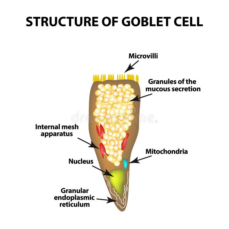
Free with trial Structure Goblet cells of the intestine. Infographics. Vector illustration on isolated background. Conjunctiva vectors Structure Goblet cells of the intestine. Infographics. Vector illustration on isolated background.
Free with trial Structure Goblet cells of the intestine. Infographics. Vector illustration on isolated background. Conjunctiva vectors Structure Goblet cells of the intestine. Infographics. Vector illustration on isolated background.
Free with trial Conjunctivitis. Redness and inflammation of the eye. Vessels in the eye. Infographics. Vector illustration on isolated background. Conjunctiva vectors Conjunctivitis. Redness and inflammation of the eye. Vessels in the eye. Infographics. Vector illustration on isolated background
Free with trial The vessel in the eye burst. Inflammation and redness. The structure of the eye. Infographics. Vector illustration on isolated background. Conjunctiva vectors The vessel in the eye burst. Inflammation and redness. The structure of the eye. Infographics. Vector illustration. On isolated background
Free with trial Conjunctivitis. Redness and inflammation of the eye. Vessels. Eye drops. Treatment. Infographics. Vector illustration on isolated background. Conjunctiva vectors Conjunctivitis. Redness and inflammation of the eye. Vessels. Eye drops. Treatment. Infographics. Vector illustration. On isolated background
Free with trial Poster for children learning. The structure of the human eye. Eye with the signed names of its parts. Learning body parts for children. Flat vector illustration. Conjunctiva vectors Poster for children learning. The structure of the human eye. Eye with the signed names of its parts. Learning body. Parts for children. Flat vector
Free with trial Girl instilling sore eyes. Vector medical illustration in cartoon style for banners or flyers design. Conjunctiva vectors Girl instilling sore eyes. Vector medical illustration in cartoon style for banners or flyers design
Free with trial Red eye and teardrop. Allergy, sickness. Cartoon design icon. Colorful flat vector illustration. Isolated on white background. Conjunctiva vectors Red eye and teardrop. Allergy, sickness. Cartoon design icon. Flat vector illustration. Isolated on white background. Red eye and teardrop. Allergy, sickness. Red eye and teardrop. Allergy, sickness. Cartoon design icon. Colorful flat vector illustration. Isolated on white background.
Free with trial Conjunctivitis symptoms. Pink eye disease, infection and allergy. Problem with vision. Isolated vector illustration in cartoon style. Conjunctiva vectors Conjunctivitis symptoms. Pink eye disease, infection and allergy. Problem with vision. Isolated vector illustration in cartoon style
Free with trial Conjunctivitis, pink eyes. Eye disease. Ophthalmology flat health illustration. Most common conjunctivitis types. Vector. Conjunctiva vectors Eye disease. Ophthalmology health illustration. Conjunctivitis, pink eyes. Eye disease. Ophthalmology flat health illustration. Most common conjunctivitis types. Conjunctivitis, pink eyes. Eye disease. Ophthalmology flat health illustration. Most common conjunctivitis types. Vector.
Free with trial Conjunctivitis, pink eyes. Eye disease. Ophthalmology flat vector health illustration. Most common conjunctivitis types. Conjunctiva vectors Eye disease. Ophthalmology health illustration. Conjunctivitis, pink eyes. Eye disease. Ophthalmology flat vector health illustration. Most common. Conjunctivitis, pink eyes. Eye disease. Ophthalmology flat vector health illustration. Most common conjunctivitis types.
Free with trial Eye disease. Ophthalmology flat health vector illustration. Healthy eye. Glaucoma chronic eye pathology. Conjunctiva vectors Eye disease. Ophthalmology health illustration. Eye disease. Ophthalmology flat health vector illustration. Healthy eye. Glaucoma chronic eye pathology. Eye disease. Ophthalmology flat health vector illustration. Healthy eye. Glaucoma chronic eye pathology.
 Sign in