Free with trial Anatomy of Asthma. Eps8, gradient and mesh printing compatible. Contraction vectors Anatomy of Asthma. Eps8, gradient and mesh printing compatible
Free with trial Muscle tissue. Medical illustration of the various types of muscle tissue. Contraction vectors Muscle tissue. Medical illustration of the various types of muscle tissue
Free with trial Skeletal muscle. Connection between skeletal muscle and motor neuron, eps8. Contraction vectors Skeletal muscle. Connection between skeletal muscle and motor neuron, eps8
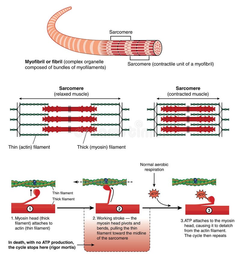
Free with trial Mechanism of muscle contraction. Detail of a muscle sarcomere showing thin and thick filaments and mechanism of mechanical contraction. Contraction vectors Mechanism of muscle contraction. Detail of a muscle sarcomere showing thin and thick filaments and mechanism of mechanical contraction
Free with trial ATP muscle contraction cycle vector illustration labeled scheme. Educational diagram with muscle, fibers and cells. Structure of myofibril with thin and thick filament. Contraction vectors ATP muscle contraction cycle vector illustration labeled educational scheme. ATP muscle contraction cycle vector illustration labeled scheme. Educational diagram. ATP muscle contraction cycle vector illustration labeled scheme. Educational diagram with muscle, fibers and cells. Structure of myofibril with thin and thick filament.
Free with trial Illustration of human arm muscles during contraction and relaxing. Contraction vectors Antagonistic musle. Illustration of human arm muscles during contraction and relaxing. Illustration of human arm muscles during contraction and relaxing.
Free with trial Muscle contraction. The subcellular components involved in muscle contration -- actin and myosin. Contraction vectors Muscle contraction. The subcellular components involved in muscle contration -- actin and myosin
Free with trial Spectacle. Black different shaped spectacle / sunglasses frame collection isolated on white. Contraction vectors Spectacle. Black different shaped spectacle / sunglasses frame collection isolated on white
Free with trial Peristalsis. Medical illustration of the peristaltic movement. Contraction vectors Peristalsis. Medical illustration of the peristaltic movement
Free with trial Arm muscles. The illustration shows the muscles that make up the arm, with its relative movements. Contraction vectors Arm muscles. The illustration shows the muscles that make up the arm, with its relative movements
Free with trial ECG explained. Connection between ECG and electrical activity of the heart, eps8. Contraction vectors ECG explained. Connection between ECG and electrical activity of the heart, eps8
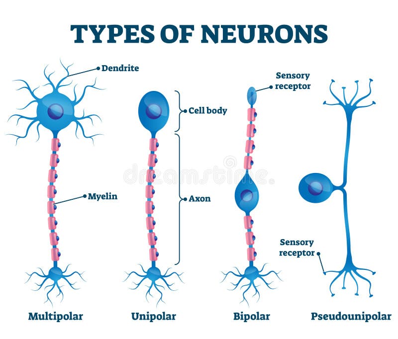
Free with trial Types Of Neurons. Basic neuron types. Unipolar, pseudo-unipolar neuron, bipolar, and multipolar Neurons. Neuron Cell Body. Different Types of Neurons. Contraction vectors Types Of Neurons. Basic neuron types. Unipolar, pseudo-unipolar neuron, bipolar, and multipolar Neurons. Neuron Cell Body. Different Types of Neurons
Free with trial Types of muscle tissue. Skeletal muscle, smooth muscle, cardiac muscle. Vector scheme. Contraction vectors Types of muscle tissue. Skeletal muscle, smooth muscle, cardiac muscle. Vector scheme
Free with trial Blood supply to the heart. Heart anatomy showing coronary arteries, sites of heart attack, eps8. Contraction vectors Blood supply to the heart. Heart anatomy showing coronary arteries, sites of heart attack, eps8
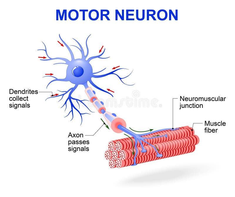
Free with trial Structure of motor neuron. Vector diagram. Include dendrites, cell body with nucleus, axon, myelin sheath, nodes of Ranvier and motor end plates. The impulses are transmitted through the motor neuron in one direction. Contraction vectors Motor neuron. Vector diagram. Structure of motor neuron. Vector diagram. Include dendrites, cell body with nucleus, axon, myelin sheath, nodes of Ranvier and. Structure of motor neuron. Vector diagram. Include dendrites, cell body with nucleus, axon, myelin sheath, nodes of Ranvier and motor end plates. The impulses are transmitted through the motor neuron in one direction
Free with trial Detail of muscle tissue showing actin and myosin, troponin complex, thin filaments and thick filaments. Created in Adobe Illustrator. Contains gradient meshes. EPS 10. Contraction vectors Detail of muscle physiology. Detail of muscle tissue showing actin and myosin, troponin complex, thin filaments and thick filaments. Created in Adobe Illustrator. Detail of muscle tissue showing actin and myosin, troponin complex, thin filaments and thick filaments. Created in Adobe Illustrator. Contains gradient meshes. EPS 10.
Free with trial Each skeletal muscle fiber has many bundles of myofilaments. Each bundle is called a myofibril. This is what gives the muscle its striated appearance. The contractile units of the cells are called sarcomeres. Contraction vectors Structure of skeletal muscle. Each skeletal muscle fiber has many bundles of myofilaments. Each bundle is called a myofibril. This is what gives the muscle its. Each skeletal muscle fiber has many bundles of myofilaments. Each bundle is called a myofibril. This is what gives the muscle its striated appearance. The contractile units of the cells are called sarcomeres.
Free with trial Structure of skeletal muscle fiber. With actin and myosin filaments, eps10. Contraction vectors Structure of skeletal muscle fiber. With actin and myosin filaments, eps10
Free with trial Colic. Medical illustration of symptoms and causes of colic. Contraction vectors Colic. Medical illustration of symptoms and causes of colic
Free with trial Types of Muscle Tissue of Human Body Diagram including cardiac skeletal smooth. Examples of heart digestive system. Along with involuntary voluntary control for medical science education. Contraction vectors Types of Muscle Tissue of Human Body Diagram. Including cardiac skeletal smooth. Examples of heart digestive system. Along with involuntary voluntary control. Types of Muscle Tissue of Human Body Diagram including cardiac skeletal smooth. Examples of heart digestive system. Along with involuntary voluntary control for medical science education
Free with trial Pneumothorax. Medical illustration of the symptoms of pneumothorax. Contraction vectors Pneumothorax. Medical illustration of the symptoms of pneumothorax
Free with trial Pneumothorax. Medical illustration of the symptoms of pneumothorax. Contraction vectors Pneumothorax. Medical illustration of the symptoms of pneumothorax
Free with trial Stages of Labor and Birth in a vaginal delivery infographic diagram including engagement descent internal complete rotation extension poster for medical science education and healthcare. Contraction vectors Stages of Labor and Birth in a vaginal delivery. Infographic diagram including engagement descent internal complete rotation extension poster for medical. Stages of Labor and Birth in a vaginal delivery infographic diagram including engagement descent internal complete rotation extension poster for medical science education and healthcare
Free with trial Cardiac arrhythmia. Schematic illustration of cardiac arrhythmia. Contraction vectors Cardiac arrhythmia. Schematic illustration of cardiac arrhythmia
Free with trial Heart valve defect. Aortic vavle defect, the most common heart valve problem, eps8. Contraction vectors Heart valve defect. Aortic vavle defect, the most common heart valve problem, eps8
Free with trial Anatomy of a muscle. From gross structure to the level of the myofibril and sarcomere. Contraction vectors Anatomy of a muscle. From gross structure to the level of the myofibril and sarcomere
Free with trial Tetralogy of Fallot. Congenital heart disease: Tetralogy of Fallot, eps8. Contraction vectors Tetralogy of Fallot. Congenital heart disease: Tetralogy of Fallot, eps8
Free with trial Diseases of heart muscle. 3 different diseases of heart muscle, eps8. Contraction vectors Diseases of heart muscle. 3 different diseases of heart muscle, eps8
Free with trial Cartoon illustration of Structure Skeletal Muscle Anatomy. Contraction vectors Cartoon illustration of Structure Skeletal Muscle Anatomy. Illustration of Structure Skeletal Muscle Anatomy
Free with trial Parkinson. A vector illustration of Old man with Parkinson symptoms difficult walking. Contraction vectors Parkinson. A vector illustration of Old man with Parkinson symptoms difficult walking
Free with trial Ventricular septal defect. Congenital heart disease: ventricular septal defect, eps8. Contraction vectors Ventricular septal defect. Congenital heart disease: ventricular septal defect, eps8
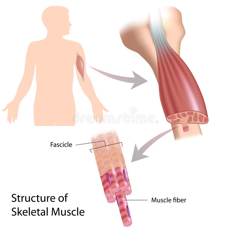
Free with trial Skeletal muscle structure. With example of the biceps, eps10. Contraction vectors Skeletal muscle structure. With example of the biceps, eps10
Free with trial Skeletal muscle cells are tubular. They have multiple nuclei. Skeletal muscle is striated, it has an alternating pattern of light and darks bands. Contraction vectors The skeletal muscle fiber. Skeletal muscle cells are tubular. They have multiple nuclei. Skeletal muscle is striated, it has an alternating pattern of light and. Skeletal muscle cells are tubular. They have multiple nuclei. Skeletal muscle is striated, it has an alternating pattern of light and darks bands.
Free with trial Smooth muscle innervation. Types of Smooth muscle innervation, eps10. Contraction vectors Smooth muscle innervation. Types of Smooth muscle innervation, eps10
Free with trial Structure motor neuron. Vector diagram. Include dendrites, cell body with nucleus, axon, myelin sheath, nodes of Ranvier and motor end plates. The impulses are transmitted through the motor neuron in one direction. Contraction vectors Motor neuron. Vector diagram. Structure motor neuron. Vector diagram. Include dendrites, cell body with nucleus, axon, myelin sheath, nodes of Ranvier and motor. Structure motor neuron. Vector diagram. Include dendrites, cell body with nucleus, axon, myelin sheath, nodes of Ranvier and motor end plates. The impulses are transmitted through the motor neuron in one direction
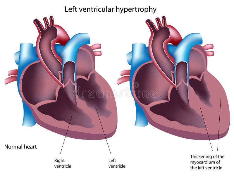
Free with trial Left ventricular hypertrophy. Normal heart and heart with left ventricular hypertrophy, eps8. Contraction vectors Left ventricular hypertrophy. Normal heart and heart with left ventricular hypertrophy, eps8
Free with trial Vector science illustration. Muscle Tissues. Contraction vectors Structure of skeletal muscle. Vector science illustration. Muscle Tissues. Vector science illustration. Muscle Tissues.
Free with trial Lung alveoli normal and asthma. Structure of terminal bronchioles and alveoli in the normal state and during asthmatic constriction. Contraction vectors Lung alveoli normal and asthma. Structure of terminal bronchioles and alveoli in the normal state and during asthmatic constriction
Free with trial Motor neuron controls muscle movement. Diagram showing neuromuscular synaptic junction. Contraction vectors Motor neuron controls muscle movement. Diagram showing neuromuscular synaptic junction
Free with trial Histamine. Allergies or allergic diseases. local immune responses. Allergic reaction. Contraction vectors Histamine. Allergies or allergic diseases. Histamine. local immune responses. Allergic reaction
Free with trial Sequence of events in voiding the bladder. Four stages of micturition, showing detrusor muscle contraction and pelvic floor relaxation. Contraction vectors Sequence of events in voiding the bladder. Four stages of micturition, showing detrusor muscle contraction and pelvic floor relaxation
Free with trial A neuron, also known as a neurone and nerve cell, is an electrically excitable cell that receives, processes, and transmits information through electrical and chemical signals. Contraction vectors Types of Neurons - sensory, intereuron, motor. A neuron, also known as a neurone and nerve cell, is an electrically excitable cell that receives, processes, and. A neuron, also known as a neurone and nerve cell, is an electrically excitable cell that receives, processes, and transmits information through electrical and chemical signals.
Free with trial Muscle tissue. Skeletal muscle, smooth in a gastrointestinal tract and cardiac muscle in a heart. Types of Muscle cells. vector for medical, educational, biologycal and science use. Contraction vectors Muscle tissue: Skeletal, smooth and cardiac. Muscle tissue. Skeletal muscle, smooth in a gastrointestinal tract and cardiac muscle in a heart. Types of Muscle. Muscle tissue. Skeletal muscle, smooth in a gastrointestinal tract and cardiac muscle in a heart. Types of Muscle cells. vector for medical, educational, biologycal and science use
Free with trial Human heart normal sinus rhythm and human heart detailed anatomy. Medical illustration. Contraction vectors Human heart normal sinus rhythm and heart anatomy. Human heart normal sinus rhythm and human heart detailed anatomy. Medical illustration. Human heart normal sinus rhythm and human heart detailed anatomy. Medical illustration.
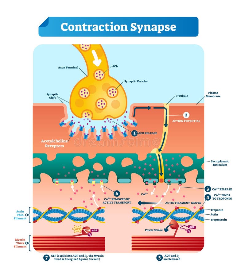
Free with trial Contraction synapse vector illustration. Labeled closeup medical structure scheme. Diagram with full cycle of ACH release, action potential, troponin bonding and filament. Contraction vectors Contraction synapse vector illustration. Labeled medical structure scheme. Contraction synapse vector illustration. Labeled closeup medical structure scheme. Contraction synapse vector illustration. Labeled closeup medical structure scheme. Diagram with full cycle of ACH release, action potential, troponin bonding and filament
Free with trial The muscle contraction as a result of Nerve impulses set off a biochemical reaction that causes myosin to stick to actin. Human. Body infographic. Contraction vectors The muscle contraction as a result of Nerve impulses set off a biochemical reaction that causes myosin to stick to actin. Human. Body infographic
Free with trial Muscle contraction and relaxation. The mechanism by which muscle contracts and relaxes due to the action of calcium ions on the troponin complex. Contraction vectors Muscle contraction and relaxation. The mechanism by which muscle contracts and relaxes due to the action of calcium ions on the troponin complex
Free with trial Structure Skeletal Muscle. myofibril with thin and thick filament. close up of a sarcomere. Muscles contract by sliding the myosin and actin filaments along each other. Biomedical Science. Mechanism of mechanical contraction. Contraction vectors Structure Skeletal Muscle. myofibril with sarcomeres. Close-up of actin and myosin. Structure Skeletal Muscle. myofibril with thin and thick filament. close up. Structure Skeletal Muscle. myofibril with thin and thick filament. close up of a sarcomere. Muscles contract by sliding the myosin and actin filaments along each other. Biomedical Science. Mechanism of mechanical contraction
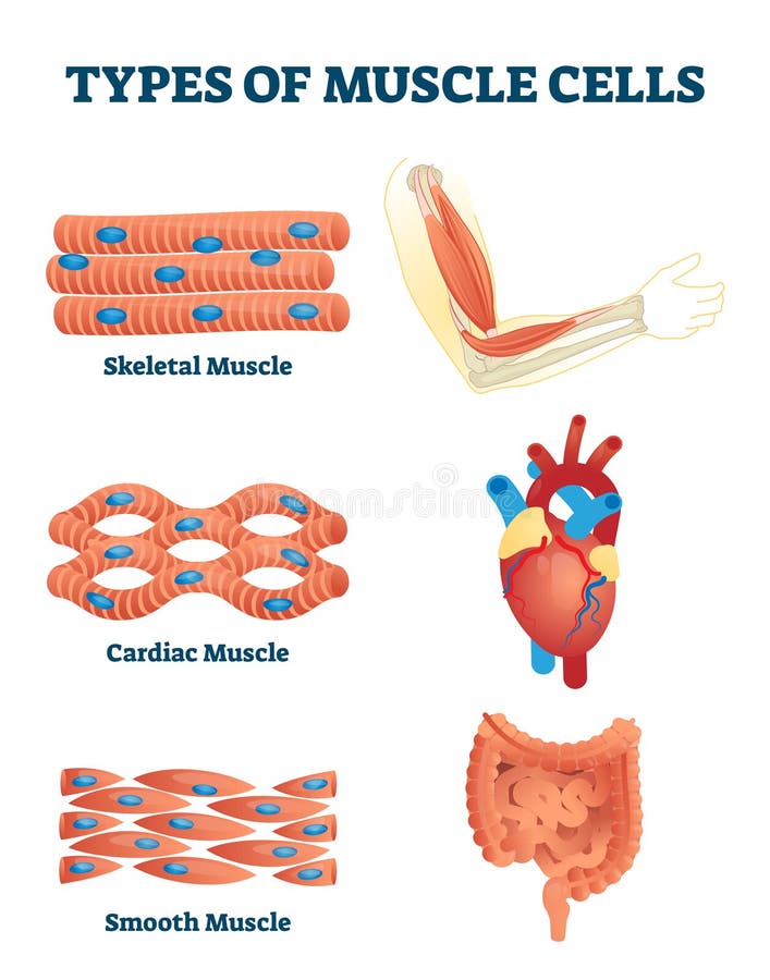
Free with trial Types of muscle cells vector illustration. Labeled various soft tissues differences explanation. Educational skeletal, cardiac and smooth contraction. Anatomical body parts example for biology handout. Contraction vectors Types of muscle cells vector illustration. Labeled soft tissues explanation. Types of muscle cells vector illustration. Labeled various soft tissues differences. Types of muscle cells vector illustration. Labeled various soft tissues differences explanation. Educational skeletal, cardiac and smooth contraction. Anatomical body parts example for biology handout
Free with trial Peristalsis is waves of alternate contraction and relaxation from Esophagus and stomach to intestine. esophageal motility. Vector illustration for medical, biological, educational and science use. Contraction vectors Peristalsis. esophageal motility. Peristalsis is waves of alternate contraction and relaxation from Esophagus and stomach to intestine. esophageal motility. Peristalsis is waves of alternate contraction and relaxation from Esophagus and stomach to intestine. esophageal motility. Vector illustration for medical, biological, educational and science use
Free with trial Hiccup. Medical illustration of the symptoms and causes of hiccup. Contraction vectors Hiccup. Medical illustration of the symptoms and causes of hiccup
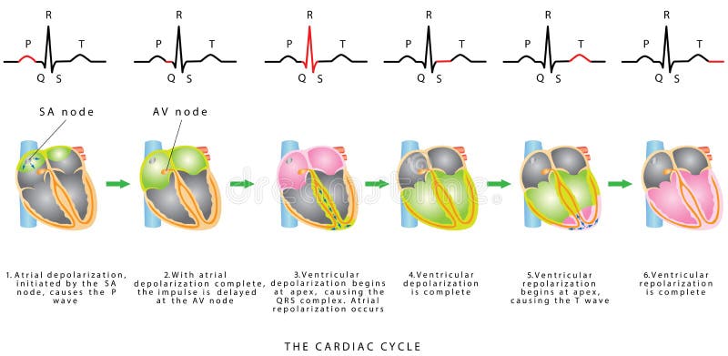
Free with trial The Cardiac Cycle. The sequence of heart excitation is associated with the deviation of ECG waves by tracing. Diagram of the phases of cardiac cycle. Contraction vectors The Cardiac Cycle. The sequence of heart excitation is associated with the deviation of ECG waves by tracing. Diagram of the phases of cardiac cycle
Free with trial Heart. Anatomy of the Heart. The Cardiac Cycle. Diastole Ventricular Relaxation and Filling. Systole Ventricular Contraction and Ejection. Contraction vectors Heart. Anatomy of the . The Cardiac Cycle. Diastole Ventricular Relaxation and Filling. Systole Ventricular Contraction and Ejection
Free with trial Sarcomere muscular biology scheme vector illustration. Myosin filaments, discs, lines and bands. Myofibril detailed labeled diagram. Sports educational health information. Muscular system anatomy. Contraction vectors Sarcomere muscular biology scheme vector illustration. Myosin filaments, discs, lines and bands. Myofibril detailed labeled diagram. Sports educational health
Free with trial Smooth muscle tissue. Anatomy of a relaxed and contracted smooth muscle cell. Contraction vectors Smooth muscle tissue. Anatomy of a relaxed and contracted smooth muscle cell
Free with trial Types of muscle tissue. Skeletal, smooth and cardiac muscle. Silhouette of a man with highlighted the Human internal organs heart, intestines, muscle and Muscle cells close up. Contraction vectors Types of muscle tissue. Skeletal, smooth and cardiac muscle. Silhouette of a man with highlighted the Human internal organs heart, intestines, muscle and
Free with trial Ouch! What a pain, doctor!. Cartoon-style illustration: a patient with a terrible backache, the doctor by his side. Contraction vectors Ouch! What a pain, doctor!. Cartoon-style illustration: a patient with a terrible backache, the doctor by his side
Free with trial Heart muscle anatomy and function. Drawing of heart muscle showing cardiac myofibres, actin, tropomyosin and troponin complex. Contraction vectors Heart muscle anatomy and function. Drawing of heart muscle showing cardiac myofibres, actin, tropomyosin and troponin complex
Free with trial How do muscles work labeled principle explanation scheme vector illustration. Anatomical and physical movement process example with biceps relaxed and triceps contracted. Educational comparison graph. Contraction vectors How do muscles work labeled principle explanation scheme vector illustration. Anatomical and physical movement process example with biceps relaxed and triceps
Free with trial Human heart normal sinus rhythm, electrocardiogram record. Medical illustration. Contraction vectors Human heart normal sinus rhythm, electrocardiogram. Record. Medical illustration
Free with trial Visual accomodation. Medical illustration of the visual accomodation. Contraction vectors Visual accomodation. Medical illustration of the visual accomodation
Free with trial Structure of a skeletal muscle. (useful for education in schools and clinics ) - vector illustration. Contraction vectors Structure of a skeletal muscle. (useful for education in schools and clinics ) - vector illustration
Free with trial Digestion and Peristalsis. Esophagus Function. Contraction vectors Digestion and Peristalsis. Esophagus Function
Free with trial Calf muscle tear. Medical illustration of the symptoms of calf muscle tear. Contraction vectors Calf muscle tear. Medical illustration of the symptoms of calf muscle tear
Free with trial Cardiac Cycle Diastole and Systole of Human Heart Anatomy infographic diagram with all stages of pumping filling in right left atrium and ventricule for medicine science education medical healthcare. Contraction vectors Cardiac Cycle Diastole and Systole of Human Heart Anatomy. Infographic diagram with all stages of pumping filling in right left atrium and ventricule for. Cardiac Cycle Diastole and Systole of Human Heart Anatomy infographic diagram with all stages of pumping filling in right left atrium and ventricule for medicine science education medical healthcare
Free with trial Types of neurons vector illustration. Labeled anatomical nerve parts comparison scheme. Synapses receptors educational closeup with multipolar, unipolar, bipolar and pseudounipola sensory receptors. Contraction vectors Types of neurons vector illustration. Labeled nerve parts comparison scheme. Types of neurons vector illustration. Labeled anatomical nerve parts comparison. Types of neurons vector illustration. Labeled anatomical nerve parts comparison scheme. Synapses receptors educational closeup with multipolar, unipolar, bipolar and pseudounipola sensory receptors.
Free with trial Business design conference concept people set presentation, planninh, meeting, building, success, agreement or partnership. with flat icons. vector illustration. Contraction vectors Business design conference concept people set presentation, plan. Ninh, meeting, building, success, agreement or partnership. with flat icons. vector
Free with trial Peristalsis, or wave-like contractions of the muscles in the outer walls of the digestive tract, carries the bolus by the esophagus. Peristalsis does not only occur in the esophagus. It continues through the stomach, small intestine, and large intestine. Vector diagram. Contraction vectors Peristalsis. involuntary wavelike muscular contrac. Peristalsis, or wave-like contractions of the muscles in the outer walls of the digestive tract, carries the. Peristalsis, or wave-like contractions of the muscles in the outer walls of the digestive tract, carries the bolus by the esophagus. Peristalsis does not only occur in the esophagus. It continues through the stomach, small intestine, and large intestine. Vector diagram
Free with trial Cardiac arrhythmia. Schematic illustration of cardiac arrhythmia. Contraction vectors Cardiac arrhythmia. Schematic illustration of cardiac arrhythmia
Free with trial Functions of proteins with anatomical roles in body outline collection set. Labeled educational list with molecules support for immune system, muscle strength and transportation vector illustration. Contraction vectors Functions of proteins with anatomical roles in body outline collection set. Labeled educational list with molecules support for immune system, muscle strength
Free with trial Types of cardiomyopathy: the walls of the ventricles thicken and become stiff; the left ventricle is enlarged and weakened; the walls of the ventricles become stiff, but not necessarily thickened. Contraction vectors Diseases of the Heart Muscle. Types of cardiomyopathy: the walls of the ventricles thicken and become stiff; the left ventricle is enlarged and weakened; the. Types of cardiomyopathy: the walls of the ventricles thicken and become stiff; the left ventricle is enlarged and weakened; the walls of the ventricles become stiff, but not necessarily thickened
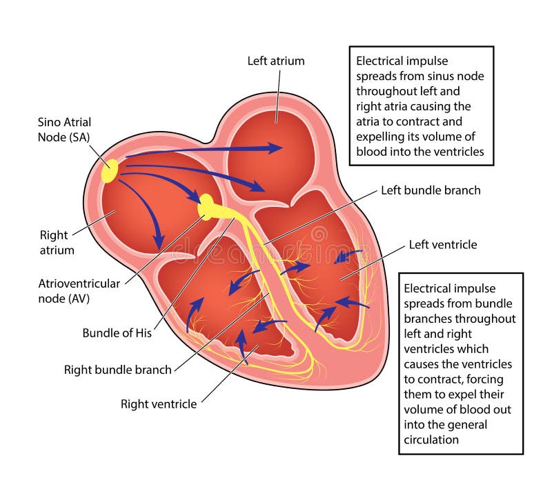
Free with trial The electrical conducting system of the heart, showing the impulse path from the sino atrial node to the atrioventricular node and the ventricles. Contraction vectors Heart electrical conducting system. The electrical conducting system of the heart, showing the impulse path from the sino atrial node to the atrioventricular. The electrical conducting system of the heart, showing the impulse path from the sino atrial node to the atrioventricular node and the ventricles
Free with trial Science diagram illustrating thermal expansion and molecular structure, showing how thermometer liquid expands when heated. Molecules respond to less heat and more heat. Contraction vectors Thermal Expansion of Liquids. Science diagram illustrating thermal expansion and molecular structure, showing how thermometer liquid expands when heated. Science diagram illustrating thermal expansion and molecular structure, showing how thermometer liquid expands when heated. Molecules respond to less heat and more heat.
Free with trial Cytoskeleton structure as complex dynamic network of interlinking protein filaments outline diagram. Labeled educational cell with microtubules and microfilaments explanation vector illustration. Contraction vectors Cytoskeleton structure as complex protein filaments network outline diagram. Cytoskeleton structure as complex dynamic network of interlinking protein filaments. Cytoskeleton structure as complex dynamic network of interlinking protein filaments outline diagram. Labeled educational cell with microtubules and microfilaments explanation vector illustration.
Free with trial Flat linear Infographic Business Education Pen Tree with Light bulb Outline concept. Vector Illustration. Contraction vectors Flat linear Infographic Business Education Pen Tree with Light. Bulb Outline concept.Vector Illustration
Free with trial Muscular dystrophy vector illustration. Compared normal and healthy muscle versus atrophied and weak. Medical scheme with explanation how time affects bicep - normal strong muscle and degeneration. Contraction vectors Vector illustration about muscular dystrophy. Compared normal muscle and atrophied muscle. Scheme with healthy and weak human. Muscular dystrophy vector. Muscular dystrophy vector illustration. Compared normal and healthy muscle versus atrophied and weak. Medical scheme with explanation how time affects bicep - normal strong muscle and degeneration.
Free with trial Gastroesophageal reflux disease. Healthy and sick. Stomach. Medicine anatomy healthy, organ and illness illustration. Vector medical GERD infographic. Contraction vectors Gastroesophageal reflux disease. Healthy and sick. Stomach. Medicine anatomy healthy, organ and illness illustration. Vector medical GERD infographic
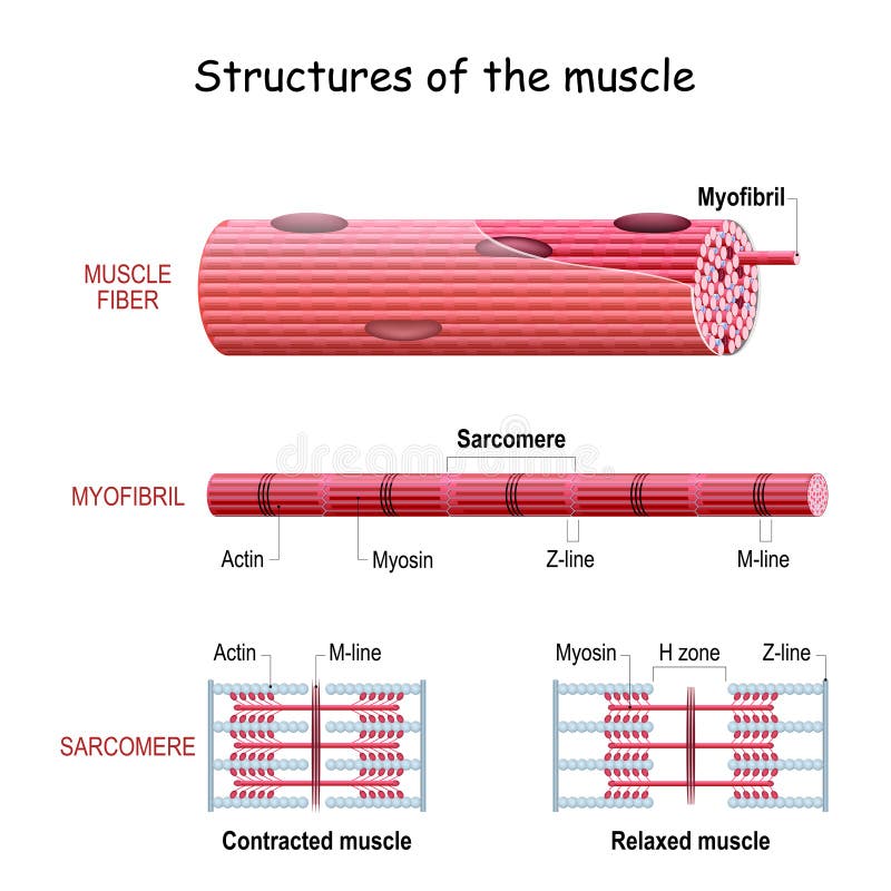
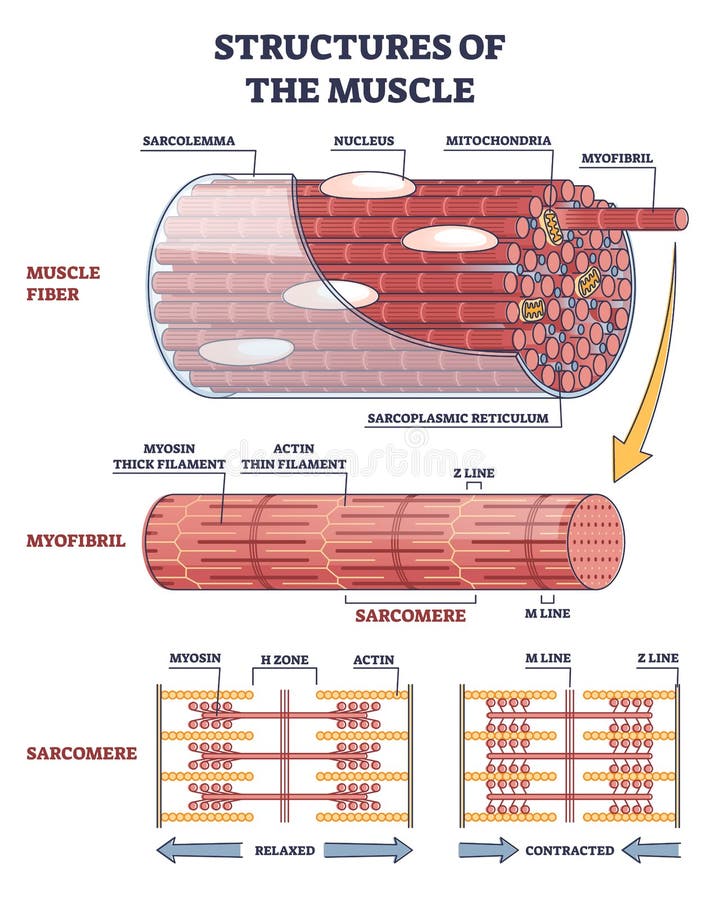
Free with trial Structures of muscle with fiber, myofibril and sarcomere contraction outline diagram. Labeled educational isolated parts closeup description from anatomical and physiology sides vector illustration. Contraction vectors Structures of muscle with fiber, myofibril and sarcomere outline diagram. Structures of muscle with fiber, myofibril and sarcomere contraction outline diagram. Structures of muscle with fiber, myofibril and sarcomere contraction outline diagram. Labeled educational isolated parts closeup description from anatomical and physiology sides vector illustration.
Free with trial Types of neurons. Structure sensory, motor neuron, astrocyte, pyromidal, Betz cell, microglia. Set. Infographics Vector illustration on background. Contraction vectors Types of neurons. Structure sensory, motor neuron, astrocyte, pyromidal, Betz cell, microglia. Set. Infographics. Vector. Types of neurons. Structure sensory. Types of neurons. Structure sensory, motor neuron, astrocyte, pyromidal, Betz cell, microglia. Set. Infographics Vector illustration on background
Free with trial The muscle contraction as a result of Nerve impulses set off a biochemical reaction that causes myosin to stick to actin. Human body infographic. Contraction vectors Muscle contraction. Human body infographic. The muscle contraction as a result of Nerve impulses set off a biochemical reaction that causes myosin to stick to. The muscle contraction as a result of Nerve impulses set off a biochemical reaction that causes myosin to stick to actin. Human body infographic
 Sign in