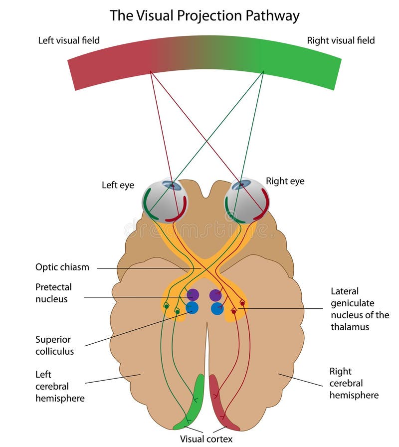
Free with trial Visual pathway medical vector illustration diagram. Eye and brain anatomical system with optic nerves and visual cortex. Educational human vision explanation scheme with visual and binocular fields. Cortex vectors Visual pathway medical vector illustration diagram. Eye and brain anatomical system with optic nerves and visual cortex. Educational human vision explanation
Free with trial Cortical homunculus diagram showing how body areas map to cortex, motor homunculus, sensory homunculus, and brain gyri emphasize exaggerated hands, lips, tongue. Outline diagram. Cortex vectors Cortical homunculus diagram showing how body areas map to cortex, ... Cortical homunculus diagram showing how body areas map to cortex, motor homunculus. Cortical homunculus diagram showing how body areas map to cortex, motor homunculus, sensory homunculus, and brain gyri emphasize exaggerated hands, lips, tongue. Outline diagram
Free with trial Amygdala medical labeled vector illustration. Anatomical scheme with visual thalamus, cortex and response to threat. Diagram with cerebrum, thalamus and corpus callosum. Cortex vectors Amygdala medical labeled vector illustration and scheme with response to threat. Amygdala medical labeled vector illustration. Anatomical scheme with visual. Amygdala medical labeled vector illustration. Anatomical scheme with visual thalamus, cortex and response to threat. Diagram with cerebrum, thalamus and corpus callosum.
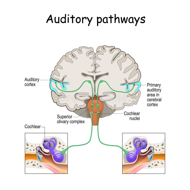
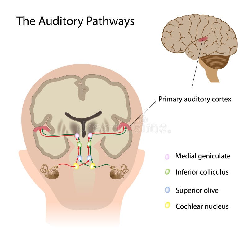
Free with trial Auditory pathways from cochlea in ear to cortex in brain. Sound Localization. anatomy of the auditory system. Structures of the Ear. Cortex vectors Auditory pathways from cochlea in ear to cortex in brain. Sound Localization. anatomy of the auditory system. Structures of the Ear
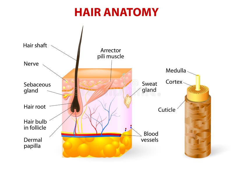
Free with trial The Structure of human hair illustration. Cuticle, cortex, and medulla. Isolated on whitebackground. Cortex vectors Layer of hair structure. The hair shaft consists of cortex,cuticle, and medulla. Hair care and beauty concept. The Structure of human hair illustration. Cuticle. The Structure of human hair illustration. Cuticle, cortex, and medulla. Isolated on whitebackground
Free with trial Olfactory cortex pathway showing how smell signals travel via olfactory bulb and tract to hippocampus, linking odor to memory and emotion. Outline diagram. Cortex vectors Olfactory cortex pathway showing how smell signals travel via olfactory ... Olfactory cortex pathway showing how smell signals travel via olfactory bulb and. Olfactory cortex pathway showing how smell signals travel via olfactory bulb and tract to hippocampus, linking odor to memory and emotion. Outline diagram
Free with trial Motor nerves originating in the leg muscles traveling via the spinal cord to the motor cortex or the brain. Created in Adobe Illustrator. EPS 10. Cortex vectors Motor nerves from leg to motor cortex. Motor nerves originating in the leg muscles traveling via the spinal cord to the motor cortex or the brain. Created in. Motor nerves originating in the leg muscles traveling via the spinal cord to the motor cortex or the brain. Created in Adobe Illustrator. EPS 10.
Free with trial Created brain function, human brain anatomy with Basal Ganglia, Cortex, Brain Stem, Cerebellum and Spinal Cord- vector eps10. Cortex vectors Brain function, human brain anatomy with Basal Ganglia, Cortex, Brain Stem, Cerebellum and Spinal Cord. Created brain function, human brain anatomy with Basal. Created brain function, human brain anatomy with Basal Ganglia, Cortex, Brain Stem, Cerebellum and Spinal Cord- vector eps10
Free with trial Brain vector illustration. Medical educational scheme with neurological cells closeup. Silhouette with cerebrum, cerebellum and stem. Cortex, lobe, respiratory, pituitary gland and wernickes diagram. Cortex vectors Brain vector illustration. Medical educational scheme with neurological cells. Silhouette with cerebrum, stem, cortex and lobe. Brain vector illustration. Brain vector illustration. Medical educational scheme with neurological cells closeup. Silhouette with cerebrum, cerebellum and stem. Cortex, lobe, respiratory, pituitary gland and wernickes diagram.
Free with trial Adrenal gland hormones diagram maps cortex layers and medulla with aldosterone, cortisol, catecholamines, key objects, adrenal gland, cortex zones, medulla. Outline diagram. Cortex vectors Adrenal gland hormones diagram maps cortex layers and medulla with ... Adrenal gland hormones diagram maps cortex layers and medulla with aldosterone, cortisol. Adrenal gland hormones diagram maps cortex layers and medulla with aldosterone, cortisol, catecholamines, key objects, adrenal gland, cortex zones, medulla. Outline diagram
Free with trial Diagram of the structure of the cerebral cortex, vector. Cortex vectors Diagram of the structure of the cerebral cortex. Vector
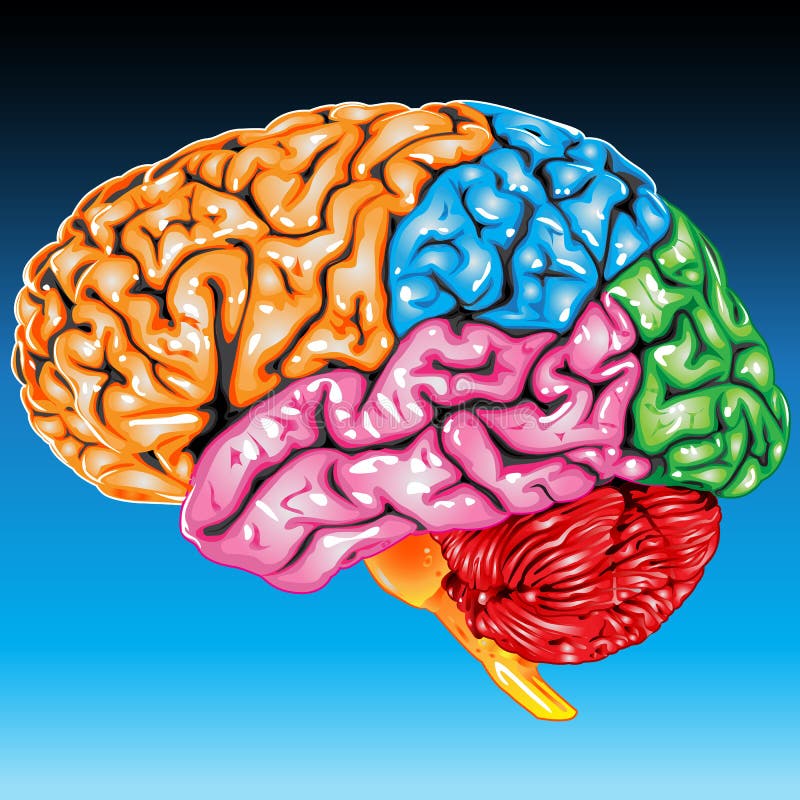
Free with trial Structure of cerebral cortex illustration. Colored anatomical regions responsible for intelligence and movement through nervous vector system. Cortex vectors Structure of cerebral cortex illustration. Colored anatomical regions responsible for intelligence and movement. Structure of cerebral cortex illustration. Structure of cerebral cortex illustration. Colored anatomical regions responsible for intelligence and movement through nervous vector system.
Free with trial Stress response system. Stress is a main cause of high levels of cortisol secretion. Cortisol is a hormone produced by the adrenal cortex. Cortex vectors Stress response system. Stress is a main cause of high levels of cortisol secretion. Cortisol is a hormone produced by the adrenal cortex
Free with trial Neurotechnology biomedical engineering schematic of a flexible hexagonal neural lace brain-computer interface (BCI) cortex mesh. Cortex vectors Neural Lace Brain Cortex Microelectrode Mesh Vector. Neurotechnology biomedical engineering schematic of a flexible hexagonal neural lace brain-computer. Neurotechnology biomedical engineering schematic of a flexible hexagonal neural lace brain-computer interface (BCI) cortex mesh
Free with trial Adrenal gland hormone secretion. Adrenal glands sit atop the kidneys and are composed of an outer cortex and an inner medulla, which produce different types of hormones. Cortex vectors Adrenal gland hormone secretion. Adrenal glands sit atop the kidneys and are composed of an outer cortex and an inner medulla, which produce different types of
Free with trial Sensory and motor nerve pathways from a stimulus to the sensory cortex and back to muscle. The mechanism for avoiding noxious stimuli. Created in Adobe Illustrator. Contains transparencies. EPS 10. Cortex vectors Sensory and motor nerve pathways. From a stimulus to the sensory cortex and back to muscle. The mechanism for avoiding noxious stimuli. Created in Adobe. Sensory and motor nerve pathways from a stimulus to the sensory cortex and back to muscle. The mechanism for avoiding noxious stimuli. Created in Adobe Illustrator. Contains transparencies. EPS 10.
Free with trial The adrenal glands consisting of two structurally different parts, the adrenal cortex and adrenal medulla. medulla secrete epinephrine and norepinephrine. Cortex vectors Adrenal gland. The adrenal glands consisting of two structurally different parts, the adrenal cortex and adrenal medulla. medulla secrete epinephrine and. The adrenal glands consisting of two structurally different parts, the adrenal cortex and adrenal medulla. medulla secrete epinephrine and norepinephrine.
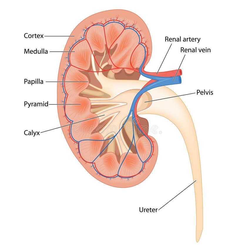
Free with trial Kidney cross section illustration displaying internal structures including the renal cortex medulla pelvis and associated vascular system for medical study. Cortex vectors Kidney Anatomy Cross Section Showing Renal Cortex and Blood Vessels. Kidney cross section illustration displaying internal structures including the renal cortex. Kidney cross section illustration displaying internal structures including the renal cortex medulla pelvis and associated vascular system for medical study
Free with trial Healthy and damaged hair. Internal structure of the Healthy and damaged hair: cuticle, cortex, medulla. Porous, Open and damaged cuticle. Cortex vectors Healthy and damaged hair. Internal structure of the Healthy and damaged hair: cuticle, cortex, medulla. Porous, Open and damaged cuticle
Free with trial Cross section of human kidney. Showing detail of blood supply, cortex, medulla, pyramids and calyx. Cortex vectors Cross section of human kidney. Showing detail of blood supply, cortex, medulla, pyramids and calyx
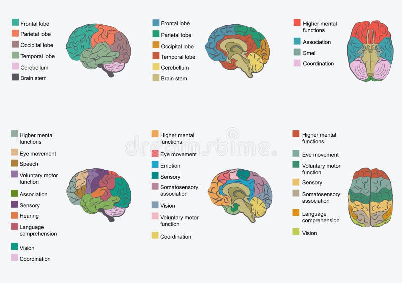
Free with trial Anatomical diagram of human Brain. Medicine, Vector illustration poster. Anatomy Medical study info graphics banner for education. Functional areas of cerebral cortex, named lobe parts. Cortex vectors Anatomical diagram of human Brain. Medicine, Vector illustration. Poster. Anatomy Medical study info graphics banner for education. Functional areas of cerebral. Anatomical diagram of human Brain. Medicine, Vector illustration poster. Anatomy Medical study info graphics banner for education. Functional areas of cerebral cortex, named lobe parts
Free with trial Brain shows a black and white illustration of auditory cortex connections with wave patterns. Cortex vectors Brain Auditory Cortex Connectivity Icon for Health Technology Presentations. Brain shows a black and white illustration of auditory cortex connections with wave. Brain shows a black and white illustration of auditory cortex connections with wave patterns
Free with trial Brain functions vector illustration. Labeled explanation head organ parts scheme. Inner side view with educational section description. Cerebral cortex, hypothalamus, spinal cord and thalamus diagram. Cortex vectors Brain functions vector illustration. Labeled explanation organ parts scheme. Brain functions vector illustration. Labeled explanation head organ parts scheme. Brain functions vector illustration. Labeled explanation head organ parts scheme. Inner side view with educational section description. Cerebral cortex, hypothalamus, spinal cord and thalamus diagram.
Free with trial Posttraumatic stress disorder. Adrenal cortex, Anterior Pituitary and Hypothalamus. Regions of the brain associated with stress and PTSD. Endocrine Aspects of PTSD: hypothalamic-pituitary-adrenal HPA axis. Cortex vectors Posttraumatic stress disorder. Adrenal cortex, Anterior Pituitary and Hypothalamus. Regions of the brain associated with stress and PTSD. Endocrine Aspects of
Free with trial Medulla oblongata vector illustration. Labeled diagram with ventral view and core structure. Midbrain and pons position. Shown left and right cerebral cortex sides. Cortex vectors Medulla oblongata vector illustration. Labeled diagram with ventral view. Medulla oblongata vector illustration. Labeled diagram with ventral view and core. Medulla oblongata vector illustration. Labeled diagram with ventral view and core structure. Midbrain and pons position. Shown left and right cerebral cortex sides.
Free with trial Lichen Structure Cross Section Labeled Diagram, Educational Mycology Poster, Lichen Anatomy Showing Upper Cortex, Algal Layer, Medulla, Lower Cortex, Rhizines and Hyphae. Composite organism. Cortex vectors Lichen Structure Cross Section Labeled Diagram, Educational Mycology Poster, Lichen Anatomy Showing Upper Cortex, Algal Layer,. Medulla, Lower Cortex, Rhizines. Lichen Structure Cross Section Labeled Diagram, Educational Mycology Poster, Lichen Anatomy Showing Upper Cortex, Algal Layer, Medulla, Lower Cortex, Rhizines and Hyphae. Composite organism
Free with trial Human brain sketch. Human brain cortex top view sketch isolated on white background vector illustration. Cortex vectors Human brain sketch. Human brain cortex top view sketch isolated on white background vector illustration
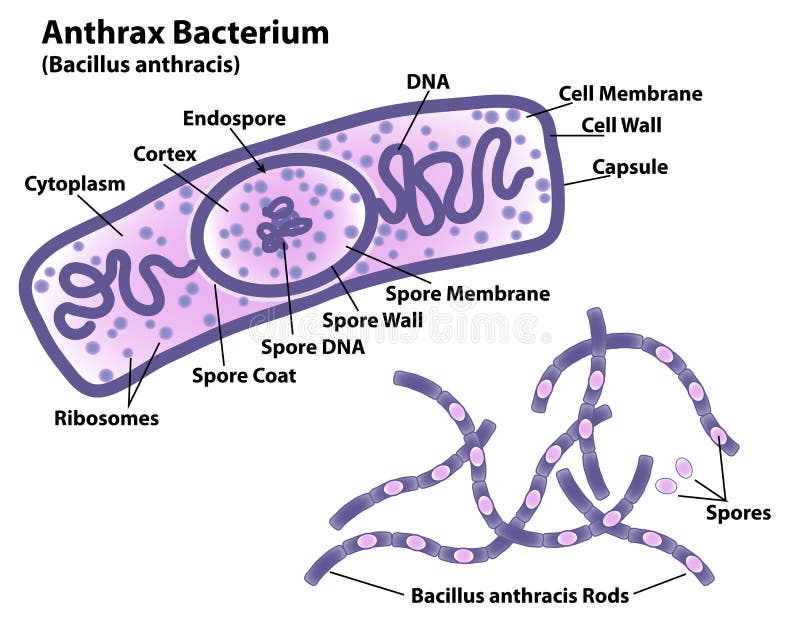
Free with trial Anthrax bacteria Bacillus anthracis with endospore labeled, rods and spores indicated, DNA, ribosomes, and cell wall. Science illustration includes labels for cell membrane, capsule, spore membrane, cytoplasm, cortex, and endospore. Cortex vectors Anthrax Bacteria Morphology and Cell. Anthrax bacteria Bacillus anthracis with endospore labeled, rods and spores indicated, DNA, ribosomes, and cell wall. Anthrax bacteria Bacillus anthracis with endospore labeled, rods and spores indicated, DNA, ribosomes, and cell wall. Science illustration includes labels for cell membrane, capsule, spore membrane, cytoplasm, cortex, and endospore.
Free with trial Medical anatomy of human Brain. Medicine, Vector illustration poster. Anatomical Medical study info graphics banner for education. Functional areas of cerebral cortex, named lobe parts. Cortex vectors Medical anatomy of human Brain. Medicine, Vector illustration po. Ster. Anatomical Medical study info graphics banner for education. Functional areas of cerebral. Medical anatomy of human Brain. Medicine, Vector illustration poster. Anatomical Medical study info graphics banner for education. Functional areas of cerebral cortex, named lobe parts
Free with trial This image showcases a detailed microscopic view of brain cortex tissue, highlighting the intricate patterns and textures of neural structures. The folds and grooves represent the complexity of the cerebral cortex, essential for cognitive functions and neural connectivity. Cortex vectors Microscopic View of Highly Detailed Brain Cortex Tissue Structure. This image showcases a detailed microscopic view of brain cortex tissue, highlighting the. This image showcases a detailed microscopic view of brain cortex tissue, highlighting the intricate patterns and textures of neural structures. The folds and grooves represent the complexity of the cerebral cortex, essential for cognitive functions and neural connectivity
Free with trial Stress response. Activation of the stress system. Stress is a main cause of high levels of adrenaline and cortisol secretion. hormones that produced by the medulla, and cortex of adrenal. Cortex vectors Stress response. Activation of the stress system. Stress is a main cause of high levels of adrenaline and cortisol secretion. Hormones that produced by the
Free with trial This vector shows functions of different part of the cortex and cerebellum. Each icon represents a function. Perfectly suits for presentations as well as educational resource. Cortex vectors Polygonal brain function infografic. This vector shows functions of different part of the cortex and cerebellum.Each icon represents a function. Perfectly suits. This vector shows functions of different part of the cortex and cerebellum.Each icon represents a function. Perfectly suits for presentations as well as educational resource.
Free with trial Visual system. Brain with optic nerve and Eyeball. Primary visual cortex. vector illustration. Cortex vectors Visual system. Brain with optic nerve and Eyeball. Primary visual cortex. vector illustration
Free with trial This image was created using Generative AI. A stylized of a human brain in purple tones showing the cortex and hemispheres. Ideal for medical, science, and education projects. Cortex vectors Purple Human Brain Icon With Cortex Details. This image was created using Generative AI. A stylized of a human brain in purple tones showing the cortex and. This image was created using Generative AI. A stylized of a human brain in purple tones showing the cortex and hemispheres. Ideal for medical, science, and education projects
Free with trial Clostridium tetani. Anatomy of the cell with terminal spore, and vegetative cell. Structure of the terminal spore: Core, cortex, and spore coat. Vector diagram for educational, medical, biological, and scientific use. Cortex vectors Clostridium tetani. Anatomy of the cell with terminal spore, and vegetative cell. Structure of the terminal spore: Core, cortex, and spore coat. Vector diagram
Free with trial Brain sketch icons of human organ. Vector isolated brain cerebellum vital organ of nervous system and cerebral cortex for medical design or surgery and body medicine symbols. Cortex vectors Human organ brain vector sketch icon. Brain sketch icons of human organ. Vector isolated brain cerebellum vital organ of nervous system and cerebral cortex for. Brain sketch icons of human organ. Vector isolated brain cerebellum vital organ of nervous system and cerebral cortex for medical design or surgery and body medicine symbols
Free with trial The structure of hair in the section. Medulla cortex and cuticle close-up vector illustration. Hair anatomy and medicine concept. Isolated on white background. Cortex vectors The structure of hair in the section. Medulla cortex and cuticle close-up vector illustration. Hair anatomy and medicine concept. Isolated on white background
Free with trial This image was created using Generative AI. Detailed of human brain anatomy with cerebral cortex and white matter. Cortex vectors Human brain anatomy with detailed cerebral cortex and white matter. This image was created using Generative AI. Detailed of human brain anatomy with cerebral. This image was created using Generative AI. Detailed of human brain anatomy with cerebral cortex and white matter
Free with trial Human brain anatomy,. Function area, mind system. Cortex vectors Human brain anatomy,. Function area, mind system
Free with trial Illustration of blood supply of the head. Cortex vectors Blood supply of the head. Illustration of blood supply of the head. Illustration of blood supply of the head.
Free with trial Brain. Human brain on white background. Cortex vectors Brain. Human brain on white background
Free with trial This image was created using Generative AI. Clean black and white line art of a human brain showing cerebral hemispheres, cortex folds, and brain stem. Ideal for medical, psychology, and neuroscience educational materials. Cortex vectors Human Brain Outline Icon with Cerebral Cortex and Brain Stem Black White Medical Design. This image was created using Generative AI. Clean black and white line. This image was created using Generative AI. Clean black and white line art of a human brain showing cerebral hemispheres, cortex folds, and brain stem. Ideal for medical, psychology, and neuroscience educational materials
Free with trial Brain function. Vector illustration of the Brain function. Cortex vectors Brain function. Vector illustration of the Brain function
Free with trial Hair structure. Human hair structure. Vector illustration. Cortex vectors Hair structure. Human hair structure. Vector illustration
Free with trial A vector drawing represents brain design. Cortex vectors Brain. A vector drawing represents brain design. A vector drawing represents brain design.
Free with trial A detailed 3D illustration of a human brain viewed from the side, highlighting the complex anatomy of the cerebral cortex. The design uses a professional blue and grey color scheme to represent medical and scientific concepts clearly. Cortex vectors Anatomical human brain model showing the side view of the cerebral cortex in blue and grey colors. A detailed 3D illustration of a human brain viewed from the. A detailed 3D illustration of a human brain viewed from the side, highlighting the complex anatomy of the cerebral cortex. The design uses a professional blue and grey color scheme to represent medical and scientific concepts clearly
Free with trial Cross section of brain showing the pituitary and pineal glands, cerebellum and brainstem. Created in Adobe Illustrator. Contains gradient fills. EPS 10. Cortex vectors Cross section of brain. Showing the pituitary and pineal glands, cerebellum and brainstem. Created in Adobe Illustrator. Contains gradient fills. EPS 10
Free with trial A vector drawing represents brain head design. Cortex vectors Brain head. A vector drawing represents brain head design. A vector drawing represents brain head design.
Free with trial The visual projection pathway. Diagram explaining how visual signals are received by the brain, eps8. Cortex vectors The visual projection pathway. Diagram explaining how visual signals are received by the brain, eps8
Free with trial Cross-section of a Longitudinal Kidney Diagram. A simple black-and-white line drawing showing the internal structure of a kidney in longitudinal cross-section, highlighting the renal pyramid and cortex. Cortex vectors Structure of a kidney in longitudinal cross-section, highlighting the renal pyramid and cortex. Generative AI. Cross-section of a Longitudinal Kidney Diagram.A. Cross-section of a Longitudinal Kidney Diagram.A simple black-and-white line drawing showing the internal structure of a kidney in longitudinal cross-section, highlighting the renal pyramid and cortex.
Free with trial The hypothalamic pituitary axes. The 3 hypothalamic pituitary axes: HPA, HPG and HPT, eps10. Cortex vectors The hypothalamic pituitary axes. The 3 hypothalamic pituitary axes: HPA, HPG and HPT, eps10
Free with trial Illustration whit main function of the brain,type. Cortex vectors Brain main function. Illustration whit main function of the brain,type. Illustration whit main function of the brain,type .
Free with trial Noxious and pain receptors in skin and the nerve pathways to the brain, via the spinal cord and thalamus. Created in Adobe Illustrator. Contains transparencies. EPS 10. Cortex vectors Nerve response to pain and touch. Noxious and pain receptors in skin and the nerve pathways to the brain, via the spinal cord and thalamus. Created in Adobe. Noxious and pain receptors in skin and the nerve pathways to the brain, via the spinal cord and thalamus. Created in Adobe Illustrator. Contains transparencies. EPS 10.
Free with trial Visualize nervous system states with this stress vs calm brain infographic. Features side by side comparison of sympathetic nervous system fight-or-flight mode versus parasympathetic nervous system rest-and-digest mode. Includes amygdala activation zone, cortisol and adrenaline visualization, prefrontal cortex shut down, breathing pattern changes, heart rate variability, digestion impact, muscle tension comparison, and switching techniques including deep breathing, vagus nerve stimulation, and mindfulness practices. Cortex vectors Stress Vs Calm Brain Infographic Fight Flight Rest Digest Comparison Vector. Visualize nervous system states with this stress vs calm brain infographic. Features. Visualize nervous system states with this stress vs calm brain infographic. Features side by side comparison of sympathetic nervous system fight-or-flight mode versus parasympathetic nervous system rest-and-digest mode. Includes amygdala activation zone, cortisol and adrenaline visualization, prefrontal cortex shut down, breathing pattern changes, heart rate variability, digestion impact, muscle tension comparison, and switching techniques including deep breathing, vagus nerve stimulation, and mindfulness practices.
Free with trial PTSD vector illustration. Labeled anatomical mental disorder causes scheme. Compared healthy and problematic brain differences set. Explained psychiatry diagnosis after disasters, abuse and accidents. Cortex vectors PTSD vector illustration. Labeled anatomical mental disorder causes scheme. Compared healthy and problematic brain differences set. Explained psychiatry
Free with trial Human brain. On a beige background, vector illustration. Cortex vectors Human brain. On a beige background, vector illustration
Free with trial Kidney logo. Illustration art of a love shape kidney logo with isolated background. Cortex vectors Kidney logo. Illustration art of a love shape kidney logo with isolated background
Free with trial Detailed anatomical illustration of the human thymus gland showing its location in the upper chest, paired lobes, blood vessels, capsule, cortex, medulla, and Hassall s corpuscles. Cortex vectors Human Thymus Gland Anatomy Location Structure and Internal Histology Medical Illustration. Thymus Anatomy Diagram Showing Lobes. Detailed anatomical illustration. Detailed anatomical illustration of the human thymus gland showing its location in the upper chest, paired lobes, blood vessels, capsule, cortex, medulla, and Hassall s corpuscles.
Free with trial Human brain, detailed illustration. Beautiful colorful design, isolated on a white background. Cortex vectors Human brain. Detailed illustration. Beautiful colorful design, isolated on a white background
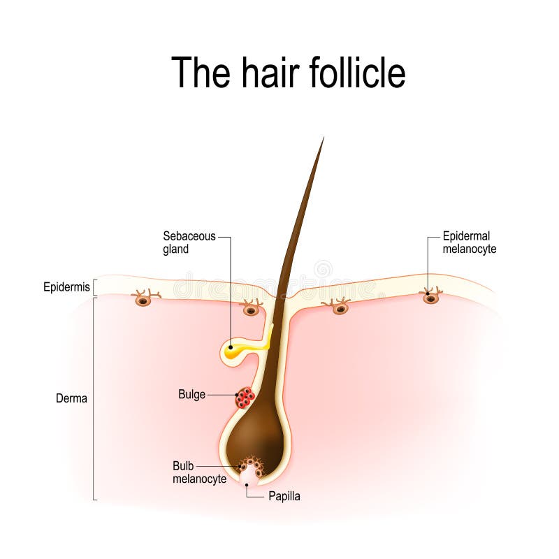
Free with trial Hair anatomy. Vector diagram. The hair shaft grows from the hair follicle consisting of transformed skin tissue. The epidermal cells transform at the command of the dermal papilla cells and generate the hair shaft. Cortex vectors Diagram of a hair follicle in a cross section of s. Hair anatomy. Vector diagram. The hair shaft grows from the hair follicle consisting of transformed skin. Hair anatomy. Vector diagram. The hair shaft grows from the hair follicle consisting of transformed skin tissue. The epidermal cells transform at the command of the dermal papilla cells and generate the hair shaft.
Free with trial Hair follicle. Anatomy of the hair follicle. distribution of differentiated and immature melanocytes is shown. Cortex vectors Hair follicle. Anatomy of the hair follicle. distribution of differentiated and immature melanocytes is shown
Free with trial Seamless pattern neurons brain hand drawn. synapse neuroplasticity, perception consciousness, cerebrum cortex neurons brain black sketch illustration. Cortex vectors Seamless pattern neurons brain sketch hand drawn. Seamless pattern neurons brain hand drawn. synapse neuroplasticity, perception consciousness, cerebrum cortex. seamless pattern neurons brain hand drawn. synapse neuroplasticity, perception consciousness, cerebrum cortex neurons brain black sketch illustration
Free with trial Anatomy of the kidney, vector illustration (for basic medical education, for clinics & Schools). Cortex vectors Anatomy of the kidney. Vector illustration (for basic medical education, for clinics & Schools
Free with trial Anatomy Of Kidney. internal and external. human anatomy. Cortex vectors Anatomy Of Kidney. Internal and external. human anatomy
Free with trial The adrenal gland. Anatomy of human adrenal gland, eps10. Cortex vectors The adrenal gland. Anatomy of human adrenal gland, eps10
Free with trial Pattern neuroplasticity brain hand drawn. perception consciousness, cerebrum cortex, neurotransmitter dopamine neuroplasticity brain vector sketch illustration. Cortex vectors Pattern neuroplasticity brain sketch hand drawn. Pattern neuroplasticity brain hand drawn. perception consciousness, cerebrum cortex, neurotransmitter dopamine. pattern neuroplasticity brain hand drawn. perception consciousness, cerebrum cortex, neurotransmitter dopamine neuroplasticity brain vector sketch illustration
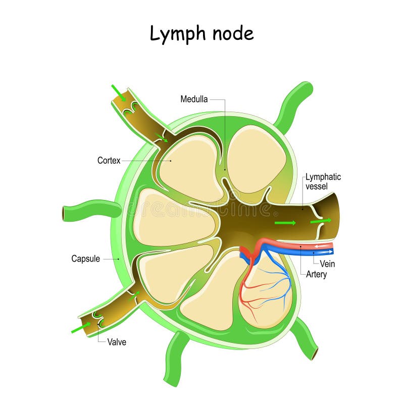
Free with trial Lymph Node Anatomy. Labeled diagram showing the flow of lymph. Afferent and efferent vessels. Vector illustration. Cortex vectors Lymph Node Anatomy. Labeled diagram showing the flow of lymph. Afferent and efferent vessels. Vector illustration
Free with trial Cortisol Functions. Cortisol is released in response to stress and low blood-glucose concentration. Cortex vectors Cortisol Functions. Cortisol is released in response to stress and low blood-glucose concentration
Free with trial The auditory system is the sensory system for the sense of hearing. It includes both the sensory organs the ears and the auditory parts of the sensory system. Health care education infographic. Vector design. Cortex vectors The auditory pathways. The auditory system is the sensory system for the sense of hearing. It includes both the sensory organs the ears and the auditory parts. The auditory system is the sensory system for the sense of hearing. It includes both the sensory organs the ears and the auditory parts of the sensory system. Health care education infographic. Vector design.
Free with trial Medical comparing a healthy brain with Alzheimer's disease, showing cerebral cortex atrophy and neurodegenerative changes, highlighting dementia, vascular dementia, Lewy body disease, frontotemporal dementia and related cognitive disorders. Cortex vectors Alzheimer's Disease Brain Atrophy Vector. Medical comparing a healthy brain with Alzheimer's disease, showing cerebral cortex atrophy and neurodegenerative. Medical comparing a healthy brain with Alzheimer's disease, showing cerebral cortex atrophy and neurodegenerative changes, highlighting dementia, vascular dementia, Lewy body disease, frontotemporal dementia and related cognitive disorders.
Free with trial Alzheimer's disease. Medical illustration of the symptoms of Alzheimer's disease. Cortex vectors Alzheimer's disease. Medical illustration of the symptoms of Alzheimer's disease
Free with trial The auditory pathways. Diagram of the auditory pathway in the brain, eps10. Cortex vectors The auditory pathways. Diagram of the auditory pathway in the brain, eps10
Free with trial Brain front view icon. Human brain front view icon. Hnternal organs symbol. Vector illustration in cartoon style isolated on white background. Cortex vectors Brain front view icon. Human brain front view icon. Hnternal organs symbol. Vector illustration in cartoon style isolated on white background
Free with trial The a monochrome of a human brain and a DNA double helix strand. The brain is depicted with detailed internal structures, including the cerebral cortex and neural pathways, shown in a solid black silhouette. The DNA strand, also in black, is composed of interconnected circles representing nucleotides. Both are set against a plain white background, emphasizing their shapes and forms. The style is. Cortex vectors Black and white of a brain and dna strand biology science. The a monochrome of a human brain and a DNA double helix strand. The brain is depicted with detailed. The a monochrome of a human brain and a DNA double helix strand. The brain is depicted with detailed internal structures, including the cerebral cortex and neural pathways, shown in a solid black silhouette. The DNA strand, also in black, is composed of interconnected circles representing nucleotides. Both are set against a plain white background, emphasizing their shapes and forms. The style is
Free with trial Stress response system. fight-or-flight response is a physiological reaction that occurs in response to threat to life. vector diagram for medical, educational and scientific use. Cortex vectors Stress response system. Fight-or-flight response is a physiological reaction that occurs in response to threat to life. vector diagram for medical, educational
Free with trial Functions of the brain. Differences in functions of the two hemispheres, eps8. Cortex vectors Functions of the brain. Differences in functions of the two hemispheres, eps8
Free with trial Human brain lateral view. Illustration body part vector, human brain lateral view. Cortex vectors Human brain lateral view. Illustration body part vector, human brain lateral view
Free with trial Scientific vector illustration of a bacterial spore showing its internal and external structural layers. The diagram labels exosporium, outer and inner spore coats, outer membrane, cortex, germ cell wall, inner membrane, and core. Suitable for microbiology education, bacteriology, laboratory training, scientific presentations, and life science learning materials. Cortex vectors Bacterial Spores Structure Diagram with Labeled Anatomical Layers. Scientific vector illustration of a bacterial spore showing its internal and external. Scientific vector illustration of a bacterial spore showing its internal and external structural layers. The diagram labels exosporium, outer and inner spore coats, outer membrane, cortex, germ cell wall, inner membrane, and core. Suitable for microbiology education, bacteriology, laboratory training, scientific presentations, and life science learning materials.
Free with trial Nerve pathways to the bladder. Drawing to show the nervous control of the urinary bladder viw the hypogastric, pelvic and pudendal nerves. Cortex vectors Nerve pathways to the bladder. Drawing to show the nervous control of the urinary bladder viw the hypogastric, pelvic and pudendal nerves
 Sign in