Free with trial The lumbar plexus. Nerve network, eps8. Cutaneous vectors The lumbar plexus. Nerve network, eps8
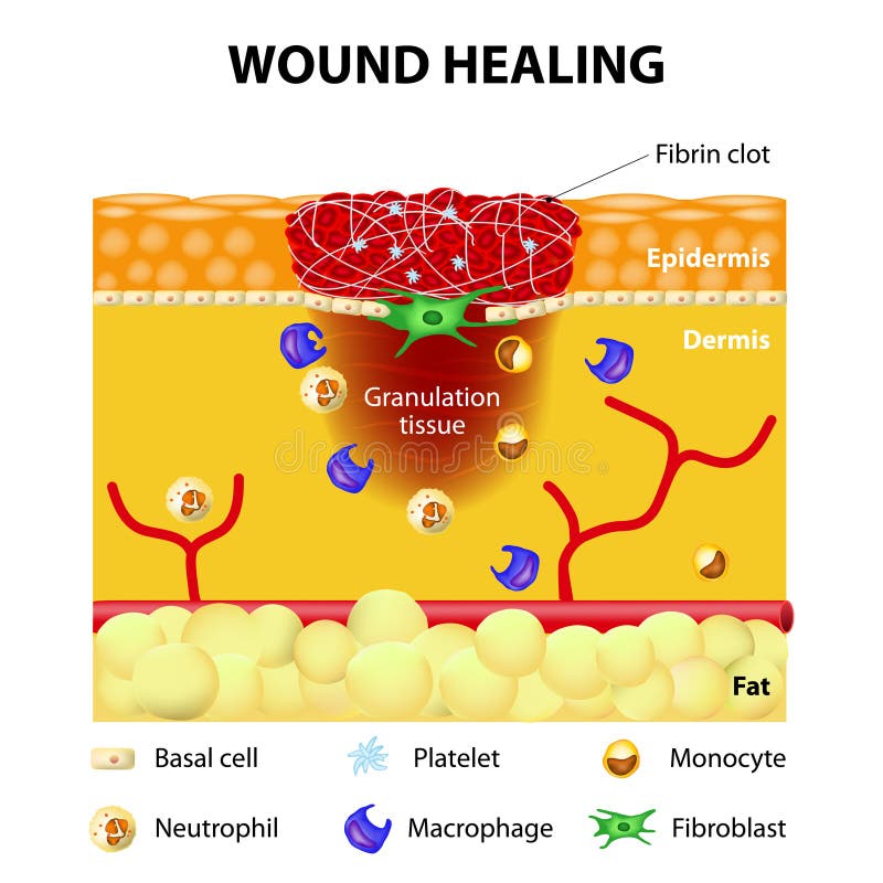
Free with trial Process of wound healing. The wound healing process. Cutaneous wound after injury. Cutaneous vectors Process of wound healing. The wound healing process. Cutaneous wound after injury
Free with trial Medically accurate vector illustration of human feet, includes nervous system, veins, arteries, etc. Cutaneous vectors Medically accurate vector illustration of human feet, includes nervous system, veins, arteries, etc.
Free with trial Cutaneous Receptors. Sense organs on the skin. Skin layers and principal cutaneous receptors. Cutaneous vectors Cutaneous Receptors. Sense organs on the skin. Skin layers and principal cutaneous receptors
Free with trial Sensory receptors in the human skin. Cutaneous Receptors. labeled. Human anatomy. Cutaneous vectors Sensory receptors in the human skin. Cutaneous Receptors. Sensory receptors in the human skin. labeled. Human anatomy
Free with trial Nerves of the foot. Vector illustration diagram of the nerves and cutaneous innervation of the human foot with palmar and dorsal view. Used transparency. Cutaneous vectors Nerves of the foot. Vector illustration diagram of the nerves and cutaneous innervation of the human foot with palmar and dorsal view. Used transparency
Free with trial The Sural nerve. Sural nerve commonly used for biopsy and graft, eps10. Cutaneous vectors The Sural nerve. Sural nerve commonly used for biopsy and graft, eps10
Free with trial Leishmania. The life cycle of the Leishmania parasite transmitted by the female phlebotomine sandfly. Cutaneous vectors Leishmania. The life cycle of the Leishmania parasite transmitted by the female phlebotomine sandfly
Free with trial Dorsal digital nerves of foot, commonly affected in diabetic neuropathy, eps10. Cutaneous vectors Dorsal digital nerves of foot. Commonly affected in diabetic neuropathy, eps10
Free with trial Candidiasis medical vector illustration diagram poster with all types of candida fungus on various human body parts and organs. Health care bacterial information. Cutaneous vectors Candidiasis medical vector illustration diagram poster with all types of candida fungus on various human body parts and organs. Health care bacterial
Free with trial Epidermis structure. Skin anatomy. Cell, and layers of a human skin. Cross section of the epidermis. Skin care. vector illustration. Cutaneous vectors Epidermis structure. Skin anatomy. Cell, and layers of a human skin. Cross section of the epidermis. Skin care. vector illustration
Free with trial Lumbar plexus. Clinical Anatomy of Spinal Nerves. overview. Vector diagram for medical, science and education use. Cutaneous vectors Lumbar plexus. Clinical Anatomy of Spinal Nerves. Overview. Vector diagram for medical, science and education use
Free with trial A medical illustration of the dermatomes of the head and neck. Cutaneous vectors Dermatomes of head and neck. A medical illustration of the dermatomes of the head and neck. A medical illustration of the dermatomes of the head and neck.
Free with trial Anthrax vector illustration. Labeled medical infection disease cycle scheme. Bacterial illness used as biological warfare weapons. Diagram of spores exposure to oxygen produce toxins and cause death. Cutaneous vectors Anthrax vector illustration. Labeled medical infection disease cycle scheme. Bacterial illness used as biological warfare weapons. Diagram of spores exposure
Free with trial Girl with pimples, acne, facial cleansing, adolescence. Skin problems. Cutaneous vectors Girl with pimples, acne, facial cleansing, adolescence. Skin problems
Free with trial Cutaneous receptors. layers of the human skin with sensory receptors. Part of the somatosensory system found in the dermis or epidermis. End-bulbs of Krause Cold, Free nerve endings pain, thermoreceptors heat, Merkel`s disc touch. Cutaneous vectors Cutaneous receptors. layers of the human skin with sensory receptors. Part of the somatosensory system found in the dermis or epidermis. End-bulbs of Krause
Free with trial Cutaneous larva migrans. Skin disease in humans caused byhookworm Ancylostoma braziliense. Vector illustration for medical, biological, science and educational use. Cutaneous vectors Cutaneous larva migrans. Skin disease in humans. Caused byhookworm Ancylostoma braziliense. Vector illustration for medical, biological, science and educational
Free with trial Skin breathing. Human cutaneous respiration. Schematic cross section illustration of layers of skin with pores and its gas exchange. Cutaneous vectors Skin Breathing Human Cutaneous Respiration. Skin breathing. Human cutaneous respiration. Schematic cross section illustration of layers of skin with pores and. Skin breathing. Human cutaneous respiration. Schematic cross section illustration of layers of skin with pores and its gas exchange.
Free with trial Medical illustration of the cutaneous innervation of the foot, with annotations. Cutaneous vectors Cutaneous innervation of the foot. Medical illustration of the cutaneous innervation of the foot, with annotations. Medical illustration of the cutaneous innervation of the foot, with annotations.
Free with trial Cutaneous respiration in frog. Gas exchange in amphibians and their circulatory system structure. Vector illustration. Cutaneous vectors Cutaneous respiration in frog. Respiratory System of Frog. Cutaneous respiration in frog. Gas exchange in amphibians and their circulatory system structure. Cutaneous respiration in frog. Gas exchange in amphibians and their circulatory system structure. Vector illustration
Free with trial Map on skin of cutaneous sensory innervation of the human body. Cutaneous vectors Skin map. Map on skin of cutaneous sensory innervation of the human body. Map on skin of cutaneous sensory innervation of the human body.
Free with trial Bacteria Infection Collection Icons Set Vector. Candida Bacteria And Plague, Cancer Cell And Thyroid Disease, Cutaneous Mucormycosis And Parasit Worms Black Contour Illustrations. Cutaneous vectors Bacteria Infection Collection Icons Set Vector. Candida Bacteria And Plague, Cancer Cell And Thyroid Disease, Cutaneous Mucormycosis And Parasit Worms Black
Free with trial Problems with skin RGB color icon. Cutaneous signs and symptoms of coronavirus. Effects of virus infections. Covid-19 diagnosis. Disease and treatment. Isolated vector illustration. Cutaneous vectors Problems with skin RGB color icon. Cutaneous signs and symptoms of coronavirus. Effects of virus infections. Covid-19 diagnosis. Disease and treatment
Free with trial Bacteria Infection Collection Icons Set Vector. Candida Bacteria And Plague, Cancer Cell And Thyroid Disease, Cutaneous Mucormycosis And Parasit Worms Line Pictograms. Contour Color Illustrations. Cutaneous vectors Bacteria Infection Collection Icons Set Vector. Candida Bacteria And Plague, Cancer Cell And Thyroid Disease, Cutaneous Mucormycosis And Parasit Worms Line
Free with trial Dorsal ramus innervation shows pathways from spinal nerve to back muscles and skin, key parts, dorsal ramus, spinal cord, nerve branch. Outline diagram. Cutaneous vectors Dorsal ramus innervation shows pathways from spinal nerve to back ... Dorsal ramus innervation shows pathways from spinal nerve to back muscles and skin, key. Dorsal ramus innervation shows pathways from spinal nerve to back muscles and skin, key parts, dorsal ramus, spinal cord, nerve branch. Outline diagram
Free with trial Arteries of the foot dorsal and plantar view of an ankle. diagram. Cutaneous vectors Arteries of the foot. Dorsal and plantar view of an ankle. diagram
Free with trial Papaya. Hand drawn vector ripe papaya fruit on light background. Cutaneous vectors Papaya. Hand drawn vector ripe papaya fruit on light background
Free with trial Medically accurate anatomy illustration of human feet legs with nervous and blood system. Educational x-ray style illustration. Cutaneous vectors Medically accurate anatomy illustration of human feet legs with nervous and blood system. Educational x-ray style illustration
Free with trial Medically accurate vector illustration of human feet, includes nervous system, veins, arteries, etc. Cutaneous vectors Medically accurate vector illustration of human back chest, includes nervous system, veins, arteries, heart, etc. Medically accurate vector illustration of. Medically accurate vector illustration of human feet, includes nervous system, veins, arteries, etc.
Free with trial Papaya slice. Hand drawn vector ripe half of, slice papaya fruit on light background. Cutaneous vectors Papaya slice. Hand drawn vector ripe half of, slice papaya fruit on light background
Free with trial The Sciatic Nerve. Sensory innervation of the sciatic nerve. Sciatica. Medical Illustration. Cutaneous vectors The Sciatic Nerve. Sensory innervation of the sciatic nerve. Sciatica. Medical Illustration
Free with trial Lumbar plexus as anatomical web of lumbosacral nerves outline diagram. Labeled educational scheme with medical location of genitofemoral, iliohypogastric and ilioinguinal nerves vector illustration. Cutaneous vectors Lumbar plexus as anatomical web of lumbosacral nerves outline diagram. Labeled educational scheme with medical location of genitofemoral, iliohypogastric and
Free with trial Central nervous system anatomical diagram. Nerves send electrical signals to and from brain and spinal cord in human body. CNS and PNS concept. Medical poster for neurology clinic vector illustration. Cutaneous vectors Central nervous system. Anatomical diagram. Nerves send electrical signals to and from brain and spinal cord in human body. CNS and PNS concept. Medical poster. Central nervous system anatomical diagram. Nerves send electrical signals to and from brain and spinal cord in human body. CNS and PNS concept. Medical poster for neurology clinic vector illustration.
Free with trial Cryptococcosis infection overview shows airborne yeast inhaled from soil to lungs, spreading via bloodstream to brain and skin, key objects, lungs, brain, yeast. Diagram. Cutaneous vectors Cryptococcosis infection overview shows airborne yeast inhaled from soil to ... Cryptococcosis infection overview shows airborne yeast inhaled from soil to. Cryptococcosis infection overview shows airborne yeast inhaled from soil to lungs, spreading via bloodstream to brain and skin, key objects, lungs, brain, yeast. Diagram
Free with trial Anatomical structure and function of skin. Cutaneous-Associated Lymphoid Tissue. Medical vector illustration. Cutaneous vectors Anatomical structure and function of skin, medical illustration. Anatomical structure and function of skin. Cutaneous-Associated Lymphoid Tissue. Medical vector. Anatomical structure and function of skin. Cutaneous-Associated Lymphoid Tissue. Medical vector illustration
Free with trial Central nervous system anatomical diagram. Nerves send electrical signals to and from brain and spinal cord in human body. CNS and PNS concept. Medical poster for neurology clinic vector illustration. Cutaneous vectors Central nervous system. Anatomical diagram. Nerves send electrical signals to and from brain and spinal cord in human body. CNS and PNS concept. Medical poster. Central nervous system anatomical diagram. Nerves send electrical signals to and from brain and spinal cord in human body. CNS and PNS concept. Medical poster for neurology clinic vector illustration.
Free with trial Leishmania amastigotes are the intracellular form of the Leishmania parasite. Leishmania is a genus of protozoan parasites that cause the disease leishmaniasis, which affects both humans and animals. The life cycle of Leishmania involves two main stages: the promastigote stage, which resides in the midgut of sandflies, and the amastigote stage, which proliferates inside the cells of the mammalian host, typically macrophages. Amastigotes are small, round, and non-flagellated forms of the parasite adapted for survival within the phagolysosomes of host cells. They replicate by binary fission within the host cells and are responsible for the pathological changes associated with leishmaniasis. Cutaneous vectors Leishmania amastigote. S are the intracellular form of the Leishmania parasite. Leishmania is a genus of protozoan parasites that cause the disease leishmaniasis. Leishmania amastigotes are the intracellular form of the Leishmania parasite. Leishmania is a genus of protozoan parasites that cause the disease leishmaniasis, which affects both humans and animals. The life cycle of Leishmania involves two main stages: the promastigote stage, which resides in the midgut of sandflies, and the amastigote stage, which proliferates inside the cells of the mammalian host, typically macrophages. Amastigotes are small, round, and non-flagellated forms of the parasite adapted for survival within the phagolysosomes of host cells. They replicate by binary fission within the host cells and are responsible for the pathological changes associated with leishmaniasis.
Free with trial Leishmania is a genus of parasitic protozoa belonging to the family Trypanosomatidae. These parasites are responsible for causing leishmaniasis, a disease that affects humans and other mammals. Cutaneous vectors Leishmania. Is a genus of parasitic protozoa belonging to the family Trypanosomatidae. These parasites are responsible for causing sis, a disease that affects. Leishmania is a genus of parasitic protozoa belonging to the family Trypanosomatidae. These parasites are responsible for causing leishmaniasis, a disease that affects humans and other mammals
Free with trial Child with ringworm tinea disease symptoms. Isolated vector sick boy with rash caused by fungal infection of skin. Dermatophytosis illness with red, itchy, scaly, circular rash on body. Cutaneous vectors Child with ringworm tinea disease sickness symptom. Child with ringworm tinea disease symptoms. Isolated vector sick boy with rash caused by fungal infection of. Child with ringworm tinea disease symptoms. Isolated vector sick boy with rash caused by fungal infection of skin. Dermatophytosis illness with red, itchy, scaly, circular rash on body
Free with trial Central nervous system anatomical diagram. Nerves send electrical signals to and from brain and spinal cord in human body. CNS and PNS concept. Medical poster for neurology clinic vector illustration. Cutaneous vectors Central nervous system. Anatomical diagram. Nerves send electrical signals to and from brain and spinal cord in human body. CNS and PNS concept. Medical poster. Central nervous system anatomical diagram. Nerves send electrical signals to and from brain and spinal cord in human body. CNS and PNS concept. Medical poster for neurology clinic vector illustration.
Free with trial Penicillium mold fungi line icon vector. penicillium mold fungi sign. isolated contour symbol black illustration. Cutaneous vectors Penicillium mold fungi line icon vector illustration. Penicillium mold fungi line icon vector. penicillium mold fungi sign. isolated contour symbol black. penicillium mold fungi line icon vector. penicillium mold fungi sign. isolated contour symbol black illustration
Free with trial Candida fungi albicans line icon vector. candida fungi albicans sign. isolated contour symbol black illustration. Cutaneous vectors Candida fungi albicans line icon vector illustration. Candida fungi albicans line icon vector. candida fungi albicans sign. isolated contour symbol black. candida fungi albicans line icon vector. candida fungi albicans sign. isolated contour symbol black illustration
Free with trial Diseases color line icons set. Pictograms for web page, mobile app, promo. UI UX GUI design element. Editable stroke. Cutaneous vectors Diseases color line icons set. Pictograms for web page. Mobile app, promo. UI UX GUI design element. Editable stroke
Free with trial Part of the body, limb icons in set collection for design. Human anatomy vector symbol stock illustration. Cutaneous vectors Part of the body, limb icons in set collection for design. Human anatomy vector symbol stock illustration
Free with trial Parasitic worms ascaris color icon vector illustration. Parasitic worms ascaris color icon vector. parasitic worms ascaris sign. isolated symbol illustration. Cutaneous vectors Parasitic worms ascaris color icon vector illustration. Parasitic worms ascaris color icon vector. parasitic worms ascaris sign. isolated symbol illustration
Free with trial Cutaneous sarcoidosis icon, pictogram. Editable vector illustration of granulomatous skin inflammation. Dermatology symbol used in medical education, autoimmune disease awareness, and diagnostics. Cutaneous vectors Cutaneous sarcoidosis icon, pictogram. Editable vector illustration. Of granulomatous skin inflammation. Dermatology symbol used in medical education. Cutaneous sarcoidosis icon, pictogram. Editable vector illustration of granulomatous skin inflammation. Dermatology symbol used in medical education, autoimmune disease awareness, and diagnostics.
Free with trial Hand with red, cutaneous anthrax skin lesion featuring dark center. Cutaneous vectors Hand with red, cutaneous anthrax skin lesion featuring dark center. Eschar, Hand with red, cutaneous anthrax skin lesion featuring dark center
Free with trial Cutaneous sarcoidosis icon, pictogram. Editable vector illustration of granulomatous skin inflammation. Dermatology symbol used in medical education, autoimmune disease awareness, and diagnostics. Cutaneous vectors Cutaneous sarcoidosis icon, pictogram. Editable vector illustration. Of granulomatous skin inflammation. Dermatology symbol used in medical education. Cutaneous sarcoidosis icon, pictogram. Editable vector illustration of granulomatous skin inflammation. Dermatology symbol used in medical education, autoimmune disease awareness, and diagnostics.
Free with trial Cutaneous sarcoidosis icon, pictogram. Editable vector illustration of granulomatous skin inflammation. Dermatology symbol used in medical education, autoimmune disease awareness, and diagnostics. Cutaneous vectors Cutaneous sarcoidosis icon, pictogram. Editable vector illustration. Of granulomatous skin inflammation. Dermatology symbol used in medical education. Cutaneous sarcoidosis icon, pictogram. Editable vector illustration of granulomatous skin inflammation. Dermatology symbol used in medical education, autoimmune disease awareness, and diagnostics.
Free with trial Porphyria is a group of rare metabolic disorders caused by problems in heme production, leading to buildup of porphyrins in the body. It can affect the skin, nervous system, or both. Cutaneous vectors Porphyria, Acute and Cutaneous Porphyrias Flashcard Illustration. Porphyria is a group of rare metabolic disorders caused by problems in heme production, leading. Porphyria is a group of rare metabolic disorders caused by problems in heme production, leading to buildup of porphyrins in the body. It can affect the skin, nervous system, or both.
Free with trial Educational dermatology and infectious disease showing the clinical stages of rash progression in monkeypox (mpox) infection in full color. The labeled medical diagram details the chronological evolution and diameter measurements of cutaneous lesions: early vesicle (3 mm diameter), small pustule (2 mm diameter), umbilicated pustule with central depression (3-4 mm diameter), umbilicated lesion (5 mm diameter), crusting of a mature lesion, and a partially removed scab lesion. High-quality infographic graphic ideal for epidemiology textbooks, public health clinic posters, dermatology reference manuals, infectious disease control guides, medical school lectures, and patient awareness presentations. Cutaneous vectors Stages of Rash in Monkeypox Mpox Skin Lesion Progression Vector Illustration. Educational dermatology and infectious disease showing the clinical stages of rash. Educational dermatology and infectious disease showing the clinical stages of rash progression in monkeypox (mpox) infection in full color. The labeled medical diagram details the chronological evolution and diameter measurements of cutaneous lesions: early vesicle (3 mm diameter), small pustule (2 mm diameter), umbilicated pustule with central depression (3-4 mm diameter), umbilicated lesion (5 mm diameter), crusting of a mature lesion, and a partially removed scab lesion. High-quality infographic graphic ideal for epidemiology textbooks, public health clinic posters, dermatology reference manuals, infectious disease control guides, medical school lectures, and patient awareness presentations.
Free with trial Educational infectious disease and epidemiology detailing monkeypox (mpox) virus infection and its clinical cutaneous manifestation in full color. The medical diagram showcases a characteristic red viral pathogen virion structure alongside a human hand presenting multiple scattered papular, vesicular, and pustular skin rash lesions on the dorsal surface and fingers. High-quality infographic graphic ideal for public health campaign posters, virology textbooks, dermatology clinical display guides, epidemic prevention manuals, medical school lectures, and patient healthcare education presentations. Cutaneous vectors Monkeypox Virus Hand Rash Lesions Mpox Infection Pathology Vector Illustration. Educational infectious disease and epidemiology detailing monkeypox (mpox) virus. Educational infectious disease and epidemiology detailing monkeypox (mpox) virus infection and its clinical cutaneous manifestation in full color. The medical diagram showcases a characteristic red viral pathogen virion structure alongside a human hand presenting multiple scattered papular, vesicular, and pustular skin rash lesions on the dorsal surface and fingers. High-quality infographic graphic ideal for public health campaign posters, virology textbooks, dermatology clinical display guides, epidemic prevention manuals, medical school lectures, and patient healthcare education presentations.
Free with trial Skin cancer, malignant melanoma outline icon. Oncological sign. Medical linear pictogram. Basal cell carcinoma. Cutaneous malignancy. Editable vector illustration isolated on white background. Cutaneous vectors Skin cancer, malignant melanoma outline icon. Oncological sign. Medical linear pictogram. Basal cell carcinoma. Cutaneous malignancy. Editable vector
Free with trial Skin cancer, malignant melanoma outline icon. Oncological sign. Medical linear pictogram. Basal cell carcinoma. Cutaneous malignancy. Editable vector illustration isolated on white background. Cutaneous vectors Skin cancer, malignant melanoma outline icon. Oncological sign. Medical linear pictogram. Basal cell carcinoma. Cutaneous malignancy. Editable vector
Free with trial Skin cancer, malignant melanoma outline icon. Oncological sign. Medical linear pictogram. Basal cell carcinoma. Cutaneous malignancy. Editable vector illustration isolated on white background. Cutaneous vectors Skin cancer, malignant melanoma outline icon. Oncological sign. Medical linear pictogram. Basal cell carcinoma. Cutaneous malignancy. Editable vector
Free with trial Bacteria Infection Collection doodle icons set vector. Candida Bacteria Plague, Cancer Cell Thyroid Disease, Cutaneous Mucormycosis Parasit Worms sketch hand drawn. Cutaneous vectors Bacteria Infection set Icons hand drawn. Bacteria Infection Collection doodle icons set vector. Candida Bacteria Plague, Cancer Cell Thyroid Disease, Cutaneous. Bacteria Infection Collection doodle icons set vector. Candida Bacteria Plague, Cancer Cell Thyroid Disease, Cutaneous Mucormycosis Parasit Worms sketch hand drawn
Free with trial Infographic illustrating symptoms and treatments for Cutaneous B-cell Lymphoma, featuring icons for medical conditions and therapies. Cutaneous vectors Cutaneous B-cell Lymphoma symptoms and treatments infographic with icons for awareness ribbon. Infographic illustrating symptoms and treatments for Cutaneous B. Infographic illustrating symptoms and treatments for Cutaneous B-cell Lymphoma, featuring icons for medical conditions and therapies
Free with trial Cutaneous larva migrans infection presenting characteristic reddish brown, raised serpiginous tracks on human foot skin, with a magnified view of migrating hookworm larvae. Cutaneous vectors Hookworm causing cutaneous larva migrans infection in human foot. Cutaneous larva migrans infection presenting characteristic reddish brown, raised serpiginous. Cutaneous larva migrans infection presenting characteristic reddish brown, raised serpiginous tracks on human foot skin, with a magnified view of migrating hookworm larvae
Free with trial Skin cell turnover. Path of the cell from the division of Epidermal stem cell in the basal layer, Spinous keratinocyte, Granular keratinocyte, to Corneocyte on the surface of the skin. Epidermal homeostasis. Vector illustration. Medical diagram. Cutaneous vectors Skin cell turnover. Epidermal homeostasis. Skin cell turnover. Path of the cell from the division of Epidermal stem cell in the basal layer, Spinous keratinocyte. Skin cell turnover. Path of the cell from the division of Epidermal stem cell in the basal layer, Spinous keratinocyte, Granular keratinocyte, to Corneocyte on the surface of the skin. Epidermal homeostasis. Vector illustration. Medical diagram
Free with trial Healthy Skin Microbiome. Friendly Bacteria Shield. Bacterial diversity. Microorganisms inhabiting the human skin and their relationship with immune function. Commensal flora and immune interaction. Skin barrier. Dermatology. Infographic diagram. Medical. Cutaneous vectors Healthy Skin Microbiome. Friendly Bacteria Shield. Bacterial diversity. Microorganisms inhabiting the human skin and their relationship with immune function
Free with trial Skin types infographic. Difference and comparison of Oily, Dry and Normal skin. Chart for skincare routine. Classification of skin types. Vector illustration. Medical diagram. Cutaneous vectors Skin types infographic. Difference and comparison of Oily, Dry and Normal skin. Chart for skincare routine. Classification of skin types. Vector illustration
Free with trial Sunburn healing process from Sunburn erythema and desquamation to Renewed skin. Cross section of human skin. Epidermal homeostasis. Skin peeling. Vector illustration. Medical diagram. Cutaneous vectors Sunburn healing process from Sunburn erythema and desquamation to Renewed skin. Cross section of human skin. Epidermal homeostasis. Skin peeling. Vector
Free with trial Skin hydration. Cross section of a human skin Before and After Deep hydration. From Dehydrated skin to Visibly plumper epidermis. Stratum granulosum, Stratum corneum, Stratum spinosum and Basal cells. Vector illustration. Medical poster. Schematic diagram. Anti-Aging Facial Rejuvenation. Cutaneous vectors Skin hydration. Cross section of a human skin Before and After Deep hydration. From Dehydrated skin to Visibly plumper epidermis. Stratum granulosum, Stratum
Free with trial Photodermatoses Skin Reaction Medical Illustration Showing Healthy Skin and Sun-Induced Inflammatory Rash Cross Section. Photodermatosis Disease. UV Triggered Inflammation and Blister Formation Anatomy. Cutaneous vectors Photodermatoses Skin Reaction Medical Illustration Showing Healthy Skin and Sun-Induced Inflammatory Rash Cross Section. Photodermatosis Disease. UV Triggered
Free with trial Skin cleaning. Cross section of human skin with clogged pore or blackhead acne. Before and after cleansing procedure. Vector illustration. Medical diagram. Cutaneous vectors Skin cleaning. Before and after cleansing procedure. Skin cleaning. Cross section of human skin with clogged pore or blackhead acne. Before and after cleansing. Skin cleaning. Cross section of human skin with clogged pore or blackhead acne. Before and after cleansing procedure. Vector illustration. Medical diagram
Free with trial Skin sensory receptors overview shows epidermis with Meissner corpuscle, Pacinian corpuscle, and hair root plexus sensing touch, pressure, and movement. Outline diagram. Cutaneous vectors Skin sensory receptors overview shows epidermis with Meissner corpuscle, Pacinian ... Skin sensory receptors overview shows epidermis with Meissner corpuscle. Skin sensory receptors overview shows epidermis with Meissner corpuscle, Pacinian corpuscle, and hair root plexus sensing touch, pressure, and movement. Outline diagram
Free with trial Medical illustration comparing young and aged human skin structure. Cross section shows collagen and elastin fibers, fibroblasts, dermis, epidermis, wrinkles and age-related changes in the extracellular matrix. Cutaneous vectors Medical illustration comparing young and aged human skin structure. Cross section shows collagen and elastin fibers, fibroblasts. Dermis, epidermis, wrinkles. Medical illustration comparing young and aged human skin structure. Cross section shows collagen and elastin fibers, fibroblasts, dermis, epidermis, wrinkles and age-related changes in the extracellular matrix.
Free with trial Apocrine gland anatomy depicts sweat gland structure and secretion, highlighting apocrine coil, hair follicle, and eccrine gland while mapping skin layers and key body sites. Outline diagram. Cutaneous vectors Apocrine gland anatomy depicts sweat gland structure and secretion, highlighting ... Apocrine gland anatomy depicts sweat gland structure and secretion. Apocrine gland anatomy depicts sweat gland structure and secretion, highlighting apocrine coil, hair follicle, and eccrine gland while mapping skin layers and key body sites. Outline diagram
Free with trial Thermoregulation process. Heat loss and heat retention. Human skin with blood vessel and thermoreceptors. Detailed Vector poster. Cutaneous vectors Thermoregulation. Human skin layers. Thermoregulation process. Heat loss and heat retention. Human skin with blood vessel and thermoreceptors. Detailed Vector. Thermoregulation process. Heat loss and heat retention. Human skin with blood vessel and thermoreceptors. Detailed Vector poster
Free with trial Melanocyte Cell Anatomy in Human Skin Cross Section Melanin Production and Epidermis Structure. Human Skin Layers with Melanocyte Close Up Dermatology, Pigmentation and Melanin Biology Diagram. Cutaneous vectors Melanocyte Cell Anatomy in Human Skin Cross Section Melanin Production and Epidermis Structure. Human Skin Layers with Melanocyte. Close Up Dermatology. Melanocyte Cell Anatomy in Human Skin Cross Section Melanin Production and Epidermis Structure. Human Skin Layers with Melanocyte Close Up Dermatology, Pigmentation and Melanin Biology Diagram
Free with trial Line disease acanthosis nigricans icon. Vector medical symbol of laser treatment applied to man underarm and neck with handheld device aimed at skin and beam marks indicating therapy for pigmentation. Cutaneous vectors Line disease acanthosis laser treatment icon. Line disease acanthosis nigricans icon. Vector medical symbol of laser treatment applied to man underarm and neck. Line disease acanthosis nigricans icon. Vector medical symbol of laser treatment applied to man underarm and neck with handheld device aimed at skin and beam marks indicating therapy for pigmentation
Free with trial Dermatology diagnostics and skin health concept with cross-section skin layers, hair follicle and mole, glowing blue neon polygonal style, ideal for skincare advertising, vector illustration. Cutaneous vectors Human skin cross section with hair follicle, mole and blood vessels, Glowing blue neon illustration. Dermatology diagnostics and skin health concept with cross. Dermatology diagnostics and skin health concept with cross-section skin layers, hair follicle and mole, glowing blue neon polygonal style, ideal for skincare advertising, vector illustration.
Free with trial Epidermis structure. Epithelium anatomy. Skin regeneration and Epidermal cells. Cross section of the epidermis. Vector illustration. Medical poster. Schematic diagram. Cutaneous vectors Epidermis structure. Epithelium anatomy. Skin regeneration and Epidermal cells. Cross section of the epidermis. Vector illustration. Medical poster. Schematic
Free with trial Layers of Skin Anatomy Illustration with Epidermis, Dermis, Hypodermis and Muscle Cross Section. Vector flat isolated on white background. Cutaneous vectors Illustration of Human Skin Layers Anatomy with Epidermis, Dermis, Hypodermis and Muscle. Layers of Skin Anatomy Illustration with Epidermis, Dermis, Hypodermis. Layers of Skin Anatomy Illustration with Epidermis, Dermis, Hypodermis and Muscle Cross Section. Vector flat isolated on white background
Free with trial Vacuum Pore Cleaner Extracting Blackhead Before and After. Blackhead Removal Process Using Vacuum Suction Tool. Cutaneous vectors Vacuum Pore Cleaner Extracting Blackhead Before and After Skin Comparison. Blackhead Removal Process Using Vacuum Suction Tool. Vacuum Pore Cleaner Extracting. Vacuum Pore Cleaner Extracting Blackhead Before and After. Blackhead Removal Process Using Vacuum Suction Tool
Free with trial Cancer cell line seamless pattern. Cancer cell vector line seamless pattern cancer cell sign illustration. Cutaneous vectors Cancer cell line seamless pattern. Cancer cell vector line seamless pattern cancer cell sign illustration
Free with trial Candida bacteria line seamless pattern. Candida bacteria vector line seamless pattern candida bacteria sign illustration. Cutaneous vectors Candida bacteria line seamless pattern. Candida bacteria vector line seamless pattern candida bacteria sign illustration
Free with trial Cancer cell color multipack icon vector. cancer cell sign. isolated symbol. Cutaneous vectors Cancer cell color multipack icon vector. Cancer cell sign. isolated symbol
Free with trial Medical showing common acne lesion categories on facial skin. Educational dermatology concept for skincare awareness, diagnosis, treatment guidance, and healthcare content. Cutaneous vectors Types of Acne Medical Vector Illustration. Medical showing common acne lesion categories on facial skin. Educational dermatology concept for skincare awareness. Medical showing common acne lesion categories on facial skin. Educational dermatology concept for skincare awareness, diagnosis, treatment guidance, and healthcare content.
Free with trial Edition thyroid cancer color icon vector. Thyroid cancer color icon vector. thyroid cancer edition sign. isolated symbol. Cutaneous vectors Edition thyroid cancer color icon vector. Thyroid cancer color icon vector. thyroid cancer edition sign. isolated symbol
 Sign in