Free with trial Dislocated shoulder. Dislocation of the humerus as a result of a sport injury, anterior and posterior types, eps8. Dislocation vectors Dislocated shoulder. Dislocation of the humerus as a result of a sport injury, anterior and posterior types, eps8
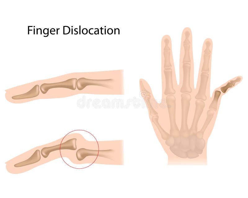
Free with trial Finger dislocation. Dislocation of bones at different finger joints, eps10. Dislocation vectors Finger dislocation. Dislocation of bones at different finger joints, eps10
Free with trial Mechanism of formation of an ankle sprain and Grades of an ankle sprain (Inversion) illustration with external and skeletal (lateral) view. Dislocation vectors Ankle sprain.Inversion. Mechanism of formation of an ankle sprain and Grades of an ankle sprain (Inversion) illustration with external and skeletal (lateral). Mechanism of formation of an ankle sprain and Grades of an ankle sprain (Inversion) illustration with external and skeletal (lateral) view
Free with trial Hip dislocation, a common orthopedic injury, eps10. Dislocation vectors Hip dislocation. A common orthopedic injury, eps10
Free with trial Patellar dislocation. Normal position of kneecap and Patella displaced. Anatomy of the Knee. Dislocation vectors Patellar dislocation. Normal position of kneecap and Patella displaced. Anatomy of the Knee
Free with trial Luxating patella. Healthy and unhealthy dog`s knee joint, the medial luxating patella or knee cap dislocation illustration. Dislocation vectors Luxating patella. Healthy and unhealthy dog`s knee joint, the medial luxating patella or knee cap dislocation illustration
Free with trial Dislocation after hip replacement surgery. Common complication after total hip replacement surgery procedure, eps10. Dislocation vectors Dislocation after hip replacement surgery. Common complication after total hip replacement surgery procedure, eps10
Free with trial Vector illustration of a healthy human foot and a foot with lisfranc injury with weight bearing and mechanism of injury, dislocation of the second metatarsal bone. Medial, top view of the foot. EPS 10. Dislocation vectors Lisfranc Injury. Vector illustration of a healthy human foot and a foot with lisfranc injury with weight bearing and mechanism of injury, dislocation of the. Vector illustration of a healthy human foot and a foot with lisfranc injury with weight bearing and mechanism of injury, dislocation of the second metatarsal bone. Medial, top view of the foot. EPS 10
Free with trial Ankle sprain. Eversion. Mechanism of formation of an ankle sprain and Grades of an ankle sprain (Eversion) illustration with external and skeletal (medial) view. Dislocation vectors Ankle sprain.Eversion. Mechanism of formation of an ankle sprain and Grades of an ankle sprain (Eversion) illustration with external and skeletal (medial) view
Free with trial Back pain and other common joints problems. Pictogram collection of back pain, neck pain, knee pain and elbow pain. Dislocation vectors Back pain and other common joints problems. Pictogram collection of back pain, neck pain, knee pain and elbow pain
Free with trial Illustration of Mechanism of formation of a High ankle sprain (Syndesmotic Sprain) and Grades of high ankle sprain with external and skeletal (lateral and front) view of an ankle. Dislocation vectors High ankle sprain. Illustration of Mechanism of formation of a High ankle sprain (Syndesmotic Sprain) and Grades of high ankle sprain with external and skeletal. Illustration of Mechanism of formation of a High ankle sprain (Syndesmotic Sprain) and Grades of high ankle sprain with external and skeletal (lateral and front) view of an ankle.
Free with trial Bone fracture healing process. Fracture and repair. Vector scheme. Dislocation vectors Bone fracture healing process. Fracture and repair. Vector scheme
Free with trial Traumatology and pain. Illustration of joint injuries and pain. Dislocation vectors Traumatology and pain. Illustration of joint injuries and pain
Free with trial Illustration of Entrapment of the ulnar nerve in the wrist in the Guyon’s canal. Used: transparency, blend mode. Dislocation vectors Entrapment of the ulnar nerve in the wrist in the Guyon’s canal 2. Illustration of Entrapment of the ulnar nerve in the wrist in the Guyon’s canal. Used. Illustration of Entrapment of the ulnar nerve in the wrist in the Guyon’s canal. Used: transparency, blend mode.
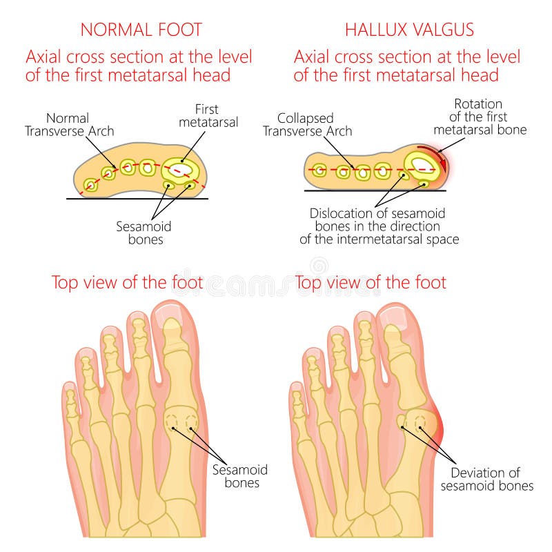
Free with trial Vector illustration of a healthy human forefoot and a foot with hallux valgus, dislocation of sesamoid bones. Top view and cross section of of the foot. For advertising, medical publications. EPS 10. Dislocation vectors Normal foot and Hallux valgus with rotation of the first mematarsal bone. Vector illustration of a healthy human forefoot and a foot with hallux valgus. Vector illustration of a healthy human forefoot and a foot with hallux valgus, dislocation of sesamoid bones. Top view and cross section of of the foot. For advertising, medical publications. EPS 10.
Free with trial Vector illustration. Anatomy, front x-ray of an arthritic knee joint and a knee after unicompartmental or partial knee replacement. For advertising and medical publications. EPS 10. Dislocation vectors Meniscus _Arthritic knee and Partial knee replacement. Vector illustration. Anatomy, front x-ray of an arthritic knee joint and a knee after unicompartmental or. Vector illustration. Anatomy, front x-ray of an arthritic knee joint and a knee after unicompartmental or partial knee replacement. For advertising and medical publications. EPS 10
Free with trial Knee Dislocations - medical vector illustration diagrams. Anatomical knee injury types scheme. Physiotherapy educational information. Dislocation vectors Knee Dislocations - medical vector illustration diagrams. Anatomical knee injury types scheme. Physiotherapy educational information
Free with trial Shoulder dislocation. A vector illustration of shoulder dislocation. Dislocation vectors Shoulder dislocation. A vector illustration of shoulder dislocation
Free with trial Knee dislocation. Lateral trauma to the knee, torn collateral ligaments, cruciate ligament injury and meniscus injury. An obvious deformity of the knee. Human anatomy. Dislocation vectors Knee dislocation. Lateral trauma to the knee, torn collateral ligaments, cruciate ligament injury and meniscus injury. An obvious deformity of the knee. Human
Free with trial Dislocation Shoulder symptoms or separated. Bone and joints to slipping out of place. Dislocation vectors Dislocation Shoulder symptoms or separated. Bone and joints to slipping out of place
Free with trial Knee dislocation and normal. Lateral trauma to the knee, torn collateral ligaments, cruciate ligament injury and meniscus injury. Human anatomy. Dislocation vectors Knee dislocation and normal. Lateral trauma to the knee, torn collateral ligaments, cruciate ligament injury and meniscus injury. Human anatomy
Free with trial Shoulder dislocation and humerus bone trauma explanation outline diagram. Labeled educational medical injury when skeletal part rotates out of scapula vector illustration. Painful arm vs healthy. Dislocation vectors Shoulder dislocation and humerus bone trauma explanation outline diagram. Labeled educational medical injury when skeletal part rotates out of scapula vector
Free with trial Vector illustration anatomy of a hip joint with dysplasia, subluxation and dislocation of the femoral head in the joint. Front view. For advertising and medical publications. EPS 10. Dislocation vectors Pelvis and Hip joint problem_Hip dysplasia and dislocation. Vector illustration anatomy of a hip joint with dysplasia, subluxation and dislocation of the femoral. Vector illustration anatomy of a hip joint with dysplasia, subluxation and dislocation of the femoral head in the joint . Front view. For advertising and medical publications. EPS 10
Free with trial Cuboid syndrome as orthopedic trauma after foot torn injury outline diagram. Labeled educational scheme with feet bone dislocation and free movement vector illustration. Cuboid subluxation problem. Dislocation vectors Cuboid syndrome as orthopedic trauma after foot torn injury outline diagram. Labeled educational scheme with feet bone dislocation and free movement vector
Free with trial A medical illustration with a normal knee and a dislocated knee. Dislocation vectors Dislocation of knee. A medical illustration with a normal knee and a dislocated knee. A medical illustration with a normal knee and a dislocated knee.
Free with trial Joint pain thin line icon. Rheumatism concept. Editable stroke. Vector. Dislocation vectors Joint pain thin line icon . Rheumatism concept . Editable stroke . Vector.
Free with trial Vector illustrations of sling, bandage, and elevation techniques treatment for broken bones and pain. Dislocation vectors First aid for fractures and injuries on different body parts stick figure icons. Vector illustrations of sling, bandage, and elevation techniques treatment for. Vector illustrations of sling, bandage, and elevation techniques treatment for broken bones and pain
Free with trial Vector illustration. Anatomy, front x-ray of an arthritic knee joint and a knee after total knee replacement. For advertising and medical publications. EPS 10. Dislocation vectors Meniscus _Arthritic knee and Total knee replacement. Vector illustration. Anatomy, front x-ray of an arthritic knee joint and a knee after total knee replacement. Vector illustration. Anatomy, front x-ray of an arthritic knee joint and a knee after total knee replacement. For advertising and medical publications. EPS 10
Free with trial Vector illustration of bursitis of the elbow joint. Dislocation vectors Bursitis of the elbow joint. Vector illustration of bursitis of the elbow joint. Vector illustration of bursitis of the elbow joint.
Free with trial Backache low back pain. man health and medicine. Pop art retro vector illustration vintage kitsch drawing. Dislocation vectors Backache low back pain. man health and medicine. Pop art retro vector illustration vintage kitsch drawing
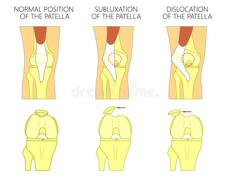
Free with trial Vector illustration of a healthy human knee joint and unhealthy knees with problem. Subluxation and dislocation of the patella or kneecap. Anatomy of human knee joint, front view of straight and bent knee. Dislocation vectors Knee joint problem_Dislocation of the patella. Vector illustration of a healthy human knee joint and unhealthy knees with problem. Subluxation and dislocation of. Vector illustration of a healthy human knee joint and unhealthy knees with problem. Subluxation and dislocation of the patella or kneecap. Anatomy of human knee joint, front view of straight and bent knee.
Free with trial Vector illustration diagram. Turf toe. Sport trauma. Mechanism of rupture of plantar plate of the first toe of the foot. External and skeletal views of an ankle. Used: blend. Dislocation vectors Turf toe. Rupture of the plantar plate of the first toe of the f. Vector illustration diagram.Turf toe. Sport trauma. Mechanism of rupture of plantar plate of. Vector illustration diagram.Turf toe. Sport trauma. Mechanism of rupture of plantar plate of the first toe of the foot. External and skeletal views of an ankle. Used: blend.
Free with trial Elbow joint pain. Vector illustration of a human elbow joint with a pain or injury. EPS 10. Dislocation vectors Elbow joint pain. Vector illustration of a human elbow joint with a pain or injury. EPS 10
Free with trial Vector illustration anatomy of a healthy human hip joint and a hip with avascular necrosis of the femoral head. Front view. For advertising and medical publications. EPS 10. Dislocation vectors Pelvis and Hip joint problem_Avascular necrosis of the femoral h. Vector illustration anatomy of a healthy human hip joint and a hip with avascular necrosis of. Vector illustration anatomy of a healthy human hip joint and a hip with avascular necrosis of the femoral head. Front view. For advertising and medical publications. EPS 10
Free with trial Illustration diagram of normal leg with a healthy knee and a leg with over pronation of the foot arch and chondromalacia patella. Used: gradient, blend mode, transparency. Dislocation vectors Knee Pain and chondromalacia patella. Illustration diagram of normal leg with a healthy knee and a leg with over pronation of the foot arch and chondromalacia. Illustration diagram of normal leg with a healthy knee and a leg with over pronation of the foot arch and chondromalacia patella. Used: gradient, blend mode, transparency.
Free with trial Vector illustration anatomy of a knee joint with healthy and torn anterior cruciate ligament. Side or medial view of straight knee with sagittal section of femur bone. For medical publications. EPS 10. Dislocation vectors Ligaments of the knee_Torn anterior cruciate ligament. Vector illustration anatomy of a knee joint with healthy and torn anterior cruciate ligament. Side or. Vector illustration anatomy of a knee joint with healthy and torn anterior cruciate ligament. Side or medial view of straight knee with sagittal section of femur bone. For medical publications. EPS 10
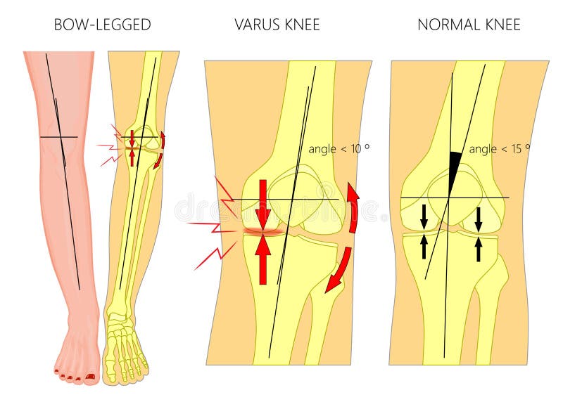
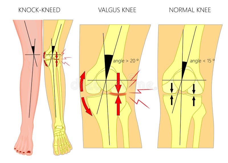
Free with trial Vector illustration diagram. Shapes of human legs. Normal and curved legs. Knock knees. Bowed legs. Genu valgum and genu varum. For advertising, medical publications. EPS 10. Dislocation vectors Shapes of the legs.Normal and curved legs.Knock knees.Bowed leg. Vector illustration diagram. Shapes of human legs. Normal and curved legs. Knock knees. Bowed. Vector illustration diagram. Shapes of human legs. Normal and curved legs. Knock knees. Bowed legs. Genu valgum and genu varum. For advertising, medical publications. EPS 10.
Free with trial Vector illustration diagram. Hammer toe. Plantar plate injury. Mechanism of rupture of plantar plate of the second toe of the foot. External and skeletal views of an ankle. Dislocation vectors Plantar plate tear. Hammer toe. Vector illustration diagram. Hammer toe. Plantar plate injury. Mechanism of rupture of plantar plate of the second toe of the. Vector illustration diagram. Hammer toe. Plantar plate injury. Mechanism of rupture of plantar plate of the second toe of the foot. External and skeletal views of an ankle.
Free with trial Rupture of the quadriceps tendon. Knee joint with patella and Tendon Tears. Dislocation vectors Rupture of the quadriceps tendon. Knee joint with patella and Tendon Tears
Free with trial Vector illustration of a healthy knee joint with intact, partial tear of anterior cruciate ligament and complete tear of ACL. Anterior or front view of flexed knee. For medical publications. EPS 10. Dislocation vectors Ligaments of the knee_Tear of the anterior cruciate ligament. Vector illustration of a healthy knee joint with intact, partial tear of anterior cruciate ligament. Vector illustration of a healthy knee joint with intact, partial tear of anterior cruciate ligament and complete tear of ACL. Anterior or front view of flexed knee. For medical publications. EPS 10.
Free with trial Vector illustration anatomy of a knee joint with healthy ligaments and sprain, tear or rupture of medial collateral ligament. Front view of straight knee. For advertising, medical publications. EPS 10. Dislocation vectors Ligaments of the knee_Tear of the medial collateral ligament. Vector illustration anatomy of a knee joint with healthy ligaments and sprain, tear or rupture of. Vector illustration anatomy of a knee joint with healthy ligaments and sprain, tear or rupture of medial collateral ligament. Front view of straight knee. For advertising, medical publications. EPS 10
Free with trial Vector illustration of a healthy knee joint and an unhealthy knee with a quadriceps tendon rupture problem. Anatomy of the human knee, side view of the bent knee. Dislocation vectors Knee problem_Quadriceps tendon rupture. Vector illustration of a healthy knee joint and an unhealthy knee with a quadriceps tendon rupture problem. Anatomy of. Vector illustration of a healthy knee joint and an unhealthy knee with a quadriceps tendon rupture problem. Anatomy of the human knee, side view of the bent knee.
Free with trial Vector illustration diagram. Deviation of the toe. Mechanism of rupture of plantar plate of the seond toe of the foot. External and skeletal views of an ankle. Dislocation vectors Plantar plate tear. Deviation of the toe. Vector illustration diagram. Deviation of the toe. Mechanism of rupture of plantar plate of the seond toe of the foot. Vector illustration diagram. Deviation of the toe. Mechanism of rupture of plantar plate of the seond toe of the foot. External and skeletal views of an ankle.
Free with trial Vector illustration diagram. Shapes of human legs. Normal and curved legs. Knock knees. Genu valgum and genu varum. For advertising, medical publications. EPS 10. Dislocation vectors Shapes of the legs.Normal and curved legs.Knock knees.Bowed leg. Vector illustration diagram. Shapes of human legs. Normal and curved legs. Knock knees. Genu. Vector illustration diagram. Shapes of human legs. Normal and curved legs. Knock knees. Genu valgum and genu varum. For advertising, medical publications. EPS 10.
Free with trial Illustration diagram of normal knee joint and a knee with chondromalacia patella. Flexed bent knee. Front view. Dislocation vectors Chondromalacia patella. Flattened femoral groove. Illustration diagram of normal knee joint and a knee with chondromalacia patella. Flexed bent knee. Front view. Illustration diagram of normal knee joint and a knee with chondromalacia patella. Flexed bent knee. Front view.
Free with trial Vector illustration x-ray of human pelvis, Healthy hip, arthritic hip joint and total hip replacement. For advertisement and medical publications. Dislocation vectors Pelvis and Hip joint problem_Healthy and arthritic hip joints and hip replacement. Vector illustration x-ray of human pelvis, Healthy hip, arthritic hip joint. Vector illustration x-ray of human pelvis, Healthy hip, arthritic hip joint and total hip replacement. For advertisement and medical publications
Free with trial Vector illustration anatomy of a knee joint with healthy and torn posterior cruciate ligament. Side or lateral view of flexed knee with sagittal section of femur bone. For medical publications. EPS 10. Dislocation vectors Ligaments of the knee_Torn posterior cruciate ligament. Vector illustration anatomy of a knee joint with healthy and torn posterior cruciate ligament. Side or. Vector illustration anatomy of a knee joint with healthy and torn posterior cruciate ligament. Side or lateral view of flexed knee with sagittal section of femur bone. For medical publications. EPS 10
Free with trial Vector illustration diagram. Shapes of human legs. Normal and curved legs. Knock knees. Bowed legs. Genu valgum and genu varum. For advertising, medical publications. EPS 10. Dislocation vectors Shapes of the legs.Normal and curved legs.Knock knees.Bowed leg. Vector illustration diagram. Shapes of human legs. Normal and curved legs. Knock knees. Bowed. Vector illustration diagram. Shapes of human legs. Normal and curved legs. Knock knees. Bowed legs. Genu valgum and genu varum. For advertising, medical publications. EPS 10.
Free with trial Medical illustration of the Cubital and Carpal Tunnel Syndrome. Compressed Median and Ulnar nerve. Vector. Dislocation vectors Medical illustration of the Cubital and Carpal Tunnel Syndrome. Compressed Median and Ulnar nerve. Vector
Free with trial Bones fracture and injuries x-ray radiology diagnostics facilities patients doctor cartoon icons collection isolated vector illustration. Dislocation vectors Bone Injury X-ray Cartoon Icons Collection. Bones fracture and injuries x-ray radiology diagnostics facilities patients doctor cartoon icons collection isolated. Bones fracture and injuries x-ray radiology diagnostics facilities patients doctor cartoon icons collection isolated vector illustration
Free with trial Dislocated knee, medial collateral ligament tear medical vector illustration isolated on white background. Eps 10. Dislocation vectors Dislocated knee, medial collateral ligament tear medical vector illustration isolated on white background. Eps 10
Free with trial Medical illustration to explain Cubital tunnel syndrome. Ulnar nerve entrapment. Fascia involved in ulnar nerve. Compression. Vector. Dislocation vectors Medical illustration to explain Cubital tunnel syndrome. Ulnar nerve entrapment. Fascia involved in ulnar nerve. Compression. Vector
Free with trial Spondylolisthesis. Spinal disease when lower vertebrae slip forward. Birth defect or injury damage. Flat vector illustration. Dislocation vectors Spondylolisthesis. Spinal disease when lower vertebrae slip forward. Birth defect or injury damage. Flat vector illustration
Free with trial Pelvis, hip fracture. hip dislocation. Vector illustration of a pelvis, hip fracture. Dislocation vectors Pelvis, hip fracture. hip dislocation. Vector illustration of a pelvis, hip fracture. hip dislocation
Free with trial A medical illustrations showing the symptoms of patellar dislocation. Dislocation vectors Patellar dislocation. A medical illustrations showing the symptoms of patellar dislocation. A medical illustrations showing the symptoms of patellar dislocation.
Free with trial Dislocation Shoulder symptoms or separated. Bone and joints to slipping out of place. Dislocation vectors Dislocation Shoulder symptoms or separated. Bone and joints to slipping out of place
Free with trial Dislocation of hip. A hip dislocation takes place when the head of the thigh bone is forced out of its socket in the pelvic bone. Dislocation vectors Dislocation of hip. A hip dislocation takes place when the head of the thigh bone is forced out of its socket in the pelvic bone
Free with trial Dislocation of the spine. Vector illustration of a dislocation of the spine. Dislocation vectors Dislocation of the spine. Vector illustration of a dislocation of the spine
Free with trial Knee, dislocation, fracture. traumatology and orthopedics. Illustration of the kneecap, dislocation and fracture. Dislocation vectors Knee, dislocation, fracture. traumatology and orthopedics. Illustration of the kneecap, dislocation and fracture. traumatology and orthopedics
Free with trial Shoulder dislocation. humerus bone trauma, Sports injuries, or Weak shoulder muscles. Human arm anatomy. Bones and joint of the Shoulder, and hand. Vector illustration. Dislocation vectors Shoulder dislocation. humerus bone trauma. Sports injuries, or Weak shoulder muscles. Human arm anatomy. Bones and joint of the Shoulder, and hand. Vector
Free with trial Finger Dislocation human medical organ vector illustration. Dislocation vectors Finger Dislocation human medical organ vector illustratio. Finger Dislocation human medical organ vector illustration. Finger Dislocation human medical organ vector illustration.
Free with trial Dislocation of the shoulder joint, medical vector illustration. Vector. Dislocation vectors Dislocation of the shoulder joint, medical vector illustration. Vector
Free with trial Finger Dislocation human medical organ vector illustration. Dislocation vectors Finger Dislocation human medical organ vector illustratio. Finger Dislocation human medical organ vector illustration. Finger Dislocation human medical organ vector illustration.
Free with trial Finger Dislocation human medical organ vector illustration. Dislocation vectors Finger Dislocation human medical organ vector illustratio. Finger Dislocation human medical organ vector illustration. Finger Dislocation human medical organ vector illustration.
Free with trial Medical Concept, Illustration of Shoulder Dislocation Caused by Sports Accident or Arthritis as A Skeletal Joint Problem. Dislocation vectors Recurrent Dislocation of Shoulder with Medical Treatment. Medical Concept, Illustration of Shoulder Dislocation Caused by Sports Accident or Arthritis as A. Medical Concept, Illustration of Shoulder Dislocation Caused by Sports Accident or Arthritis as A Skeletal Joint Problem.
Free with trial Dislocation of the foot forward, vintage engraved illustration. Usual Medicine Dictionary - Paul Labarthe - 1885. Dislocation vectors Dislocation of the foot forward, vintage engraving. Dislocation of the foot forward, vintage engraved illustration. Usual Medicine Dictionary - Paul Labarthe. Dislocation of the foot forward, vintage engraved illustration. Usual Medicine Dictionary - Paul Labarthe - 1885
Free with trial Fractures. A triple illustration of types of lesions in arms, fracture, luxation, dislocation and sprain. Dislocation vectors Fractures. A triple illustration of types of lesions in arms, fracture, luxation, dislocation and sprain
Free with trial Dislocation of the lens of the eye. Vector illustration of a dislocated lens of the eye. Dislocation vectors Dislocation of the lens of the eye. Vector illustration of a dislocated lens of the eye
Free with trial Vector illustration of Peroneal Tendon Injuries. Peroneal tendon dislocation or subluxation when the superior peroneal retinaculum or retinacula is torn. Lateral ankle injury. Dislocation vectors Peroneal Tendon Injuries_Peroneal tendon dislocation. Vector illustration of Peroneal Tendon Injuries. Peroneal tendon dislocation or subluxation when the. Vector illustration of Peroneal Tendon Injuries. Peroneal tendon dislocation or subluxation when the superior peroneal retinaculum or retinacula is torn. Lateral ankle injury.
Free with trial Vector illustration of a healthy human hip joint and bones, osteoporosis of femur and femoral neck fracture with dislocation. For advertising and medical publications. EPS 10. Dislocation vectors Osteoporosis1_with femur fracture and dislocation. Vector illustration of a healthy human hip joint and bones, osteoporosis of femur and femoral neck fracture. Vector illustration of a healthy human hip joint and bones, osteoporosis of femur and femoral neck fracture with dislocation. For advertising and medical publications. EPS 10.
Free with trial Vector illustration of a healthy human thigh bone, osteoporosis of femur and femoral neck fracture with dislocation isolated on white background. For advertising and medical publications. EPS 10. Dislocation vectors Osteoporosis 3_with femur fracture and dislocation. Vector illustration of a healthy human thigh bone, osteoporosis of femur and femoral neck fracture with. Vector illustration of a healthy human thigh bone, osteoporosis of femur and femoral neck fracture with dislocation isolated on white background. For advertising and medical publications. EPS 10.
Free with trial Vector illustration of a healthy human hip joint and bones, osteoporosis of femur and femoral neck fracture without dislocation. For advertising and medical publications. EPS 10. Dislocation vectors Osteoporosis1_with femur fracture and without dislocation 1. Vector illustration of a healthy human hip joint and bones, osteoporosis of femur and femoral neck. Vector illustration of a healthy human hip joint and bones, osteoporosis of femur and femoral neck fracture without dislocation. For advertising and medical publications. EPS 10.
Free with trial Vector illustration of healthy human hip and femoral neck fracture with dislocation. For advertising and medical publications. EPS 10. Dislocation vectors Pain in the hip joint_femoral neck fracture with dislocation. Vector illustration of healthy human hip and femoral neck fracture with dislocation. For. Vector illustration of healthy human hip and femoral neck fracture with dislocation. For advertising and medical publications. EPS 10
Free with trial Anterior and posterior shoulder dislocation vector illustration. Illustration of the displaced humerus. Dislocation vectors Anterior and posterior shoulder dislocation vector illustration. Illustration of the displaced humerus
Free with trial Vector illustration x-ray Pelvis and Hip joint, dislocation after total hip replacement. For advertisement and medical publications. Dislocation vectors Pelvis and Hip joint problem_Dislocation after total hip replacement. Vector illustration x-ray Pelvis and Hip joint, dislocation after total hip replacement. Vector illustration x-ray Pelvis and Hip joint, dislocation after total hip replacement. For advertisement and medical publications
Free with trial Vector illustration of healthy human hip and subtrochanteric femur fracture with dislocation. For advertising and medical publications. EPS 10. Dislocation vectors Pain in the hip joint_subtrochanteric femur fracture with dislocation. Vector illustration of healthy human hip and subtrochanteric femur fracture with. Vector illustration of healthy human hip and subtrochanteric femur fracture with dislocation. For advertising and medical publications. EPS 10
Free with trial Vector illustration of a healthy human thigh bone, osteoporosis of femur and femoral neck fracture without dislocation isolated on white background. For advertising and medical publications. EPS 10. Dislocation vectors Osteoporosis 3_with femur fracture and without dislocation. Vector illustration of a healthy human thigh bone, osteoporosis of femur and femoral neck fracture. Vector illustration of a healthy human thigh bone, osteoporosis of femur and femoral neck fracture without dislocation isolated on white background. For advertising and medical publications. EPS 10.
Free with trial Vector illustration of healthy human hip and intertrochanteric femur fracture with dislocation. For advertising and medical publications. EPS 10. Dislocation vectors Pain in the hip joint_intertrochanteric femur fracture with dislocation. Vector illustration of healthy human hip and intertrochanteric femur fracture with. Vector illustration of healthy human hip and intertrochanteric femur fracture with dislocation. For advertising and medical publications. EPS 10
Free with trial Different types of knee disease types poster design set collection. Knee injury. Labeled bursitis, bakers cyst, knee injection, knee dislocation rheumatoid arthritis synovitis bursae ruptured tendon. Dislocation vectors Different types of knee disease types poster design set collection. Knee injury. Labeled bursitis, bakers cyst, knee injection,. Knee dislocation rheumatoid. Different types of knee disease types poster design set collection. Knee injury. Labeled bursitis, bakers cyst, knee injection, knee dislocation rheumatoid arthritis synovitis bursae ruptured tendon
Free with trial Traumatology shoulder injury bruise pain or ache isolated male body vector arm joint dislocation or trauma arthritis or rheumatism osteoporosis bone fracture skeleton treatment and medicine disease. Dislocation vectors Shoulder injury bruise pain or ache isolated male body. Traumatology shoulder injury bruise pain or ache isolated male body vector arm joint dislocation or. Traumatology shoulder injury bruise pain or ache isolated male body vector arm joint dislocation or trauma arthritis or rheumatism osteoporosis bone fracture skeleton treatment and medicine disease.
 Sign in