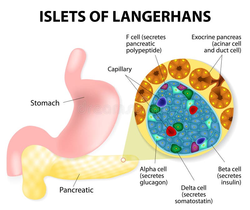
Free with trial The islets of Langerhans are responsible for the endocrine function of the pancreas. Each islet contains beta, alpha, and delta cells that are responsible for the secretion of a hormones. Duodenal vectors Islets of Langerhans. The islets of Langerhans are responsible for the endocrine function of the pancreas. Each islet contains beta, alpha, and delta cells that. The islets of Langerhans are responsible for the endocrine function of the pancreas. Each islet contains beta, alpha, and delta cells that are responsible for the secretion of a hormones.
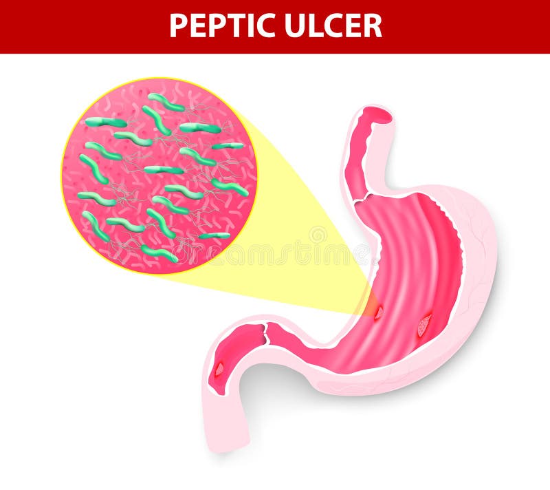
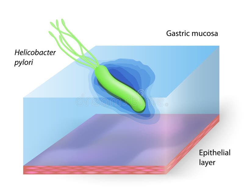
Free with trial Research shows that most ulcers of stomach ulcers and the duodenum develop because of infection with Helicobacter pylori. Bacteria weakens the protective mucous layer of the stomach. This allows acid to seep in and injure the underlying stomach cells. Duodenal vectors Helicobacter pylori and ulcers disease. Research shows that most ulcers of stomach ulcers and the duodenum develop because of infection with Helicobacter pylori. Research shows that most ulcers of stomach ulcers and the duodenum develop because of infection with Helicobacter pylori. Bacteria weakens the protective mucous layer of the stomach. This allows acid to seep in and injure the underlying stomach cells.
Free with trial Human pancreas, Lymphatics and stomach. Duodenal vectors Human pancreas. Lymphatics and stomach
Free with trial Stomach ache,. Man pain form stomach ache. Duodenal vectors Stomach ache,. Man pain form stomach ache
Free with trial Peptic ulcer. Two types of peptic ulcer, eps8, gradient and mesh printing compatible. Duodenal vectors Peptic ulcer. Two types of peptic ulcer, eps8, gradient and mesh printing compatible
Free with trial Upper gastrointestinal endoscopy. Used for examining the lining of upper digestive organs, eps8. Duodenal vectors Upper gastrointestinal endoscopy. Used for examining the lining of upper digestive organs, eps8
Free with trial Gastroparesis or delayed gastric emptying. Visual comparison of healthy gastric and stomach with Gastroparesis. Duodenal vectors Gastroparesis. Or delayed gastric emptying. Visual comparison of healthy gastric and stomach with. Gastroparesis or delayed gastric emptying. Visual comparison of healthy gastric and stomach with Gastroparesis.
Free with trial Drawing of bariatric surgery, showing a duodenal switch operation where a small gastric pouch is connected to the large intestine via the duodenum. Duodenal vectors Duodenal switch bariatric surgery. Drawing of bariatric surgery, showing a duodenal switch operation where a small gastric pouch is connected to the large. Drawing of bariatric surgery, showing a duodenal switch operation where a small gastric pouch is connected to the large intestine via the duodenum
Free with trial Woman stomach ache,. Woman sitting because stomach ache. Duodenal vectors Woman stomach ache,. Woman sitting because stomach ache
Free with trial Peptic Ulcer disease, also known as a peptic ulcer or stomach ulcer, is a break in the lining of the stomach, first part of the small intestine, or occasionally the lower esophagus. An ulcer in the stomach is known as a gastric ulcer while that in the first part of the intestines is known as a duodenal ulcer. Health care education infographic. Duodenal vectors Stomach peptic ulcer. Peptic Ulcer disease, also known as a peptic ulcer or stomach ulcer, is a break in the lining of the stomach, first part of the small. Peptic Ulcer disease, also known as a peptic ulcer or stomach ulcer, is a break in the lining of the stomach, first part of the small intestine, or occasionally the lower esophagus.An ulcer in the stomach is known as a gastric ulcer while that in the first part of the intestines is known as a duodenal ulcer. Health care education infographic.
Free with trial Peptic ulcer. A peptic ulcer is a sore on the lining of the stomach or duodenum. Cause of peptic ulcer is bacterial infection. Helicobacter pylori. Vector. Duodenal vectors Peptic ulcer. A peptic ulcer is a sore on the lining of the stomach or duodenum. Cause of peptic ulcer is bacterial infection. Helicobacter pylori. Vector
Free with trial Bacteria Helicobacter pylori. Vector. Bacteria Helicobacter pylori is a Gram-negative microaerophilic bacterium found in the stomach. Duodenal vectors Bacteria Helicobacter pylori. Vector. Bacteria Helicobacter pylori is a Gram-negative microaerophilic bacterium found in the stomach. Vector
Free with trial Сholecystitis. Inflammation of the gallbladder and bile ducts. Gallstones. Cholelithiasis. Calculous cholecystitis. Duodenal vectors Сholecystitis. Cholelithiasis. Сholecystitis. Inflammation of the gallbladder and bile ducts. Gallstones. Cholelithiasis. Calculous cholecystitis. Сholecystitis. Inflammation of the gallbladder and bile ducts. Gallstones. Cholelithiasis. Calculous cholecystitis.
Free with trial Treatment of Helicobacter pylori. Medications. Antacids, proton pump blockers and H2-histamine blockers. Infographics. Vector illustration on isolated background. Duodenal vectors Treatment of Helicobacter pylori. Medications. Antacids, proton pump blockers and H2-histamine blockers. Infographics. Vector illustration on isolated
Free with trial Peptic ulcer and helicobacter pylori. Helicobacter pylori infecting stomach, causing mucosal damage and peptic ulcer, medical illustration. Duodenal vectors Peptic ulcer and helicobacter pylori. Helicobacter pylori infecting stomach, causing mucosal damage and peptic ulcer, medical illustration
Free with trial Gallbladder. Bile duct. Duodenum. The structure of the gallbladder. Duodenal vectors Gallbladder. Bile duct. Duodenum. The structure of the gallbladder
Free with trial EGD, diagnosis of gastric ulcer. Vector illustration of EGD, diagnosis of gastric ulcer. Duodenal vectors EGD, diagnosis of gastric ulcer. Vector illustration of EGD, diagnosis of gastric ulcer
Free with trial Helicobacter pylori - Ulcer-causing bacterium. Duodenal vectors Helicobacter pylori. Ulcer-causing bacterium
Free with trial Peptic ulcer disease PUD, stomach ulcer or gastric ulcer. Human Stomach with endoscope and close-up view of bacterium Helicobacter pylori which causes ulcers. vector diagram. human anatomy. scheme for textbooks. Duodenal vectors Peptic ulcer disease PUD, stomach ulcer or gastric ulcer. Human Stomach with endoscope and close-up view of bacterium Helicobacter pylori which causes ulcers
Free with trial Esophagus ulcer affected. Ulcer of esophagus. Stomach ulcer affected. Gastric ulcer. Duodenum ulcer affected. Duodenal ulcer. Vector illustration on a background of isolation. Duodenal vectors Esophagus ulcer affected. Ulcer of esophagus. Stomach ulcer affected. Gastric ulcer. Duodenum ulcer affected. Duodenal ulcer. Vector illustration on a
Free with trial Helicobacter pylori in the stomach. Infographics. Vector illustration. On background. Duodenal vectors Helicobacter pylori in the stomach. Infographics. Vector illustration. On background
Free with trial Helicobacter h. pylori illustration microaerophilic bacterium which infects various areas of the stomach and duodenum. Duodenal vectors Helicobacter pylori illustration microaerophilic bacterium which infects various areas of the stomach and duodenum. Helicobacter h.pylori illustration. Helicobacter h.pylori illustration microaerophilic bacterium which infects various areas of the stomach and duodenum
Free with trial Duodenal sounding stomach, gaster, liver. Illustration of duodenal of the stomach. diagnosis of liver diseases. Duodenal vectors Duodenal sounding stomach, gaster, liver. Illustration of duodenal of the stomach. diagnosis of liver diseases
Free with trial Peptic ulcer disease. Gastric and duodenal ulcer. Stress ulcer is a mucosal defect. This ulcers can be located anywhere within the stomach and proximal duodenum. Human anatomy. Vector illustration for medical use. Duodenal vectors Peptic ulcer disease. Gastric and duodenal ulcer. Stress ulcer is a mucosal defect. This ulcers can be located anywhere within the stomach and proximal
Free with trial Acute pancreatitis. Pancreas inflammation. Realistic image of abdominal organs Gallbladder, Duodenum, Stomach, and Pancreas. Close-up of a Gallstone that blocked of pancreatic duct and duodenal papilla. Duodenal vectors Acute pancreatitis. Pancreas inflammation. Realistic image of abdominal organs Gallbladder, Duodenum, Stomach, and Pancreas. Close-up of a Gallstone that
Free with trial Pancreas anatomy. Close-up of Cell Structure of islet langerhans. gallbladder, bile duct, pancreatic duct, and major duodenal papilla. spleen and splenic artery. Vector illustration. Duodenal vectors Pancreas anatomy. Cell Structure of islet of langerhans. Pancreas anatomy. Close-up of Cell Structure of islet langerhans. gallbladder, bile duct, pancreatic. Pancreas anatomy. Close-up of Cell Structure of islet langerhans. gallbladder, bile duct, pancreatic duct, and major duodenal papilla. spleen and splenic artery. Vector illustration
Free with trial Duodenum ulcer affected. Duodenal ulcer. Vector illustration on a background of isolation. Duodenal vectors Duodenum ulcer affected. Duodenal ulcer. Vector illustration on a background of isolation.
Free with trial Biliopancreatic diversion with duodenal switch. BPD stomach surgery, weight loss gastric procedure. Internal organs before and after operation. Overweight and obesity problem flat vector illustration. Duodenal vectors Biliopancreatic diversion procedure. Biliopancreatic diversion with duodenal switch. BPD stomach surgery, weight loss gastric procedure. Internal organs before. Biliopancreatic diversion with duodenal switch. BPD stomach surgery, weight loss gastric procedure. Internal organs before and after operation. Overweight and obesity problem flat vector illustration
Free with trial Helicobacter pylori. Ulcers. The disease of the stomach and duodenum. Сaricature, cartoon. Duodenal vectors Helicobacter pylori. Ulcers. The disease of the stomach and duodenum. Сaricature, cartoon
Free with trial Helicobacter pylori. Ulcers. The disease of the stomach and duodenum. Сaricature, cartoon. Duodenal vectors Helicobacter pylori. Ulcers. The disease of the stomach and duodenum. Сaricature, cartoon
Free with trial Duodenum. Anatomy. anatomical locations labelled. Human silhouette with highlighted internal organs. Duodenal vectors Duodenum. Anatomy. anatomical locations labelled. Human silhouette with highlighted internal organs
Free with trial Location of peptic ulcer. Human digestive system: esophagus, duodenum, stomach. Cross section of the human stomach. Vector illustration for medical, biological, educational and science use. Duodenal vectors Location of peptic ulcer. Human digestive system: esophagus, duodenum, stomach. Cross section of the human stomach. Vector illustration for medical, biological
Free with trial Gastroscopy, research of a stomach gastroscope. Helicobacter pylori. Ulcers. The disease of the stomach and duodenum. Сaricature, cartoon. Duodenal vectors Gastroscopy, research of a stomach gastroscope. Helicobacter pylori. Ulcers. The disease of the stomach and duodenum. Сaricature, cartoon
Free with trial Duodenitis is inflammation of the duodenum. Symptoms and causes. humans silhouette with red highlight duodenum. Bacteria, viruses, very spicy food chili pepper and poisonous substance international pictogramme that cause of this disease. Duodenal vectors Duodenitis. Symptoms and causes. Duodenitis is inflammation of the duodenum. Symptoms and causes. humans silhouette with red highlight duodenum. Bacteria. Duodenitis is inflammation of the duodenum. Symptoms and causes. humans silhouette with red highlight duodenum. Bacteria, viruses, very spicy food chili pepper and poisonous substance international pictogramme that cause of this disease.
Free with trial Duodenitis. Stomach and duodenum with inflammation. Human anatomy. Vector illustration for medical, scientific, and educational use. Duodenal vectors Duodenitis. Stomach and duodenum with inflammation. Human anatomy. Vector illustration for medical, scientific, and educational use
Free with trial Peptic ulcer and bacterial infection. This diagram illustrates mechanism by which Helicobacter Pylori gets across the mucus gel to epithelial cells. bacterium secretes substance that elevating the pH. Vector illustration for educational, Microbiological, biological, medical and science use. Duodenal vectors Peptic ulcer and bacterial infection. Helicobacter Pylori. Peptic ulcer and bacterial infection. This diagram illustrates mechanism by which Helicobacter Pylori. Peptic ulcer and bacterial infection. This diagram illustrates mechanism by which Helicobacter Pylori gets across the mucus gel to epithelial cells. bacterium secretes substance that elevating the pH. Vector illustration for educational, Microbiological, biological, medical and science use
Free with trial Peptic ulcer. human silhouette. close up of stomach with ulcer. Stress ulcer is a mucosal defect. Human anatomy. Vector illustration for medical use. Duodenal vectors Peptic ulcer. Human silhouette. close up of stomach with ulcer. Stress ulcer is a mucosal defect. Human anatomy. Vector illustration for medical use
Free with trial Gastroscopy procedure. Human stomach with Peptic ulcer, and endoscopic device. On black background. Illustration for medical use. Duodenal vectors Gastroscopy procedure. Human stomach with Peptic ulcer, and endoscopic device. On black background. Illustration for medical use
Free with trial EGD, diagnosis of gastric ulcer. Vector illustration of EGD, diagnosis of gastric ulcer. Duodenal vectors EGD, diagnosis of gastric ulcer. Vector illustration of EGD, diagnosis of gastric ulcer
Free with trial Gastroscopy, research of a stomach gastroscope. Helicobacter pylori. Ulcers. The disease of the stomach and duodenum. Duodenal vectors Gastroscopy, research of a stomach gastroscope. Helicobacter pylori. Ulcers. The disease of the stomach and duodenum
Free with trial Bile ducts. Bile is secreted by the liver into gallbladder and form the common bile duct which then opens into the intestine. Human`s digestive system. vector diagram. Duodenal vectors Bile ducts. digestive system. Bile ducts. Bile is secreted by the liver into gallbladder and form the common bile duct which then opens into the intestine. Human. Bile ducts. Bile is secreted by the liver into gallbladder and form the common bile duct which then opens into the intestine. Human`s digestive system. vector diagram
Free with trial Gastrointestinal tract of an amphibian frog information graphic. Vector illustration. Duodenal vectors Gastrointestinal tract of an amphibian frog information graphic. Vector illustration
Free with trial Pancreas with pancreatic duct, Duodenum, and Small Intestine on a white background. human anatomy. Part of a Human digestive system. vector Illustration. Duodenal vectors Pancreas with pancreatic duct, Duodenum, and Small Intestine on a white background. Human anatomy. Part of a Human digestive system. vector Illustration
Free with trial The structure of Helicobacter pylori. Infographics. Vector illustration on isolated background. Duodenal vectors The structure of Helicobacter pylori. Infographics. Vector illustration on isolated background
Free with trial The structure of Helicobacter pylori. Infographics. Vector illustration an background. Duodenal vectors The structure of Helicobacter pylori. Infographics. Vector illustration. An background
Free with trial Illustration of the digestive system, specifically highlighting components related to dumping syndrome. The liver is shown in brown, with the gallbladder in green nestled beneath it. The stomach, in pink, connects to the duodenum, which leads to the small intestine. The pancreas is displayed in yellow, with its pancreatic duct marked. Labels indicate the major and minor duodenal papilla, common bile duct, and the duodenum. Text explains that dumping syndrome involves rapid gastric emptying, causing symptoms like cramps, nausea, diarrhea, and dizziness. Duodenal vectors Dumping Syndrome (Rapid Gastric Emptying). Illustration of the digestive system, specifically highlighting components related to dumping syndrome. The liver is. Illustration of the digestive system, specifically highlighting components related to dumping syndrome. The liver is shown in brown, with the gallbladder in green nestled beneath it. The stomach, in pink, connects to the duodenum, which leads to the small intestine. The pancreas is displayed in yellow, with its pancreatic duct marked. Labels indicate the major and minor duodenal papilla, common bile duct, and the duodenum. Text explains that dumping syndrome involves rapid gastric emptying, causing symptoms like cramps, nausea, diarrhea, and dizziness.
Free with trial Stomach. Helicobacter pylori. Vector illustration on isolated background. Duodenal vectors Stomach. Helicobacter pylori. Vector illustration on isolated background. Stomach. Helicobacter pylori. Vector illustration on isolated background
Free with trial Pancreas of human. Digestive system. Realistic design. Isolated. Vector illustration. Duodenal vectors Pancreas of human . Digestive system . Realistic design . Isolated . Vector illustration
Free with trial Helicobacter pylori. Diseases caused by Helicobacter pylori. Helicobacter infection. Infographics. Vector. Duodenal vectors Helicobacter pylori. Diseases caused by Helicobacter pylori. Helicobacter infection. Infographics. Vector
Free with trial Metaphor function of human pancreas is to produce insulin hormone vector illustration sketch hand drawn with black lines, on white background. Education Medical concept. Duodenal vectors Metaphor function of human pancreas is to produce insulin hormone vector illustration sketch hand drawn with black lines, isolate. Metaphor function of human. Metaphor function of human pancreas is to produce insulin hormone vector illustration sketch hand drawn with black lines, on white background. Education Medical concept.
Free with trial Medical illustration of the human digestive system. scheme for textbooks. internal organs. Duodenal vectors Medical illustration of the human digestive system. scheme for textbooks. Internal organs
Free with trial Types of hernias in the abdominal cavity with a description. Medical infographic with human torso. Medical poster. Vector illustration. Duodenal vectors Types of hernias in the abdominal cavity with a description. Medical infographic with human torso. Medical poster. Vector illustration
Free with trial 3D Isometric Flat Vector Conceptual Illustration of Peptic Ulcer Stomach Disease, Gastritis and Inflammation. Duodenal vectors 3D Isometric Flat Vector Conceptual Illustration of Peptic Ulcer Stomach Disease. Gastritis and Inflammation
Free with trial Types of bariatric surgery. Healthy stomach and internal organs after operation, weight loss gastric procedure. Abdomen laparoscopy concept. Overweight and obesity problem flat vector illustration. Duodenal vectors Bariatric surgery poster. Types of bariatric surgery. Healthy stomach and internal organs after operation, weight loss gastric procedure. Abdomen laparoscopy. Types of bariatric surgery. Healthy stomach and internal organs after operation, weight loss gastric procedure. Abdomen laparoscopy concept. Overweight and obesity problem flat vector illustration
Free with trial Types of bariatric surgery. Healthy stomach and internal organs after operation, weight loss gastric procedure. Abdomen laparoscopy concept. Overweight and obesity problem flat vector illustration. Duodenal vectors Bariatric surgery poster. Types of bariatric surgery. Healthy stomach and internal organs after operation, weight loss gastric procedure. Abdomen laparoscopy. Types of bariatric surgery. Healthy stomach and internal organs after operation, weight loss gastric procedure. Abdomen laparoscopy concept. Overweight and obesity problem flat vector illustration
Free with trial Esophagogastroduodenoscopy EGD Procedure Showing Duodenal Ulcer Medical Illustration. Upper Endoscopy Examination of Duodenum with. Peptic Ulcer Diagram. Duodenal vectors Esophagogastroduodenoscopy EGD Procedure Showing Duodenal Ulcer Medical Illustration. Upper Endoscopy Examination of Duodenum with. Peptic Ulcer Diagram
Free with trial Scientific medical showing peptic ulcer disease affecting the upper digestive tract, with clearly labeled esophageal, gastric, and duodenal ulcers. Suitable for medical education, anatomy textbooks, healthcare publications, gastroenterology resources, biology, clinical studies, academic presentations, and scientific educational materials. Duodenal vectors Peptic Ulcer Disease Medical Anatomy Illustration. Scientific medical showing peptic ulcer disease affecting the upper digestive tract, with clearly labeled. Scientific medical showing peptic ulcer disease affecting the upper digestive tract, with clearly labeled esophageal, gastric, and duodenal ulcers. Suitable for medical education, anatomy textbooks, healthcare publications, gastroenterology resources, biology, clinical studies, academic presentations, and scientific educational materials.
Free with trial Scientific medical showing peptic ulcer disease affecting the upper digestive tract, with clearly labeled esophageal, gastric, and duodenal ulcers. Suitable for medical education, anatomy textbooks, healthcare publications, gastroenterology resources, biology, clinical studies, academic presentations, and scientific educational materials. Duodenal vectors Peptic Ulcer Disease Medical Anatomy Illustration Outline. Scientific medical showing peptic ulcer disease affecting the upper digestive tract, with clearly. Scientific medical showing peptic ulcer disease affecting the upper digestive tract, with clearly labeled esophageal, gastric, and duodenal ulcers. Suitable for medical education, anatomy textbooks, healthcare publications, gastroenterology resources, biology, clinical studies, academic presentations, and scientific educational materials.
Free with trial Scientific medical showing the three main types of peptic ulcer disease, including esophageal ulcers, gastric ulcers, and duodenal ulcers. Clear anatomical visuals and labels make this suitable for medical education, anatomy textbooks, healthcare publications, gastroenterology resources, biology, academic presentations, and scientific study materials. Duodenal vectors Types of Peptic Ulcer Disease Medical Illustration Outline. Scientific medical showing the three main types of peptic ulcer disease, including esophageal ulcers. Scientific medical showing the three main types of peptic ulcer disease, including esophageal ulcers, gastric ulcers, and duodenal ulcers. Clear anatomical visuals and labels make this suitable for medical education, anatomy textbooks, healthcare publications, gastroenterology resources, biology, academic presentations, and scientific study materials.
Free with trial Medical illustration showing a portion of the human digestive system, including the gallbladder, liver, pancreas, and small intestine. Annotations highlight structures such as the common bile duct, pancreatic duct, minor and major duodenal papillae. Gallstones are depicted in the gallbladder. A silhouette of a person indicates pain under the right ribcage, associated with the digestive organs illustrated. Arrows and labels describe components related to biliary colic and gallbladder anatomy. Duodenal vectors Biliary Colic Pancrea Illustration. Medical illustration showing a portion of the human digestive system, including the gallbladder, liver, pancreas, and small. Medical illustration showing a portion of the human digestive system, including the gallbladder, liver, pancreas, and small intestine. Annotations highlight structures such as the common bile duct, pancreatic duct, minor and major duodenal papillae. Gallstones are depicted in the gallbladder. A silhouette of a person indicates pain under the right ribcage, associated with the digestive organs illustrated. Arrows and labels describe components related to biliary colic and gallbladder anatomy.
Free with trial Diagram illustrating cholecystitis, showing inflamed gallbladder with gallstones in green. The common bile duct and pancreatic duct are highlighted. The pancreas is depicted in yellow with lobular structure, labeled as head, body, and tail. The small intestine is shown in pink with major and minor duodenal papillae. The image effectively demonstrates the anatomical connection between the gallbladder, ducts, and digestive organs. Duodenal vectors Cholecystitis Gallbladder Inflammation Illustration. Diagram illustrating cholecystitis, showing inflamed gallbladder with gallstones in green. The common bile. Diagram illustrating cholecystitis, showing inflamed gallbladder with gallstones in green. The common bile duct and pancreatic duct are highlighted. The pancreas is depicted in yellow with lobular structure, labeled as head, body, and tail. The small intestine is shown in pink with major and minor duodenal papillae. The image effectively demonstrates the anatomical connection between the gallbladder, ducts, and digestive organs.
Free with trial Scientific medical showing the three main types of peptic ulcer disease, including esophageal ulcers, gastric ulcers, and duodenal ulcers. Clear anatomical visuals and labels make this suitable for medical education, anatomy textbooks, healthcare publications, gastroenterology resources, biology, academic presentations, and scientific study materials. Duodenal vectors Types of Peptic Ulcer Disease Medical Illustration. Scientific medical showing the three main types of peptic ulcer disease, including esophageal ulcers, gastric. Scientific medical showing the three main types of peptic ulcer disease, including esophageal ulcers, gastric ulcers, and duodenal ulcers. Clear anatomical visuals and labels make this suitable for medical education, anatomy textbooks, healthcare publications, gastroenterology resources, biology, academic presentations, and scientific study materials.
Free with trial Biliary tree anatomy diagram showing bile flow pathways, liver, gallbladder, and common bile duct labeled to explain duct connections and pancreatic entry. Outline diagram. Duodenal vectors Biliary tree anatomy diagram showing bile flow pathways, liver, gallbladder, ... Biliary tree anatomy diagram showing bile flow pathways, liver, gallbladder. Biliary tree anatomy diagram showing bile flow pathways, liver, gallbladder, and common bile duct labeled to explain duct connections and pancreatic entry. Outline diagram
Free with trial A cartoon character of a flame coming out from the stomach, showing the heat in the stomach caused by acid reflux. Duodenal vectors Acid reflux symptoms shown through a cute cartoon illustration. A cartoon character of a flame coming out from the stomach, showing the heat in the stomach. A cartoon character of a flame coming out from the stomach, showing the heat in the stomach caused by acid reflux
Free with trial Stomach acid. An illustration of stomach acid on a gray background. Duodenal vectors Stomach acid. An illustration of stomach acid on a gray background
Free with trial This vector illustration shows acid reflux or heartburn with a cartoon stomach and fire character on a light gray background to highlight the burning sensation. Duodenal vectors Acid reflux or heartburn. This vector illustration shows acid reflux or heartburn with a cartoon stomach and fire character on a light gray background to. This vector illustration shows acid reflux or heartburn with a cartoon stomach and fire character on a light gray background to highlight the burning sensation
Free with trial A cute cartoon-style illustration of a stomach breathing out strong flames, representing the burning sensation caused by acid reflux. Duodenal vectors Cute cartoon stomach breathing fire to show acid reflux. A cute cartoon-style illustration of a stomach breathing out strong flames, representing the burning. A cute cartoon-style illustration of a stomach breathing out strong flames, representing the burning sensation caused by acid reflux
Free with trial This vector illustration shows acid reflux or heartburn with a cartoon stomach and fire character on a light gray background to highlight the burning sensation. Duodenal vectors A cartoon stomach character with a fire character on it is used to show the symptom of acid reflux. This vector illustration shows acid reflux or heartburn with. This vector illustration shows acid reflux or heartburn with a cartoon stomach and fire character on a light gray background to highlight the burning sensation
Free with trial A compelling illustration of a stomach, hot with chili and flames, powerfully conveys the internal irritation and searing pain that spicy cuisine can unleash, often igniting acid reflux. Duodenal vectors A fiery stomach inflamed by chili a striking image of how spicy food can trigger acid reflux. A compelling illustration of a stomach, hot with chili and flames. A compelling illustration of a stomach, hot with chili and flames, powerfully conveys the internal irritation and searing pain that spicy cuisine can unleash, often igniting acid reflux
Free with trial Zollinger–Ellison Syndrome (ZES) is a rare disorder caused by a gastrin-secreting neuroendocrine tumor (gastrinoma), leading to excessive stomach acid production, recurrent peptic ulcers, and chronic diarrhea. Duodenal vectors Zollinger–Ellison Syndrome (ZES). Is a rare disorder caused by a gastrin-secreting neuroendocrine tumor (gastrinoma), leading to excessive stomach acid. Zollinger–Ellison Syndrome (ZES) is a rare disorder caused by a gastrin-secreting neuroendocrine tumor (gastrinoma), leading to excessive stomach acid production, recurrent peptic ulcers, and chronic diarrhea.
Free with trial This vector illustration shows acid reflux (heartburn) with a cartoon stomach and fire character on a light gray background to highlight the burning sensation. Duodenal vectors A cartoon stomach character with a fire character on it is used to show the symptom of acid reflux. This vector illustration shows acid reflux (heartburn) with a. This vector illustration shows acid reflux (heartburn) with a cartoon stomach and fire character on a light gray background to highlight the burning sensation
Free with trial This vector illustration shows acid reflux (heartburn) with a cartoon stomach and fire character on a light gray background to highlight the burning sensation. Duodenal vectors A cartoon stomach character with a fire character on it is used to show the symptom of acid reflux. This vector illustration shows acid reflux (heartburn) with a. This vector illustration shows acid reflux (heartburn) with a cartoon stomach and fire character on a light gray background to highlight the burning sensation
Free with trial Science and laboratory elements collection in a flat style, featuring icons of scientific equipment, research tools, chemistry, biology, and educational science, isolated on a white background. Duodenal vectors Types of Bariatric Surgery: Gastric Band, Sleeve Gastrectomy and Gastric Bypass. Science and laboratory elements collection in a flat style, featuring icons of. Science and laboratory elements collection in a flat style, featuring icons of scientific equipment, research tools, chemistry, biology, and educational science, isolated on a white background.
Free with trial This medical illustration depicts gallstones within the gallbladder, alongside inflammation of the gallbladder, known as cholecystitis, and its impact on surrounding pancreatic structures. Duodenal vectors Diagram illustrating gallstones and cholecystitis affecting the gallbladder and pancreas anatomy. This medical illustration depicts gallstones within the. This medical illustration depicts gallstones within the gallbladder, alongside inflammation of the gallbladder, known as cholecystitis, and its impact on surrounding pancreatic structures
Free with trial Isolated human stomach cartoon character suffering from a gastric peptic ulcer vector illustration graphic icon symbol. Duodenal vectors Isolated cartoon character human stomach suffering from a gastric ulcer graphic icon. Isolated human stomach cartoon character suffering from a gastric peptic. Isolated human stomach cartoon character suffering from a gastric peptic ulcer vector illustration graphic icon symbol
Free with trial Isolated human stomach cartoon character suffering from a gastric peptic ulcer vector illustration graphic icon symbol. Duodenal vectors Isolated cartoon character human stomach suffering from a gastric ulcer graphic icon. Isolated human stomach cartoon character suffering from a gastric peptic. Isolated human stomach cartoon character suffering from a gastric peptic ulcer vector illustration graphic icon symbol
Free with trial 3D Isometric Flat Vector Illustration of Bariatric Stomach Reducation, Overweight and Obesity Problem Solution. Duodenal vectors 3D Isometric Flat Vector Illustration of Bariatric Stomach Reducation. Overweight and Obesity Problem Solution
Free with trial Red and yellow human pancreas and duodenum. Healthcare medical and anatomical concept. Futuristic low poly style. Geometric background. Wireframe light connection structure. Modern 3d graphic. Vector. Duodenal vectors Red and yellow human pancreas and duodenum. Healthcare medical and anatomical concept. Futuristic low poly style. Geometric background. Wireframe light
Free with trial Helicobacter pylori vector illustration. Hand-drawn stomach, bacteria. Bright informational medical image. Disease of the stomach, duodenum, esophagus. Cartoon anatomy of abdominal organs. Be healthy. Duodenal vectors Helicobacter pylori vector illustration. Cartoon stomach, bacteria. Helicobacter pylori vector illustration. Hand-drawn stomach, bacteria. Bright informational. Helicobacter pylori vector illustration. Hand-drawn stomach, bacteria. Bright informational medical image. Disease of the stomach, duodenum, esophagus. Cartoon anatomy of abdominal organs. Be healthy
Free with trial Peptic ulcer stomach disease infographic poster. Endoscopic image of stomach with duodenal and gastric PUD. Digestive tract organ disorder. Health care and medical concept. Vector illustration. Duodenal vectors Duodenal and gastric types of peptic ulcer stomach disease. Peptic ulcer stomach disease infographic poster. Endoscopic image of stomach with duodenal and. Peptic ulcer stomach disease infographic poster. Endoscopic image of stomach with duodenal and gastric PUD. Digestive tract organ disorder. Health care and medical concept. Vector illustration
 Sign in