Free with trial Osteoporosis in femur bone with microscopic view. Femur vectors Osteoporosis. In femur bone with microscopic view
Free with trial Osteoporosis 3. Medical illustration of the effects of osteoporosis in the course of a woman's life. Femur vectors Osteoporosis 3. Medical illustration of the effects of osteoporosis in the course of a woman's life
Free with trial Osteoporosis. Medical illustration of the effects of the osteoporosis. Femur vectors Osteoporosis. Medical illustration of the effects of the osteoporosis
Free with trial Osteoporosis in femur bone, human bone anatomy. Femur vectors Osteoporosis in femur bone. Human bone anatomy
Free with trial Right tight bone- Femur. Bone of the lower extremity. From above, behind and within. Detailed medical illustration. Isolated on a white background. Bright and clean design. Femur vectors Tight bone- Femur. Right tight bone- Femur. Bone of the lower extremity. From above, behind and within. Detailed medical illustration. Isolated on a white. Right tight bone- Femur. Bone of the lower extremity. From above, behind and within. Detailed medical illustration. Isolated on a white background. Bright and clean design.
Free with trial Bone Marrow & Blood Cells infographic diagram including femur reproduction of red white blood cells platelets lymphocyte monocyte esinophill basophill neurophill for medical science education. Femur vectors Bone Marrow & Blood Cells. Infographic diagram including femur reproduction of red white blood cells platelets lymphocyte monocyte esinophill basophill. Bone Marrow & Blood Cells infographic diagram including femur reproduction of red white blood cells platelets lymphocyte monocyte esinophill basophill neurophill for medical science education
Free with trial Types of bone fractures. Illustrated with the femur (hip bone), eps8. Femur vectors Types of bone fractures. Illustrated with the femur (hip bone), eps8. Types of bone fractures illustrated with the femur (hip bone), eps8
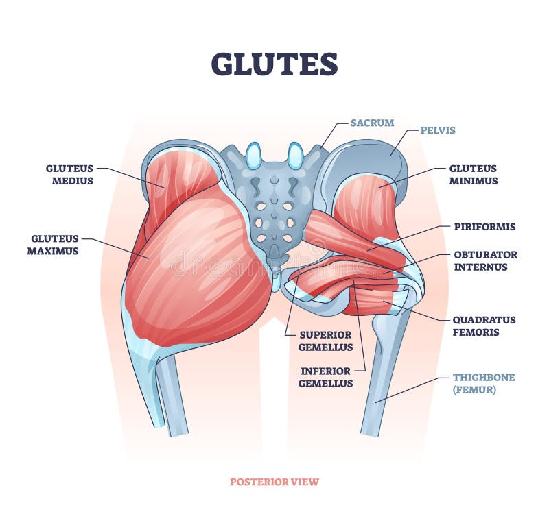
Free with trial Glutes as gluteal body muscles for human buttocks strength outline concept. Labeled educational anatomical scheme with physical skeletal and gluteus medius, maximus and minimus vector illustration. Femur vectors Glutes as gluteal body muscles for human buttocks strength outline concept. Labeled educational anatomical scheme with physical skeletal and gluteus medius
Free with trial Osteoarthritis. Arthritis or pain within a joint. degenerative joint disease. Cartilage becomes worn. This results in inflammation, swelling, and pain in the joint. Femur vectors Osteoarthritis or arthritis. Osteoarthritis. Arthritis or pain within a joint. degenerative joint disease. Cartilage becomes worn. This results in inflammation. Osteoarthritis. Arthritis or pain within a joint. degenerative joint disease. Cartilage becomes worn. This results in inflammation, swelling, and pain in the joint.
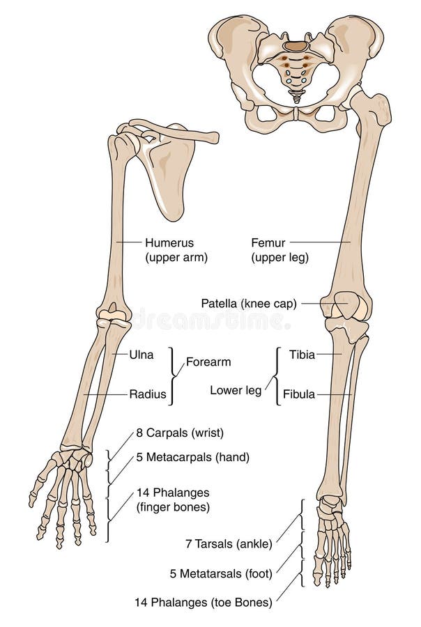
Free with trial Limb bones. Bones of upper and lower human limbs. Femur vectors Limb bones. Bones of upper and lower human limbs
Free with trial Human Knee Anatomy. Human Knee joint anatomy. Vector. Femur vectors Human Knee Anatomy. Human Knee joint anatomy. Vector
Free with trial Hip joint structure. In a whole and in cross section, eps8. Femur vectors Hip joint structure. In a whole and in cross section, eps8
Free with trial Arthrosis of the knee. Medical illustration of the symptoms of arthrosis of the knee. Femur vectors Arthrosis of the knee. Medical illustration of the symptoms of arthrosis of the knee
Free with trial Typical bone fractures. Vector scheme. Femur vectors Typical bone fractures. Vector scheme
Free with trial Rickets and Osteomalacia. Vitamine D deficiency bone disease in adults and children, eps10. Femur vectors Rickets and Osteomalacia. Vitamine D deficiency bone disease in adults and children, eps10
Free with trial Osteoporosis. health problems. medicine in medical infographics. elements and icons for design. concept. illness illustration. Femur vectors Osteoporosis infographic. Osteoporosis. health problems. medicine in medical infographics. elements and icons for design. concept. illness illustration. Osteoporosis. health problems. medicine in medical infographics. elements and icons for design. concept. illness illustration.
Free with trial Anatomy of the Right Knee. Medical illustration. Femur vectors Anatomy of the Right Knee. Medical illustration
Free with trial Total hip replacement surgery. Steps of Total hip replacement surgical procedure, eps10. Femur vectors Total hip replacement surgery. Steps of Total hip replacement surgical procedure, eps10
Free with trial Knee sprain. Ligaments involved in knee sprains, eps8. Femur vectors Knee sprain. Ligaments involved in knee sprains, eps8
Free with trial Osteoporosis - is a disease of bones that leads to an increased risk of fracture. Vector. Femur vectors Osteoporosis. Is a disease of bones that leads to an increased risk of fracture. Vector
Free with trial Hip fracture. Diagram showing the site of common hip injury as a result of osteoporosis or other bone diseases, eps8. Femur vectors Hip fracture. Diagram showing the site of common hip injury as a result of osteoporosis or other bone diseases, eps8
Free with trial Osteoporosis. Hip bone healthy and with osteoporosis, eps8, gradient and mesh printing compatible. Femur vectors Osteoporosis. Hip bone healthy and with osteoporosis, eps8, gradient and mesh printing compatible
Free with trial Osteoporosis. Illustration of a section of femur with osteoporosis. Femur vectors Osteoporosis. Illustration of a section of femur with osteoporosis
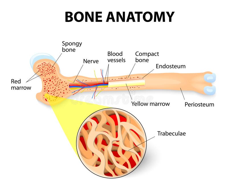
Free with trial Anatomy of the Long Bone. Periosteum, endosteum, bone marrow and trabeculae. Femur vectors Bone anatomy. Anatomy of the Long Bone. Periosteum, endosteum, bone marrow and trabeculae. Anatomy of the Long Bone. Periosteum, endosteum, bone marrow and trabeculae.
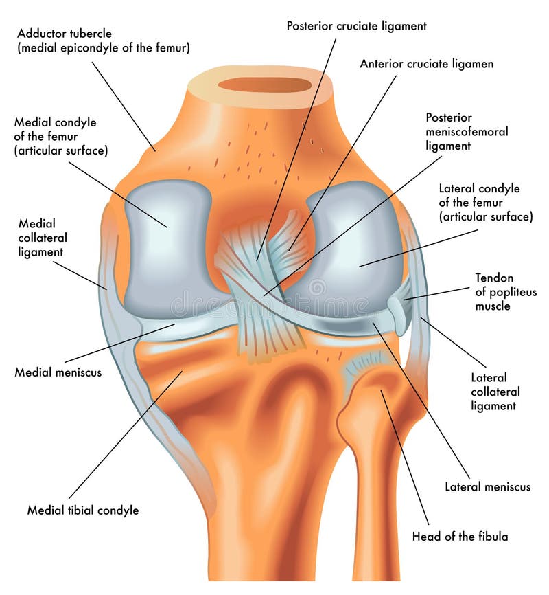
Free with trial Posterior view of the right knee in extension. Anatomy of the posterior of the right knee in extension. Femur vectors Posterior view of the right knee in extension. Anatomy of the posterior of the right knee in extension
Free with trial Arthritic knee. Medical illustration of the symptoms of arthritic knee. Femur vectors Arthritic knee. Medical illustration of the symptoms of arthritic knee
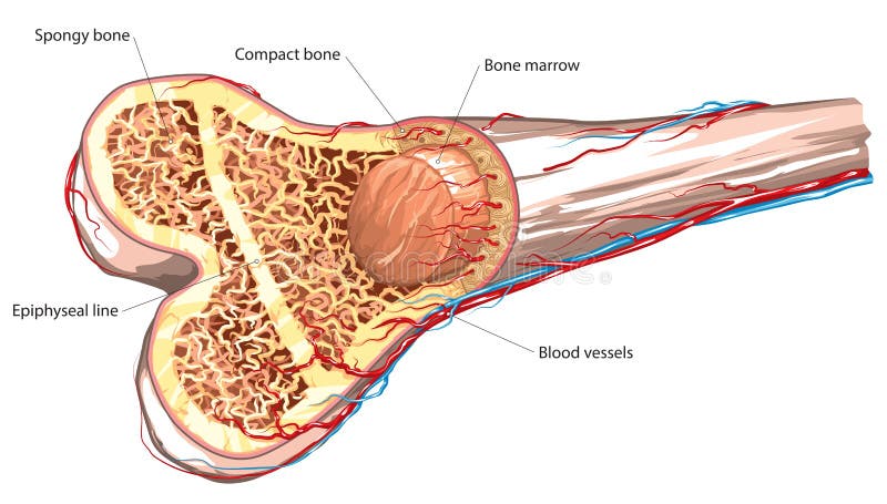
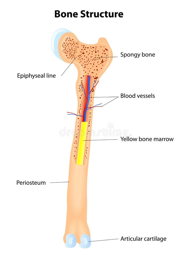
Free with trial Vector illustration of long bone basic structure. Femur vectors Bone structure. Vector illustration of long bone basic structure. Vector illustration of long bone basic structure.
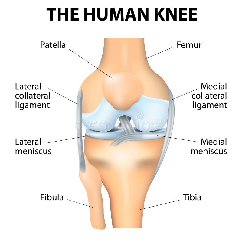
Free with trial Human knee joint. Anterior view of Human knee joint, eps8. Femur vectors Human knee joint. Anterior view of Human knee joint, eps8
Free with trial Bone marrow cells. Medical illustration of the composition of bone marrow cells. Femur vectors Bone marrow cells. Medical illustration of the composition of bone marrow cells
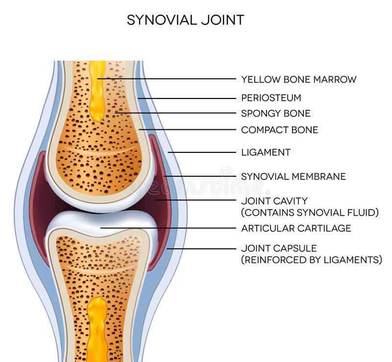
Free with trial Labeled joint anatomy. Normal joint illustration. Femur vectors Labeled joint anatomy. Normal joint illustration
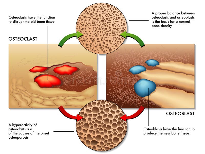
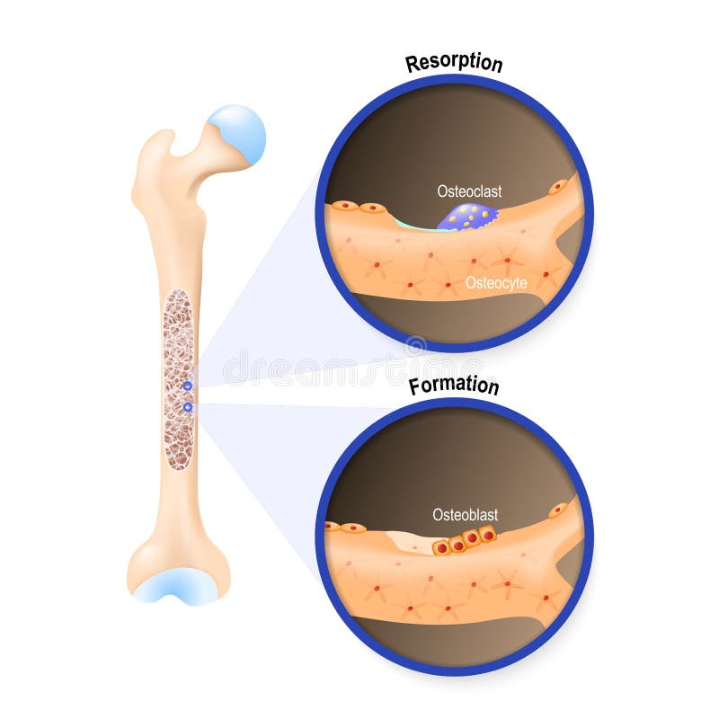
Free with trial Osteoblast & osteoclast. Medical illustration of the function of osteoblasts and osteoclasts in the regeneration of bone mass. Femur vectors Osteoblast & osteoclast. Medical illustration of the function of osteoblasts and osteoclasts in the regeneration of bone mass
Free with trial Bone structure details. Long Bone Anatomy. Vector scheme. illustration of human bone anatomy. Femur vectors Bone structure details. Long Bone Anatomy. Vector scheme. illustration of human bone anatomy
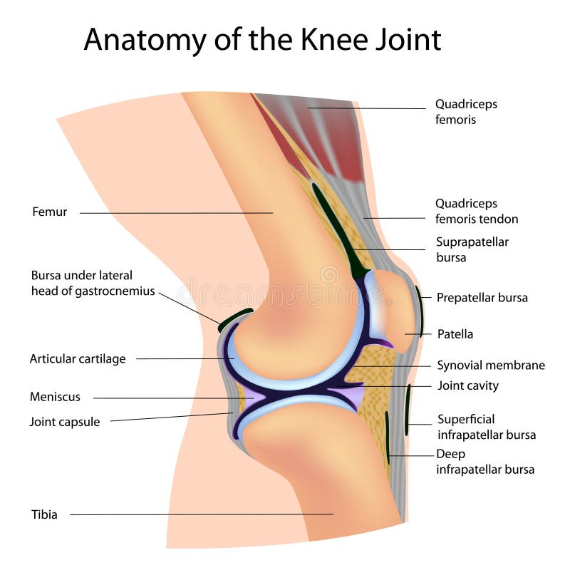
Free with trial Anatomy of the Knee Joint. Anatomy of human Knee Joint, eps8. Femur vectors Anatomy of the Knee Joint. Anatomy of human Knee Joint, eps8
Free with trial Arthritis of the hip joint, healthy joint and unhealthy joint with damaged cartilage and osteophytes. Femur vectors Arthritis of the hip joint. Healthy joint and unhealthy joint with damaged cartilage and osteophytes
Free with trial Osteoarthritis of hip joint. With cartilage breakdown and bone spurring, eps8. Femur vectors Osteoarthritis of hip joint. With cartilage breakdown and bone spurring, eps8
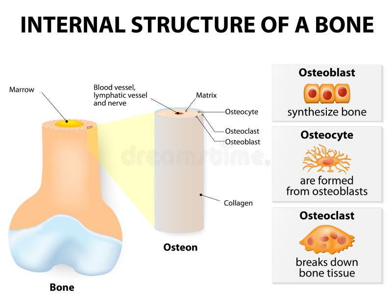
Free with trial Internal structure of a bone. 3 types of cells are found within bone tissue: Osteoblasts, Osteocytes, and Osteoclasts. Femur vectors Internal structure of a bone. 3 types of cells are found within bone tissue: Osteoblasts, Osteocytes, and Osteoclasts
Free with trial Normal synovial joint anatomy. Healthy joint detailed illustration. Femur vectors Normal synovial joint anatomy. Healthy joint detailed illustration
Free with trial Total knee replacement surgery. Total knee replacement surgical procedure, eps10. Femur vectors Total knee replacement surgery. Total knee replacement surgical procedure, eps10
Free with trial Synovial bursa of the human knee. Synovial bursa is a sac filled with lubricating fluid, located between tissues such as bone, muscle, tendons, and skin, that decreases rubbing. Femur vectors Synovial bursa of the human knee. Synovial bursa is a sac filled with lubricating fluid, located between tissues such as bone, muscle, tendons, and skin, that
Free with trial Anterior Cruciate Ligament injury. Torn Anterior Cruciate Ligament, a common sport injury. Femur vectors Anterior Cruciate Ligament injury. Torn Anterior Cruciate Ligament, a common sport injury
Free with trial Rheumatoid arthritis of hip joint. With synovial membrane inflammation and cartilage and bones erosion, eps8. Femur vectors Rheumatoid arthritis of hip joint. With synovial membrane inflammation and cartilage and bones erosion, eps8
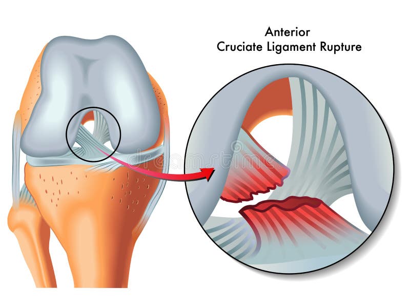
Free with trial Anterior cruciate ligament rupture. Medical Illustration of anterior cruciate ligament rupture. Femur vectors Anterior cruciate ligament rupture. Medical Illustration of anterior cruciate ligament rupture
Free with trial Osteoporosis examples vector illustration diagram with bone density. Medical scheme and educational information. Femur vectors Osteoporosis examples vector illustration diagram with bone density. Medical scheme and educational information
Free with trial Posterior cruciate ligament rupture. Medical Illustration of posterior cruciate ligament ruptur. Femur vectors Posterior cruciate ligament rupture. Medical Illustration of posterior cruciate ligament ruptur
Free with trial Front view of the right knee in flexion. Anatomy of the front of the right knee in flexion. Femur vectors Front view of the right knee in flexion. Anatomy of the front of the right knee in flexion
Free with trial Anatomy of the knee, illustration (for basic medical education, for clinics & Schools). Femur vectors Anatomy of the knee. Illustration (for basic medical education, for clinics & Schools
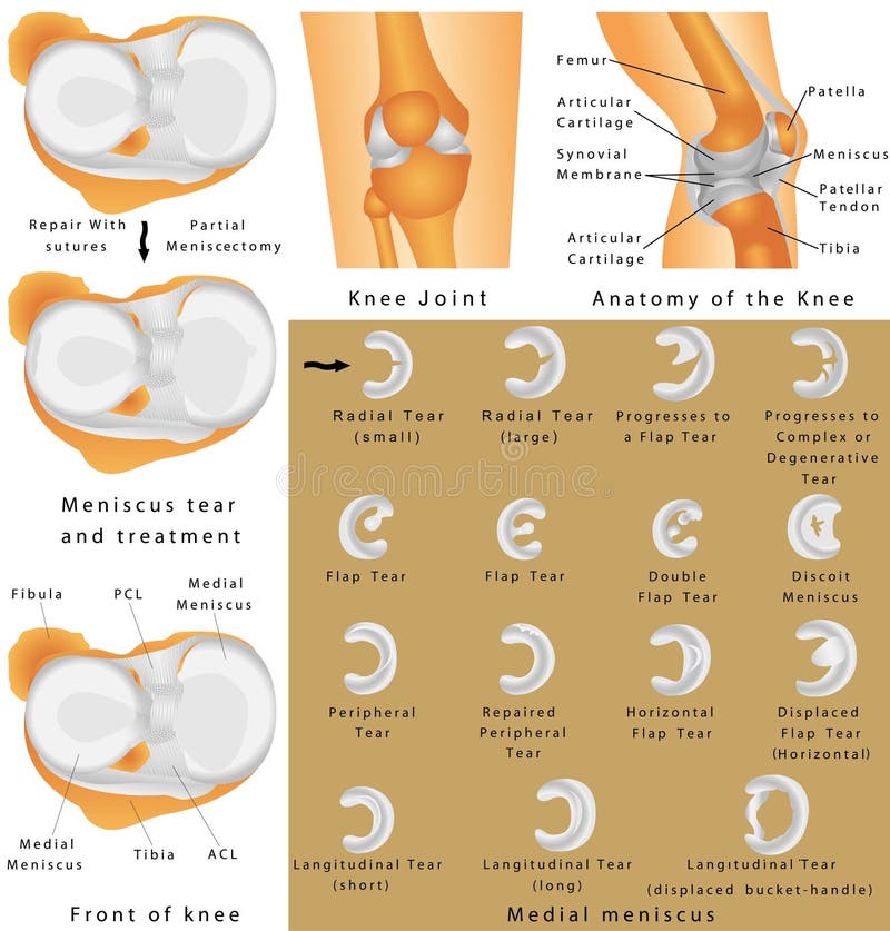
Free with trial Anatomy of the Knee. Human Knee Joint. Menisci of the knee. Medial meniscus. Lateral meniscus. Meniscus tear and surgery. Femur vectors Anatomy of the Knee. Human Knee Joint. Anatomy of the Knee. Menisci of the knee. Medial meniscus. Lateral meniscus. Meniscus tear and surgery
Free with trial Vector illustration of a human pelvis and hip with pain in the hip joint and trochanteric bursitis. Front view. For advertising and medical publications. EPS 10. Femur vectors Pain in the hip joint_trochanteric bursitis. Vector illustration of a human pelvis and hip with pain in the hip joint and trochanteric bursitis. Front view. For. Vector illustration of a human pelvis and hip with pain in the hip joint and trochanteric bursitis. Front view. For advertising and medical publications. EPS 10.
Free with trial Anatomy. Knee Joint Cross Section Showing the major parts which made the knee joint For Basic Medical Education Also for clinics. Femur vectors Anatomy. Structure knee joint vector. Anatomy. Knee Joint Cross Section Showing the major parts which made the knee joint For Basic Medical Education Also for. Anatomy. Knee Joint Cross Section Showing the major parts which made the knee joint For Basic Medical Education Also for clinics
Free with trial Fractures of Femur. Hemmed fracture with distorted alignment of the femur. Fixation of Femur Fracture with Placement of Intramedullary Rod. Types of Arthritis - Osteoarthritis, Rheumatoid Arthritis. Femur vectors Fractures of Femur. Hemmed fracture with distorted alignment of the femur. Fixation of Femur Fracture with Placement of Intramedullary Rod. Types of Arthritis
Free with trial Osteoporosis. Of femur (hip bone), eps8. Femur vectors Osteoporosis. Of femur (hip bone), eps8
Free with trial Gluteus maximus muscle as medical hip and leg medical anatomy outline diagram. Labeled educational human iliotibial tract, gluteal tuberosity of femur and groin skeletal structure vector illustration. Femur vectors Gluteus maximus muscle as medical hip and leg medical anatomy outline diagram. Labeled educational human iliotibial tract, gluteal tuberosity of femur and
Free with trial Shin splints vector illustration. Leg muscle sport trauma and bone pain labeled diagram. Isolated femur, patella, fibula, tibia and foot bones with shown injury location. Femur vectors Shin splints vector illustration. Leg muscle sport trauma and bone pain. Shin splints vector illustration. Leg muscle sport trauma and bone pain labeled diagram. Shin splints vector illustration. Leg muscle sport trauma and bone pain labeled diagram. Isolated femur, patella, fibula, tibia and foot bones with shown injury location.
Free with trial Psoas muscle as deep body muscular system for spine health outline diagram. Labeled educational scheme with ilium, femur and lesser trochanter bones vector illustration. Skeletal hip backbone anatomy. Femur vectors Psoas muscle as deep body muscular system for spine health outline diagram. Labeled educational scheme with ilium, femur and lesser trochanter bones vector
Free with trial Piriformis muscle with hip skeleton and muscular system outline diagram. Labeled educational scheme with medical human anatomy and trochanter of femur in lateral or anterior view vector illustration. Femur vectors Piriformis muscle with hip skeleton and muscular system outline diagram. Labeled educational scheme with medical human anatomy and trochanter of femur in
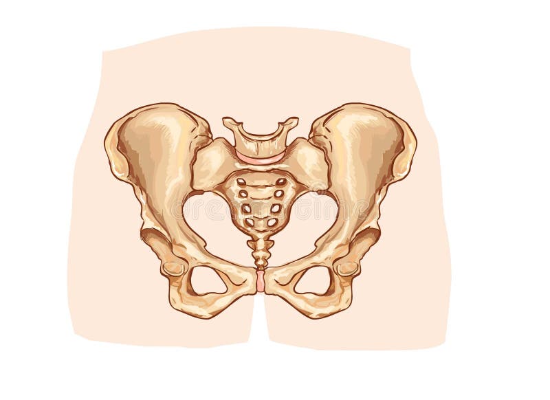
Free with trial Diagram of the pelvic girdle labeled. A vector illustration of Diagram of the pelvic girdle labeled. Femur vectors Diagram of the pelvic girdle labeled. A vector illustration of Diagram of the pelvic girdle labeled
Free with trial Hip joint. Hip fracture. Femoral Neck Fracture. Osteoarthritis of hip joint. Arthritic Hip. Osteoarthritis Hip Replacement on a white background. Femur vectors Hip joint. Hip fracture. Femoral Neck Fracture. Osteoarthritis of . Arthritic Hip. Osteoarthritis Hip Replacement on a white background
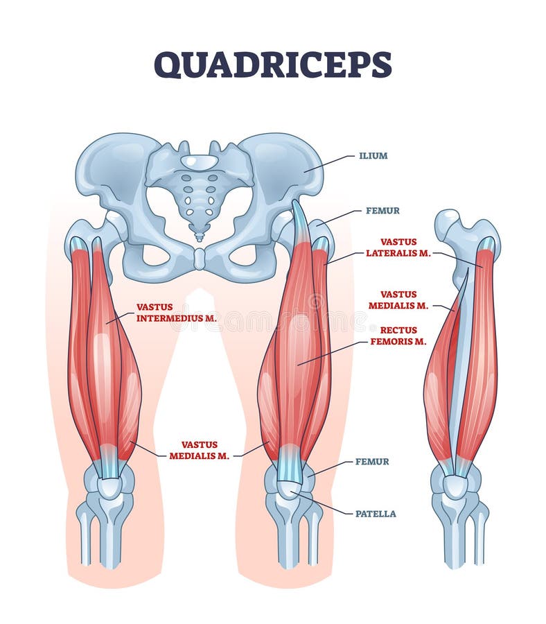
Free with trial Quadriceps muscle and quads leg muscular or bone anatomy outline diagram. Labeled educational medical scheme with vastus intermedius, medialis, lateralis or rectus femoris location vector illustration. Femur vectors Quadriceps muscle or quads leg muscular anatomical structure outline diagram. Quadriceps muscle and quads leg muscular or bone anatomy outline diagram. Labeled. Quadriceps muscle and quads leg muscular or bone anatomy outline diagram. Labeled educational medical scheme with vastus intermedius, medialis, lateralis or rectus femoris location vector illustration
Free with trial Osteoarthritis detailed illustration. From healthy joint to damaged joint, cartilage destruction. Femur vectors Osteoarthritis detailed illustration. From healthy joint to damaged joint, cartilage destruction
Free with trial Bursitis is the inflammation of bursae. Bursitis. inflammation of bursae synovial fluid. Prepatellar bursitis housemaid`s knee and Infrapatellar bursitis. Femur vectors Bursitis is the inflammation of bursae. Bursitis. inflammation of bursae synovial fluid. Prepatellar bursitis housemaid`s knee and Infrapatellar bursitis
Free with trial Osteoarthritis, unhealthy joint. Osteoarthritis, destruction of cartilage. Isolated on white. Femur vectors Osteoarthritis, unhealthy joint. Osteoarthritis, destruction of cartilage. Isolated on white
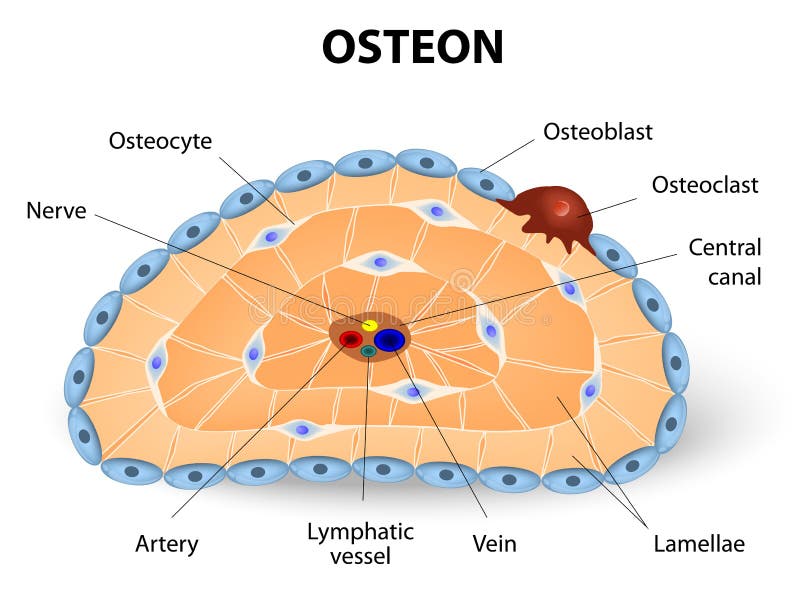
Free with trial Osteon development and structure. Osteoblast, osteocyte, and osteoclast. Femur vectors Osteon development and structure. Osteoblast, osteocyte, and osteoclast
Free with trial Burning knee, painful knee and normal knee joint, abstract design. Human legs on a blue checkered background. Femur vectors Burning knee. Painful knee and normal knee joint, abstract design. Human legs on a blue checkered background
Free with trial Osteoporosis, human bone anatomy. Medical illustration healthy bone and unhealthy bone. Femur vectors Osteoporosis. Human bone anatomy. Medical illustration healthy bone and unhealthy bone
Free with trial Meniscal Tear. A torn meniscus, a painful condition. Femur vectors Meniscal Tear. A torn meniscus, a painful condition
Free with trial Pulled Muscle. Muscle Strain degree and Treatment isolated on white background. Vector illustrations. Femur vectors Pulled Muscle. Muscle Strain degree and Treatment isolated on white background.Vector illustrations
Free with trial Synovial bursa. Synovial Joint. Human anatomy. vector. Femur vectors Synovial bursa. Synovial Joint. Human anatomy. vector
Free with trial Osteoarthritis. Medical Infographic set with icons and other elements. medical symbols for design. Femur vectors Osteoarthritis. Medical Infographic set with icons and other elements. medical symbols for design
Free with trial Bone pain symptoms. Woman with backache pain colored in red isolated on gray background illustration. Femur vectors Bone pain symptoms. Woman with backache pain colored in red isolated on gray background illustration
Free with trial Knee. Art illustration of leg with a highlight for the kneecap (patella. Femur vectors Knee. Art illustration of leg with a highlight for the kneecap (patella
Free with trial Osteoblast and osteoclast. The bone remodeling process. In a healthy body, osteoclasts and osteoblasts work together to maintain the balance between bone loss and bone formation. Femur vectors Osteoblast and osteoclast. The bone remodeling process. In a healthy body, osteoclasts and osteoblasts work together to maintain the balance between bone loss
Free with trial Knee joint. Human anatomy. knee joint on a white background. Femur vectors Knee joint. Human anatomy. knee joint on a white background
Free with trial Joint pain, joint anatomy beautiful abstract background. Femur vectors Joint pain. Joint anatomy beautiful abstract background
Free with trial Knee Joint Labeled Diagram. A knee joint with detailed labels. Femur vectors Knee Joint Labeled Diagram. A knee joint with detailed labels
Free with trial Groin strain trauma and pulled or torn muscle injury anatomy outline diagram. Labeled educational medical sport problem explanation with body hips and leg overstretching condition vector illustration. Femur vectors Groin strain trauma and pulled or torn muscle injury anatomy outline diagram. Labeled educational medical sport problem explanation with body hips and leg
Free with trial Bone Structure. Vector scheme. Long Bone Anatomy. Femur vectors Bone Structure. Vector scheme. Long Bone Anatomy. Vector scheme
Free with trial Osteoporosis - is a disease of bones that leads to an increased. Risk of fracture. Three bones: healthy, bone with Osteoporosis, and Severe osteoporosis. Femur vectors Osteoporosis - is a disease of bones that leads to an increased. Risk of fracture. Three bones: healthy, bone with Osteoporosis, and Severe osteoporosis
Free with trial Knee pain abstract background. Healthy joint and unhealthy painful joint with osteoarthritis. Femur vectors Knee pain abstract background. Healthy joint and unhealthy painful joint with osteoarthritis
 Sign in