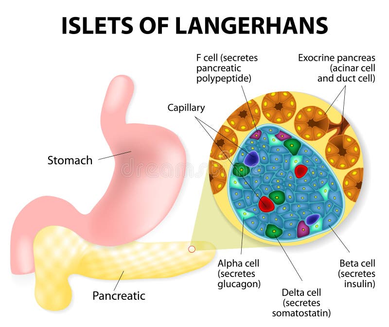
Free with trial The islets of Langerhans are responsible for the endocrine function of the pancreas. Each islet contains beta, alpha, and delta cells that are responsible for the secretion of a hormones. Glandular vectors Islets of Langerhans. The islets of Langerhans are responsible for the endocrine function of the pancreas. Each islet contains beta, alpha, and delta cells that. The islets of Langerhans are responsible for the endocrine function of the pancreas. Each islet contains beta, alpha, and delta cells that are responsible for the secretion of a hormones.
Free with trial Human pancreas, Lymphatics and stomach. Glandular vectors Human pancreas. Lymphatics and stomach
Free with trial Tonsillitis bacterial. Angina, pharyngitis and tonsillitis. Infection of tonsils caused by virus or bacteria. Recurring and persistent infection of tonsils. Glandular vectors Tonsillitis bacterial. Angina, pharyngitis and tonsillitis. Infection of tonsils caused by virus or bacteria. Recurring and persistent infection of tonsils
Free with trial Mononucleosis. Signs and symptoms of infectious mononucleosis. Human silhouette with internal organs. glandular fever, Pfeiffer's disease or Filatov's disease. Glandular vectors Infectious mononucleosis. Mononucleosis. Signs and symptoms of infectious mononucleosis. Human silhouette with internal organs. glandular fever, Pfeiffer's. Mononucleosis. Signs and symptoms of infectious mononucleosis. Human silhouette with internal organs. glandular fever, Pfeiffer's disease or Filatov's disease
Free with trial Pancreas. Beautiful colorful illustration white background. Glandular vectors Pancreas. Beautiful colorful illustration white background
Free with trial Synapse vector illustration. Labeled diagram with neuromuscular junction, glandular and other neirons example. Closeup with isolated axon, cleft and dendrite structure. Glandular vectors Synapse vector illustration. Labeled diagram with neuromuscular example. Synapse vector illustration. Labeled diagram with neuromuscular junction, glandular and. Synapse vector illustration. Labeled diagram with neuromuscular junction, glandular and other neirons example. Closeup with isolated axon, cleft and dendrite structure.
Free with trial Anatomy of cervical lymph nodes. Vector illustration of anatomy of cervical lymph nodes. Glandular vectors Anatomy of cervical lymph nodes. Vector illustration of anatomy of cervical lymph nodes
Free with trial Tonsillitis bacterial. A vector illustration of Tonsillitis bacterial. Glandular vectors Tonsillitis bacterial. A vector illustration of Tonsillitis bacterial
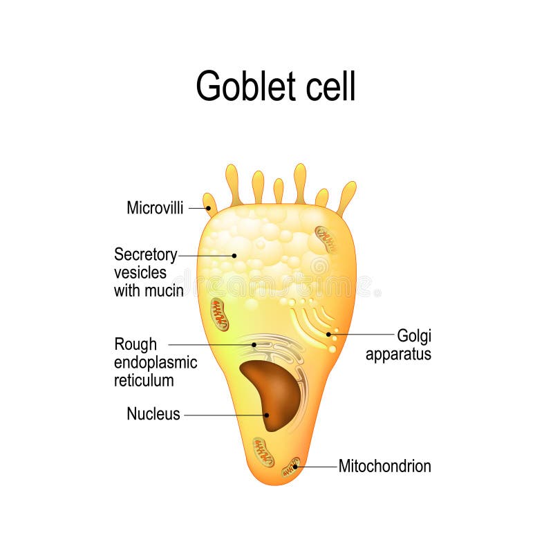
Free with trial Goblet cell. simple columnar epithelial cell for secrete mucus. They are found inside the trachea, bronchi, small and large intestine, and conjunctiva in the eyes. Structure cell nucleus and other organelles. Glandular vectors Goblet cell. Structure cell nucleus and other organelles. Goblet cell. simple columnar epithelial cell for secrete mucus. They are found inside the trachea. Goblet cell. simple columnar epithelial cell for secrete mucus. They are found inside the trachea, bronchi, small and large intestine, and conjunctiva in the eyes. Structure cell nucleus and other organelles.
Free with trial Sjogrens syndrome vector illustration. Dry body autoimmune disease. Labeled medical explanation with moisture producing glands. Symptoms, diagnosis and consequences. Glandular vectors Sjogrens syndrome vector illustration. Dry body autoimmune disease. Labeled medical explanation with moisture producing glands. Symptoms, diagnosis and
Free with trial Human anatomy icons set Circle Series - eps. 10. Glandular vectors Human anatomy icons set Circle Series. Eps.10
Free with trial Hepatitis a b c liver disease human organ virus medicine. Vector. Glandular vectors Hepatitis a b c liver disease human organ virus medicine. Vector
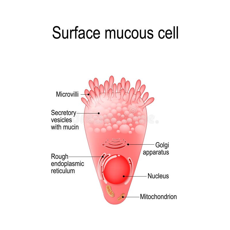
Free with trial Surface mucous cell is a foveolar mucus-producing cell that covering the inside of the stomach. Structure cell: golgi apparatus, secretory vesicle, mucin, nucleus, mitochondrion, microvilli. Glandular vectors Surface mucous cell. Is a foveolar mucus-producing cell that covering the inside of the stomach. Structure cell: golgi apparatus, secretory vesicle, mucin. Surface mucous cell is a foveolar mucus-producing cell that covering the inside of the stomach. Structure cell: golgi apparatus, secretory vesicle, mucin, nucleus, mitochondrion, microvilli
Free with trial Pancreas parts and surrounding organs, gallbladder, small intestine and spleen detailed illustration with description. Beautiful colorful design. Glandular vectors Pancreas parts and surrounding organs. Gallbladder, small intestine and spleen detailed illustration with description. Beautiful colorful design
Free with trial Human Organs Icons Set. Vector. Human Organs Icons Set on White Background. Vector illustration. Glandular vectors Human Organs Icons Set. Vector. Human Organs Icons Set on White Background. Vector illustration
Free with trial Vector illustration of the increase in lymph nodes in lymphoma. Glandular vectors Swollen lymph nodes in lymphoma. Oncology. Vector illustration of the increase in lymph nodes in lymphoma. Vector illustration of the increase in lymph nodes in lymphoma.
Free with trial Mononucleosis. Medical illustration of the symptoms of infectious mononucleosis. Glandular vectors Mononucleosis. Medical illustration of the symptoms of infectious mononucleosis
Free with trial Pancreatitis. Illustration, inflammation of pancreas on a white background. Glandular vectors Pancreatitis. Illustration, inflammation of pancreas on a white background
Free with trial Pancreas and surrounding organs, gallbladder, small intestine and spleen detailed illustration with description. Beautiful colorful design. Glandular vectors Pancreas and surrounding organs. Gallbladder, small intestine and spleen detailed illustration with description. Beautiful colorful design
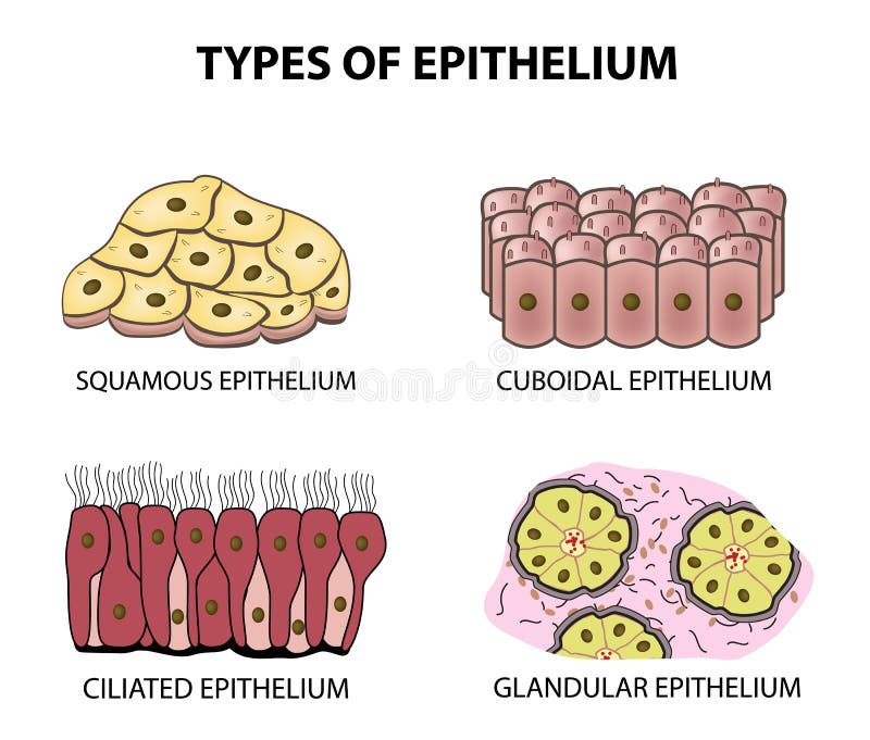
Free with trial Vector illustration of epithelial tissue. types of epithelium. Glandular vectors Epithelial tissue. types of epithelium. Vector illustration of epithelial tissue. types of epithelium. Vector illustration of epithelial tissue. types of epithelium.
Free with trial Infectious mononucleosis. mouth with pharyngitis. Epstein-Barr virus and reactive lymphocytes in blood. Glandular vectors Infectious mononucleosis. Mouth with pharyngitis. Epstein-Barr virus and reactive lymphocytes in blood
Free with trial Pancreas abstract design. Pancreas anatomy illustration on an abstract light grey scientific background with DNA chain and molecules. Glandular vectors Pancreas abstract design. Pancreas anatomy illustration on an abstract light grey scientific background with DNA chain and molecules
Free with trial Pancreatitis illustration, inflammation of pancreas on a white background. Human silhouette and internal organs. Glandular vectors Pancreatitis illustration. Inflammation of pancreas on a white background. Human silhouette and internal organs
Free with trial Melatonin and Circadian rhythm regulation. Brain with pineal gland and suprachiasmatic nucleus. sleep-wake cycle. Human anatomy. vector illustration. What does melatonin do. Glandular vectors Melatonin and Circadian rhythm regulation. Brain with pineal gland. And suprachiasmatic nucleus. sleep-wake cycle. Human anatomy. vector illustration. What does
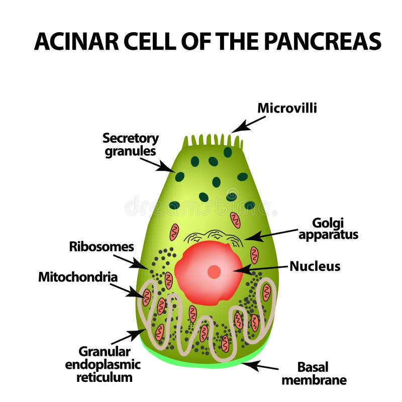
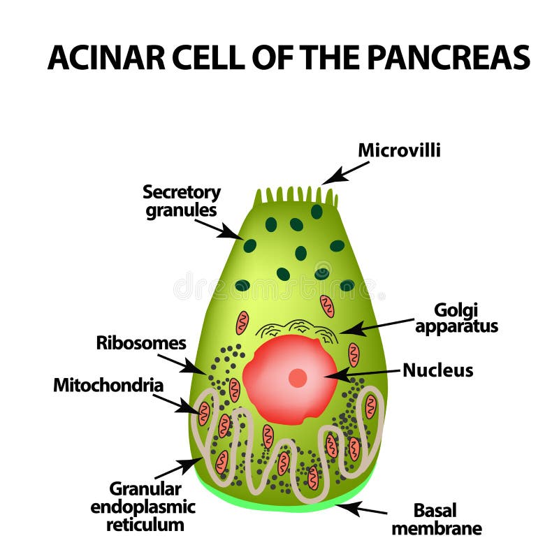
Free with trial Acinar cell of the pancreas. Acinus. Infographics. Vector illustration on isolated background. Glandular vectors Acinar cell of the pancreas. Acinus. Infographics. Vector illustration on isolated background.
Free with trial Types of epithelium. squamous, cubic, ciliated, glandular. Set Infographics Vector illustration on background. Glandular vectors Types of epithelium. squamous, cubic, ciliated, glandular. Set. Infographics. Vector illustration on background. Types of epithelium. squamous, cubic, ciliated. Types of epithelium. squamous, cubic, ciliated, glandular. Set Infographics Vector illustration on background
Free with trial Glandular carcinoma of the liver, vintage engraving. Glandular carcinoma of the liver, vintage engraved illustration. Glandular vectors Glandular carcinoma of the liver, vintage engraving. Glandular carcinoma of the liver, vintage engraved illustration
Free with trial Trichome and bristle types comparison and division groups outline diagram. Labeled educational biological categories with plant hair differences vector illustration. Tapering, glandular and stellate. Glandular vectors Trichome and bristle types comparison and division groups outline diagram. Labeled educational biological categories with plant hair differences vector
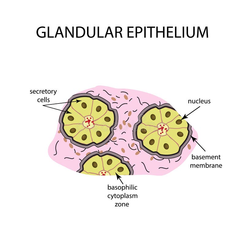
Free with trial The structure of the glandular epithelium. Infographics Vector illustration on background. Glandular vectors The structure of the glandular epithelium. Infographics. Vector illustration on background. The structure of the glandular epithelium. Infographics Vector. The structure of the glandular epithelium. Infographics Vector illustration on background
Free with trial Hormone feedback loop illustrating TRH TSH control of thyroid and negative feedback, key objects, hypothalamus, pituitary, thyroid, concept, endocrine regulation of metabolism. Outline diagram. Glandular vectors Hormone feedback loop illustrating TRH TSH control of thyroid and ... Hormone feedback loop illustrating TRH TSH control of thyroid and negative feedback, key. Hormone feedback loop illustrating TRH TSH control of thyroid and negative feedback, key objects, hypothalamus, pituitary, thyroid, concept, endocrine regulation of metabolism. Outline diagram
Free with trial Endocrine vs exocrine glands brief comparison shows hormone release to capillaries vs enzyme flow through ducts, key objects, duct, capillary, secretory cells. Outline diagram. Glandular vectors Endocrine vs exocrine glands brief comparison shows hormone release to ... Endocrine vs exocrine glands brief comparison shows hormone release to capillaries vs. Endocrine vs exocrine glands brief comparison shows hormone release to capillaries vs enzyme flow through ducts, key objects, duct, capillary, secretory cells. Outline diagram
Free with trial Histology vs Pathology shows colon tissue comparison, normal crypts with goblet cells versus adenocarcinoma tumor mass. Main objects, colon, crypts, tumor cells. Outline diagram. Glandular vectors Histology vs Pathology shows colon tissue comparison, normal crypts with ... Histology vs Pathology shows colon tissue comparison, normal crypts with goblet. Histology vs Pathology shows colon tissue comparison, normal crypts with goblet cells versus adenocarcinoma tumor mass. Main objects, colon, crypts, tumor cells. Outline diagram
Free with trial HPA axis regulation is shown with the brain, pituitary gland, and adrenal glands connected by arrows and labeled hormones. Outline diagram. Glandular vectors HPA axis regulation is shown with the brain, pituitary gland, ... HPA axis regulation is shown with the brain, pituitary gland, and adrenal glands connected by. HPA axis regulation is shown with the brain, pituitary gland, and adrenal glands connected by arrows and labeled hormones. Outline diagram
Free with trial Pancreas. Abstract medical illustration, background. Glandular vectors Pancreas. Abstract medical illustration, background
Free with trial Anatomical overview of the major salivary glands in the human head and their respective ducts, which secrete saliva into the oral cavity. Glandular vectors Salivary glands in the human head. Anatomical overview of the major salivary glands in the human head and their respective ducts, which secrete saliva into the. Anatomical overview of the major salivary glands in the human head and their respective ducts, which secrete saliva into the oral cavity
Free with trial Exocrine gland. Method of secretion. Apocrine gland cells secrete by budding off a portion of their cytoplasm. Merocrine gland cells excrete their substances by exocytosis. Vector illustration. Medical poster. Schematic diagram. Glandular vectors Exocrine gland. Apocrine gland cells and Merocrine cells. Exocrine gland. Method of secretion. Apocrine gland cells secrete by budding off a portion of their. Exocrine gland. Method of secretion. Apocrine gland cells secrete by budding off a portion of their cytoplasm. Merocrine gland cells excrete their substances by exocytosis. Vector illustration. Medical poster. Schematic diagram
Free with trial Paneth cell it is the epithelium cells of the small intestine. their granules consist of anti-microbial compounds for important in immunity. Structure of the Paneth cell nucleus, mitochondria and other organelles. Glandular vectors Paneth cell. It is the epithelium cells of the small intestine. their granules consist of anti-microbial compounds for important in immunity. Structure of the. Paneth cell it is the epithelium cells of the small intestine. their granules consist of anti-microbial compounds for important in immunity. Structure of the Paneth cell nucleus, mitochondria and other organelles.
Free with trial Adrenal insufficiency diagram shows hormone pathways, highlighting brain, adrenal glands, and hormone flow. Outline diagram. Glandular vectors Adrenal insufficiency diagram shows hormone pathways, highlighting brain, adrenal glands, ... Adrenal insufficiency diagram shows hormone pathways, highlighting. Adrenal insufficiency diagram shows hormone pathways, highlighting brain, adrenal glands, and hormone flow. Outline diagram
Free with trial Algorithm programming concept. Flat design illustration concepts for brainstorming, coding, programming, and planning. Glandular vectors Algorithm programming concept. Flat design illustration concepts for brainstorming, coding, programming, and planning
Free with trial Pancreas parts. Detailed colorful illustration on a white. Glandular vectors Pancreas parts. Detailed colorful illustration on a white
Free with trial Microscopic view of the parotid gland's secretory and ductal system, culminating in saliva delivery to the oral cavity. Glandular vectors Culminating in saliva delivery to the oral cavity. Microscopic view of the parotid gland's secretory and ductal system, culminating in saliva delivery to the. Microscopic view of the parotid gland's secretory and ductal system, culminating in saliva delivery to the oral cavity
Free with trial Pancreas and surrounding organs, gallbladder, small intestine and spleen detailed illustration on a white background. Beautiful colorful design. Glandular vectors Pancreas on white. Pancreas and surrounding organs, gallbladder, small intestine and spleen detailed illustration on a white background. Beautiful colorful. Pancreas and surrounding organs, gallbladder, small intestine and spleen detailed illustration on a white background. Beautiful colorful design.
Free with trial Infectious mononucleosis. humans silhouette with highlight liver, and viruses that cause of this disease. Epstein-Barr virus. Glandular vectors Infectious mononucleosis. Human herpevirus. Infectious mononucleosis. humans silhouette with highlight liver, and viruses that cause of this disease. Epstein. Infectious mononucleosis. humans silhouette with highlight liver, and viruses that cause of this disease. Epstein-Barr virus.
Free with trial Epstein–Barr virus EBV. human herpesvirus 4 HHV. diagram of the structure of human herpes virus. the cause of infectious mononucleosis and cancer. Glandular vectors Diagram of the structure of EBV. Epstein–Barr virus EBV. human herpesvirus 4 HHV. diagram of the structure of human herpes virus. the cause of infectious. Epstein–Barr virus EBV. human herpesvirus 4 HHV. diagram of the structure of human herpes virus. the cause of infectious mononucleosis and cancer.
Free with trial Pancreas scientific background. Pancreas anatomy illustration on an abstract scientific technology background. Glandular vectors Pancreas scientific background. Pancreas anatomy illustration on an abstract scientific technology background
Free with trial Pancreas anatomy and treatment concept, human silhouette with internal organs, gallbladder, intestine, stomach and spleen on a beautiful abstract background with normal cardiogram at the bottom. Glandular vectors Pancreas. Anatomy and treatment concept, human silhouette with internal organs, gallbladder, intestine, stomach and spleen on a beautiful abstract background. Pancreas anatomy and treatment concept, human silhouette with internal organs, gallbladder, intestine, stomach and spleen on a beautiful abstract background with normal cardiogram at the bottom.
Free with trial Pancreas anatomy illustration. On an abstract scientific blue background. Glandular vectors Pancreas anatomy illustration. On an abstract scientific blue background
Free with trial Cross-Sectional Anatomy of Tongue Mucosa with Papillae and Nerves. Educational Medical Diagram of Tongue Tissue Layers and Papillae Types. Glandular vectors Cross-Sectional Anatomy of Tongue Mucosa with Papillae and Nerves. Human Tongue Structure Showing Filiform, Fungiform, and. Cross-Sectional Anatomy of Tongue. Cross-Sectional Anatomy of Tongue Mucosa with Papillae and Nerves. Educational Medical Diagram of Tongue Tissue Layers and Papillae Types
Free with trial Human brain with Part of limbic system, and Cerebral Cortex, Suprachiasmatic Nucleus, Optic Chiasm, Hypothalamus, and Pineal Gland. regulation of circadian rhythms and the sleep-wake cycle in the brain. Human anatomy. vector illustration. Glandular vectors Brain and regulation of circadian rhythms. Human brain with Part of limbic system, and Cerebral Cortex, Suprachiasmatic Nucleus, Optic Chiasm, Hypothalamus, and. Human brain with Part of limbic system, and Cerebral Cortex, Suprachiasmatic Nucleus, Optic Chiasm, Hypothalamus, and Pineal Gland. regulation of circadian rhythms and the sleep-wake cycle in the brain. Human anatomy. vector illustration
Free with trial Acinar cell of the pancreas. Acinus. Infographics. Vector illustration on isolated background. Glandular vectors Acinar cell of the pancreas. Acinus. Infographics. Vector illustration on isolated background.
Free with trial Vintage flowers. Black and white illustration in the style of engravings. Glandular vectors Botanical plant illustration.handrawn. Vintage flowers. Black and white illustration in the style of engravings. Vintage flowers. Black and white illustration in the style of engravings.
Free with trial EBV Epstein–Barr virus written in a notebook paper- vector illustration. Glandular vectors EBV Epstein–Barr virus written in a notebook paper. Vector illustration
Free with trial Neuron line icon. Healthy nerve cell symbol. And sign illustration design. Isolated on white background. Glandular vectors Neuron line icon. Healthy nerve cell symbol. And sign illustration design. Isolated on white background
Free with trial Cirrhosis illustration with liver and medicine and alcohol as the real cause vector illustration. Glandular vectors Cirrhosis illustration with liver medicine alcohol as the real cause. Cirrhosis illustration with liver and medicine and alcohol as the real cause vector. Cirrhosis illustration with liver and medicine and alcohol as the real cause vector illustration
Free with trial Mammary gland. Vector illustration showing cross section of female breast with the names of the constituent parts isolated on. White background. Glandular vectors Mammary gland. Vector illustration showing cross section of female breast with the names of the constituent parts isolated on. White background
Free with trial Mikulicz syndrome. Enlargement of the lacrimal and salivary glands. Vector illustration of Mikulicz syndrome. Glandular vectors Mikulicz syndrome. Enlargement of the lacrimal and salivary glands. Vector illustration of Mikulicz syndrome. Enlargement of the lacrimal and salivary glands
Free with trial Tall and slender cells with oval-shaped nuclei. Lines most organs of the digestive tract like stomach, intestines. Medical illustration. Vector. Glandular vectors Simple columnar epithelium. Epithelial tissue types. Tall and slender cells with oval-shaped nuclei. Lines most organs of the digestive tract like stomach. Tall and slender cells with oval-shaped nuclei. Lines most organs of the digestive tract like stomach, intestines. Medical illustration. Vector.
Free with trial Tonsillectomy. Acute pharyngitis treatment, surgical removal of the tonsils,. Two oval-shaped pads of tissue at the back of the throat. Flat vector illustration. Glandular vectors Tonsillectomy. Acute pharyngitis treatment, surgical removal of the tonsils,. Two oval-shaped pads of tissue at the back of the throat. Flat vector illustration
Free with trial Mammary gland. Vector illustration showing cross section of female breast with the names of the constituent parts isolated on. White background. Glandular vectors Mammary gland. Vector illustration showing cross section of female breast with the names of the constituent parts isolated on. White background
Free with trial Illustration of a glandular trichome from Artemisia annua, the plant known for producing artemisinin. A colorful drawing on a white background. Glandular vectors Glandular Trichome. Illustration of a glandular trichome from Artemisia annua, the plant known for producing artemisinin. A colorful drawing on a white. Illustration of a glandular trichome from Artemisia annua, the plant known for producing artemisinin. A colorful drawing on a white background
Free with trial Illustration of a glandular trichome from Artemisia annua, the plant known for producing artemisinin. The diagram shows the basal cell, subapical (stalk) cell, and secretory apical cell. Glandular vectors Glandular Trichome of Artemisia annua â€\' Herbal Microscopic Anatomy. Illustration of a glandular trichome from Artemisia annua, the plant known for. Illustration of a glandular trichome from Artemisia annua, the plant known for producing artemisinin. The diagram shows the basal cell, subapical (stalk) cell, and secretory apical cell
Free with trial The image shows a detailed diagram of a glandular secretory trichome from the Artemisia annua plant, highlighting its various cell structures. Glandular vectors Glandular Secretory Trichome Of Artemisia Annua Plant. The image shows a detailed diagram of a glandular secretory trichome from the Artemisia annua plant. The image shows a detailed diagram of a glandular secretory trichome from the Artemisia annua plant, highlighting its various cell structures
Free with trial This illustration shows the detailed structure of a glandular secretory trichome from Artemisia annua, highlighting its various cellular components. Glandular vectors Glandular Secretory Trichome Structure Of Artemisia Annua. This illustration shows the detailed structure of a glandular secretory trichome from Artemisia annua. This illustration shows the detailed structure of a glandular secretory trichome from Artemisia annua, highlighting its various cellular components
Free with trial A detailed diagram of a glandular trichome, a type of plant cell structure, illustrating its various components such as secretory apical, subapical cell, stalk cell, and basal cell, which are essential for plant biology and education. Glandular vectors Glandular Trichome Diagram Showing Plant Cell Structure. A detailed diagram of a glandular trichome, a type of plant cell structure, illustrating its various. A detailed diagram of a glandular trichome, a type of plant cell structure, illustrating its various components such as secretory apical, subapical cell, stalk cell, and basal cell, which are essential for plant biology and education
Free with trial Different types of Trichome and bristle types plant hair. Tapering, glandular, stellate, unicellular uniseriate multiseriate pilate capitate malphigian sessile dendrite, peltate, barbed,plumose. Glandular vectors Different types of Trichome and bristle types plant hair. Tapering, glandular, stellate, unicellular, uniseriate, multiseriate,. Different types of Trichome and. Different types of Trichome and bristle types plant hair. Tapering, glandular, stellate, unicellular uniseriate multiseriate pilate capitate malphigian sessile dendrite, peltate, barbed,plumose
Free with trial A detailed diagram of a glandular trichome, a type of plant cell structure, showcasing its various components such as secretory apical, subapical cell, stalk cell, and basal cell, which are essential for plant biology and education. Glandular vectors Glandular Trichome Diagram Showing Plant Cell Structure. A detailed diagram of a glandular trichome, a type of plant cell structure, showcasing its various. A detailed diagram of a glandular trichome, a type of plant cell structure, showcasing its various components such as secretory apical, subapical cell, stalk cell, and basal cell, which are essential for plant biology and education
Free with trial Different types of exocrine gland type. Glandular epithelium. Epithelial tissue types. Labeled straight, branched, tubular, acinar, compound tubuloacinar. Produce and release different secretory. Glandular vectors Different types of exocrine gland type. Glandular epithelium. Epithelial tissue types. Labeled straight, branched, tubular, acinar. Compound tubuloacinar. Different types of exocrine gland type. Glandular epithelium. Epithelial tissue types. Labeled straight, branched, tubular, acinar, compound tubuloacinar. Produce and release different secretory
Free with trial Educational endocrinology detailing the microscopic mechanism of hormone secretion by an endocrine gland in full color. The labeled medical diagram illustrates ductless glandular architecture, clearly highlighting the secretory endocrine cells, surrounding blood capillaries, and direct release of hormones into the bloodstream for systemic circulation. High-quality infographic graphic ideal for endocrinology textbooks, physiology study guides, biology curriculum charts, medical school lectures, clinic posters, and patient healthcare education presentations. Glandular vectors Endocrine Gland Anatomy Hormone Secretion Bloodstream Capillaries Vector Illustration. Educational endocrinology detailing the microscopic mechanism of hormone. Educational endocrinology detailing the microscopic mechanism of hormone secretion by an endocrine gland in full color. The labeled medical diagram illustrates ductless glandular architecture, clearly highlighting the secretory endocrine cells, surrounding blood capillaries, and direct release of hormones into the bloodstream for systemic circulation. High-quality infographic graphic ideal for endocrinology textbooks, physiology study guides, biology curriculum charts, medical school lectures, clinic posters, and patient healthcare education presentations.
Free with trial This high-quality 3D medical visualization depicts the human pancreas in vivid anatomical detail. The organ is shown in its natural orientation, characterized by its lobulated, glandular structure and yellowish hue. Intricate networks of red arteries are clearly visible, demonstrating the complex vascular supply to the pancreas and duodenum. Glandular vectors A detailed 3D medical showing the human pancreas with surrounding connected blood vessels. This high-quality 3D medical visualization depicts the human pancreas. This high-quality 3D medical visualization depicts the human pancreas in vivid anatomical detail. The organ is shown in its natural orientation, characterized by its lobulated, glandular structure and yellowish hue. Intricate networks of red arteries are clearly visible, demonstrating the complex vascular supply to the pancreas and duodenum
Free with trial This high-resolution biological asset features an American Bullfrog (Lithobates catesbeianus) in a robust lateral-oblique profile. The illustration meticulously renders the species' characteristic olive-green dorsal skin, interspersed with dark brown maculations and a distinctively pale, creamy throat. Meticulous attention is paid to the large, prominent golden-rimmed eyes and the massive, circular tympanum (external ear) located just behind the eye, which is larger than the eye itself in this mature specimen. The asset highlights the powerful, muscular hind limbs adapted for explosive jumping and the moist, glandular texture of the integument. This professional-grade cutout is an ideal resource for herpetological studies, North American aquatic ecology education, and high-end digital amphibian illustration. Glandular vectors American Bullfrog Lateral Perspective, High-Fidelity Lithobates catesbeianus Asset, Transparent Background. This high-resolution biological asset features an. This high-resolution biological asset features an American Bullfrog (Lithobates catesbeianus) in a robust lateral-oblique profile. The illustration meticulously renders the species' characteristic olive-green dorsal skin, interspersed with dark brown maculations and a distinctively pale, creamy throat. Meticulous attention is paid to the large, prominent golden-rimmed eyes and the massive, circular tympanum (external ear) located just behind the eye, which is larger than the eye itself in this mature specimen. The asset highlights the powerful, muscular hind limbs adapted for explosive jumping and the moist, glandular texture of the integument. This professional-grade cutout is an ideal resource for herpetological studies, North American aquatic ecology education, and high-end digital amphibian illustration.
Free with trial Adrenal Gland Stress Response on Intestine Movement. This diagram illustrates how stress signals stimulate the adrenal glands to release glucocorticoids, which inhibit the Enteric Nervous System ENS. Glandular vectors Adrenal Gland Stress Response on Intestine Movement. This diagram illustrates how stress signals stimulate the adrenal glands to release glucocorticoids, which
Free with trial The Parotid Glands and Saliva Production. This image illustrates the location and function of the parotid gland, one of the major salivary glands in the human body. Glandular vectors The Parotid Glands and Saliva Production. This image illustrates the location and function of the parotid gland, one of the major salivary glands in the human
Free with trial Adrenal gland function is shown with hormone secretion, adrenal glands, and brain in a clear outline style. Outline style collection. Glandular vectors Adrenal gland function is shown with hormone secretion, adrenal glands, ... Adrenal gland function is shown with hormone secretion, adrenal glands, and brain in. Adrenal gland function is shown with hormone secretion, adrenal glands, and brain in a clear outline style. Outline style collection
Free with trial Cow Mammary Gland Anatomy and Milk Duct Structure. Cross-Section of Bovine Udder with Alveoli, Milk Sinus and Blood Vessels. Veterinary Diagram. Glandular vectors Cow Mammary Gland Anatomy and Milk Duct Structure. Cross-Section of Bovine Udder with Alveoli, Milk Sinus and Blood Vessels. Veterinary Diagram
Free with trial Detailed Anatomy of the Cross-Sectional Cow Mammary Gland with Alveoli and Milk Ducts. Scientific Diagram of Bovine Udder. Structure and Milk Production System. Glandular vectors Detailed Anatomy of the Cross-Sectional Cow Mammary Gland with Alveoli and Milk Ducts. Scientific Diagram of Bovine Udder. Structure and Milk Production System
Free with trial Detailed anatomical illustration of human skin showing all main layers epidermis, dermis, and hypodermis. Visible structures include hair shafts, hair follicles, sebaceous glands, blood vessels, nerves, and connective tissue. Glandular vectors Anatomical illustration of human skin showing all main layers epidermis, dermis, and hypodermis. Detailed anatomical illustration of human skin showing all main. Detailed anatomical illustration of human skin showing all main layers epidermis, dermis, and hypodermis. Visible structures include hair shafts, hair follicles, sebaceous glands, blood vessels, nerves, and connective tissue.
 Sign in