Free with trial Foot deformation - Valgus and varus defect. Normal human foot and the foot with pronation or flatfoot, with hindfoot deformity. Hindfoot vectors Foot deformation. Valgus and varus defect. Normal human foot and the foot with pronation or flatfoot, with hindfoot deformity
Free with trial Too many toes. Vector illustration of the normal human foot and the foot with pronation or flatfoot, with hindfoot deformity. Too many toes sign. Hindfoot vectors Too many toes. Vector illustration of the normal human foot and the foot with pronation or flatfoot, with hindfoot deformity. Too many toes sign
Free with trial In the dorsal (posterior) view of the bones of the feet, the tarsal bones are visible, including the talus, calcaneus, navicular, cuboid, and the three cuneiform bones. The metatarsal bones, numbering from one to five, can also be observed, extending towards the toes. Additionally, the phalanges, consisting of proximal, middle, and distal phalanges, complete the skeletal structure of the feet in the dorsal view. Hindfoot vectors Bones of feet, dorsal (posterior) view. In the dorsal (posterior) view of the bones of the feet, the tarsal bones are visible, including the talus, calcaneus. In the dorsal (posterior) view of the bones of the feet, the tarsal bones are visible, including the talus, calcaneus, navicular, cuboid, and the three cuneiform bones. The metatarsal bones, numbering from one to five, can also be observed, extending towards the toes. Additionally, the phalanges, consisting of proximal, middle, and distal phalanges, complete the skeletal structure of the feet in the dorsal view.
Free with trial Vector illustration of a human leg with denominations of the bones of the foot. Anatomy of lateral or side view of the foot. For advertising or medical publications. EPS 10. Hindfoot vectors Anatomy_bones of the human foot lateral view. Vector illustration of a human leg with denominations of the bones of the foot. Anatomy of lateral or side view of. Vector illustration of a human leg with denominations of the bones of the foot. Anatomy of lateral or side view of the foot. For advertising or medical publications. EPS 10.
Free with trial Vector illustration of a human leg with denominations of the bones of the foot. Anatomy of medial or side view of the foot. For advertising or medical publications. EPS 10. Hindfoot vectors Anatomy_bones of the human foot medial view. Vector illustration of a human leg with denominations of the bones of the foot. Anatomy of medial or side view of. Vector illustration of a human leg with denominations of the bones of the foot. Anatomy of medial or side view of the foot. For advertising or medical publications. EPS 10.
Free with trial Vector illustration of a human leg with denominations of the bones of the foot. Anatomy of dorsal and plantar views of the foot. For advertising or medical publications. EPS 10. Hindfoot vectors Anatomy_bones of the human foot dorsal and plantar view. Vector illustration of a human leg with denominations of the bones of the foot. Anatomy of dorsal and. Vector illustration of a human leg with denominations of the bones of the foot. Anatomy of dorsal and plantar views of the foot. For advertising or medical publications. EPS 10.
Free with trial Shoes company logo design vector illustration template. Hindfoot vectors Shoes company logo design vector illustration template. Shoes company icon logo design vector illustration template
Free with trial Shoes company logo design vector illustration template. Hindfoot vectors Shoes company logo design vector illustration template. Shoes company icon logo design vector illustration template
Free with trial Vector illustration. Sinus tarsi syndrome of the human foot, area of pain. Section of the ankle and subtalar joint bones. For medical publications. Hindfoot vectors Sinus tarsi syndrome of the human foot. Vector illustration. Sinus tarsi syndrome of the human foot, area of pain. Section of the ankle and subtalar joint bones. Vector illustration. Sinus tarsi syndrome of the human foot, area of pain. Section of the ankle and subtalar joint bones. For medical publications
Free with trial Vector illustration. Sinus tarsi syndrome of the human foot. Orthotic Manipulation of the Rearfoot Alignment for pain reduction, insole for pronated foot or flatfoot. Section of the ankle and subtalar joint bones. For medical publications. Hindfoot vectors Sinus tarsi syndrome pain reduction. Orthotic Manipulation of the Rearfoot Alignment. Vector illustration. Sinus tarsi syndrome of the human foot. Orthotic. Vector illustration. Sinus tarsi syndrome of the human foot. Orthotic Manipulation of the Rearfoot Alignment for pain reduction, insole for pronated foot or flatfoot. Section of the ankle and subtalar joint bones. For medical publications
Free with trial Illustration of biology and physiology, Foot shapes, Human feet are highly individual, Arches are usually characterized as either low or flat, medium or high. Hindfoot vectors Illustration of biology and physiology, Foot shapes. Human feet are highly individual, Arches are usually characterized as either low or flat, medium or high
Free with trial Illustration of biology and physiology, Foot shapes, Human feet are highly individual, Arches are usually characterized as either low or flat, medium or high. Hindfoot vectors Illustration of biology and physiology, Foot shapes, Human feet are highly individual. Arches are usually characterized as either low or flat, medium or high
Free with trial Illustration of biology and medical, Foot arches, metatarsal bones, the ligaments and tendons in the foot, the foot to support the weight of the body. Hindfoot vectors Illustration of biology and medical, Foot arches, metatarsal bones. The ligaments and tendons in the foot, the foot to support the weight of the body
Free with trial The forefoot contains the phalanges and metatarsals. The midfoot consists of the five tarsal bones, three cuneiforms, the navicular, and the cuboid. The hindfoot is composed of two tarsal bones, the calcaneus and the talus. Hindfoot vectors Diagram or infographic of the bones that make up a foot from an aerial view with names. The forefoot contains the phalanges and metatarsals. The midfoot consists. The forefoot contains the phalanges and metatarsals. The midfoot consists of the five tarsal bones, three cuneiforms, the navicular, and the cuboid. The hindfoot is composed of two tarsal bones, the calcaneus and the talus.
Free with trial The forefoot contains the phalanges and metatarsals. The midfoot consists of the five tarsal bones, three cuneiforms, the navicular, and the cuboid. The hindfoot is composed of two tarsal bones, the calcaneus and the talus. Hindfoot vectors Diagram or infographic of the bones that make up a foot from an aerial view. The forefoot contains the phalanges and metatarsals. The midfoot consists of the. The forefoot contains the phalanges and metatarsals. The midfoot consists of the five tarsal bones, three cuneiforms, the navicular, and the cuboid. The hindfoot is composed of two tarsal bones, the calcaneus and the talus.
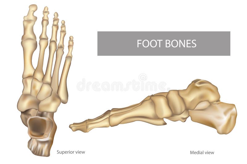
Free with trial Anatomy Bones of the Feet. OrthDiagram. Orthotics for foot Superior view and Medial view. Diagram. Hindfoot vectors Anatomy Bones of the Feet. OrthDiagram. Anatomy Bones of the Feet. Orthotics for foot Superior view and Medial view. Diagram
Free with trial The skeleton of the foot is low and a high heel. Illustration of the skeleton of the foot on a low and a high heel. Hindfoot vectors The skeleton of the foot is low and a high heel. Illustration of the skeleton of the foot on a low and a high heel
Free with trial Bones of the human foot. Illustration of the bones of the human. Hindfoot vectors Bones of the human foot. Illustration of the bones of the human
Free with trial The human foot's skeletal structure is composed of 26 bones, including the tarsal, metatarsal, and phalangeal bones. These bones form the arches of the foot, providing support, shock absorption, and flexibility for walking, running, and maintaining balance. Hindfoot vectors Bones of the right foot, dorsal view. The human foot's skeletal structure is composed of 26 bones, including the tarsal, metatarsal, and phalangeal bones. These. The human foot's skeletal structure is composed of 26 bones, including the tarsal, metatarsal, and phalangeal bones. These bones form the arches of the foot, providing support, shock absorption, and flexibility for walking, running, and maintaining balance.
Free with trial Shoes company logo design vector illustration template. Hindfoot vectors Shoes company logo design vector illustration template. Shoes company icon logo design vector illustration template
Free with trial Shoes company logo design vector illustration template. Hindfoot vectors Shoes company logo design vector illustration template. Shoes company icon logo design vector illustration template
Free with trial Shoes company logo design vector illustration template. Hindfoot vectors Shoes company logo design vector illustration template. Shoes company icon logo design vector illustration template
Free with trial Shoes company logo design vector illustration template. Hindfoot vectors Shoes company logo design vector illustration template. Shoes company icon logo design vector illustration template
Free with trial Shoes company logo design vector illustration template. Hindfoot vectors Shoes company logo design vector illustration template. Shoes company icon logo design vector illustration template
Free with trial Shoes company logo design vector illustration template. Hindfoot vectors Shoes company logo design vector illustration template. Shoes company icon logo design vector illustration template
Free with trial Shoes company logo design vector illustration template. Hindfoot vectors Shoes company logo design vector illustration template. Shoes company icon logo design vector illustration template
Free with trial Shoes company logo design vector illustration template. Hindfoot vectors Shoes company logo design vector illustration template. Shoes company icon logo design vector illustration template
 Sign in