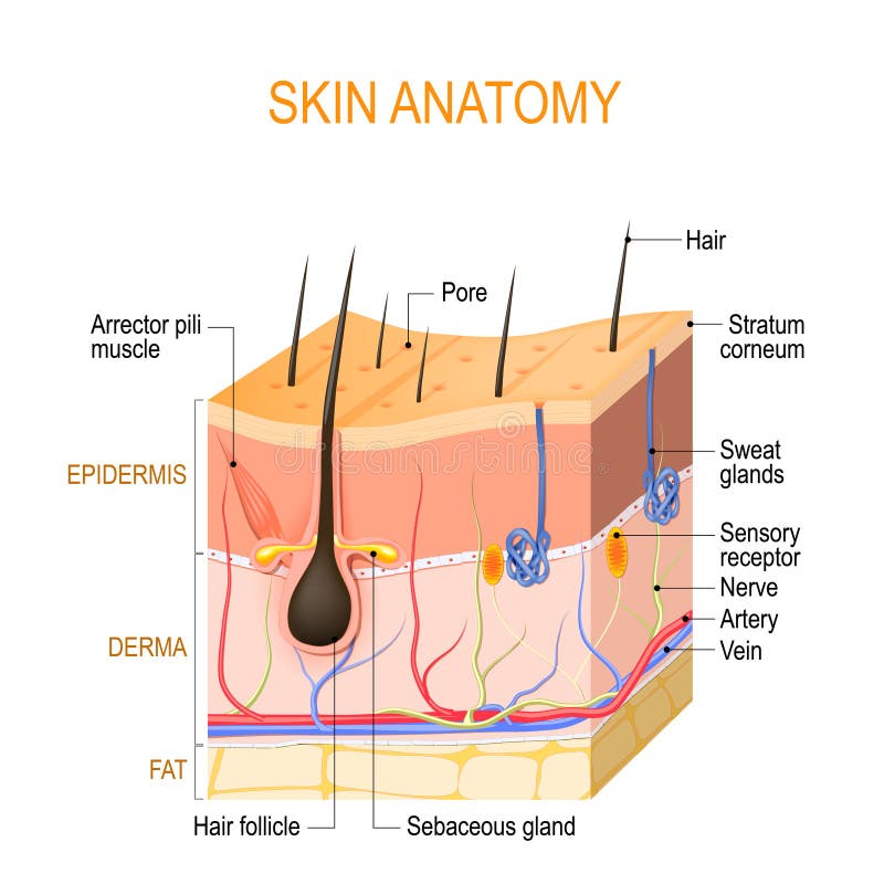
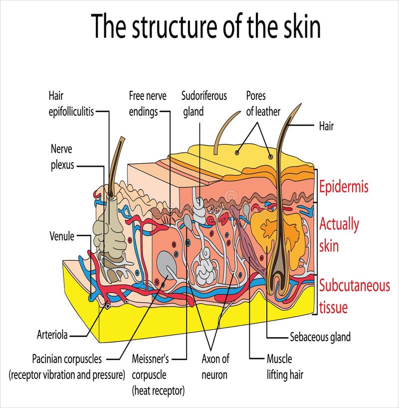
Free with trial Skin anatomy. Layers: epidermis with hair follicle, sweat and sebaceous glands, derma and fat hypodermis. Vector diagram for educational, medical, biological, and scientific use. Integument vectors Skin anatomy. Layers: epidermis with hair follicle, sweat and sebaceous glands, derma and fat hypodermis. Vector diagram for educational, medical, biological
Free with trial The Layer of Human Skin in vector style and components information. Illustration about medical and health. Integument vectors Components of human Skin layer in vector style. The Layer of Human Skin in vector style and components information. Illustration about medical and health. The Layer of Human Skin in vector style and components information. Illustration about medical and health.
Free with trial Human skin texture. Seamless pattern. Integument vectors Human skin. Texture. Seamless pattern
Free with trial Anatomy of Skin layer, fat cell and muscle layer in vector style. Illustration about medical and health. Integument vectors Anatomy of Skin layer, fat cell and muscle layer in vector style. Illustration about medical and health
Free with trial Parts of flower with english titles. Cross section of typical angiosperm flower at time of double fertilization. Integument vectors Parts of flower with titles. Cross section of typical angiosperm flower at time of double fertilization. Parts of flower with english titles. Cross section of. Parts of flower with english titles. Cross section of typical angiosperm flower at time of double fertilization
Free with trial Caveman. Vector image of an cartoon caveman and the fire. Integument vectors Caveman. Vector image of an cartoon caveman and the fire
Free with trial Gymnosperm vs angiosperm vector illustration. Labeled educational scheme with plants fertilization style and differences. Pine versus flower diagram and inner inside structure explanation graphic. Integument vectors Gymnosperm vs angiosperm vector illustration. Labeled educational scheme. Gymnosperm vs angiosperm vector illustration. Labeled educational scheme with plants. Gymnosperm vs angiosperm vector illustration. Labeled educational scheme with plants fertilization style and differences. Pine versus flower diagram and inner inside structure explanation graphic.
Free with trial Structure of green female seed cone and megasporangium of pine with titles. Integument vectors Structure of green female seed cone and megasporangium of pine with titles. Structure of green female seed cone and megasporangium of pine with english titles
Free with trial Structure of ovule of angiosperm plants with megagametophyte. Isolated on white background. Integument vectors Structure of ovule of angiosperm plants with megagametophyte. Isolated on white background
Free with trial Stages of pollination and fertilization of pine. Sexual process of pine from formation of female gametophyte, to fertilization. And formation of embryo. Integument vectors Stages of pollination and fertilization of pine. Sexual process of pine from formation of female gametophyte, to fertilization. And formation of embryo
Free with trial Uvb uva rays from sun penetrate into epidermis of skin cross-section of human skin layers structure skincare medical. Concept flat vector illustration. Integument vectors Uvb uva rays from sun penetrate into epidermis of skin cross-section of human skin layers structure skincare medical. Concept flat vector illustration
Free with trial Uv ray from sun penetrate into epidermis of skin cross-section of human skin layers structure skincare medical concept. Flat vector illustration. Integument vectors Uv ray from sun penetrate into epidermis of skin cross-section of human skin layers structure skincare medical concept. Flat vector illustration
Free with trial Layer of Healthy Human Skin in vector style and components information. Illustration about medical diagram. Integument vectors Components of human Skin layer in vector style. Layer of Healthy Human Skin in vector style and components information. Illustration about medical diagram. Layer of Healthy Human Skin in vector style and components information. Illustration about medical diagram.
Free with trial Human skin texture. Seamless pattern. Vector. Integument vectors Human skin texture. Seamless pattern. Vector. Human skin texture. Seamless pattern. Vector
Free with trial Deep skin texture background. Human deep skin texture background. Integument vectors Deep skin texture background. Human deep skin texture background
Free with trial Human skin anatomy infographic diagram cross section structure and parts layers epidermis dermis subcutaneous tissue sweat gland hair blood vessel for biology physiology science education vector. Integument vectors Human skin anatomy infographic diagram. Cross section structure and parts layers epidermis dermis subcutaneous tissue sweat gland hair blood vessel for biology. Human skin anatomy infographic diagram cross section structure and parts layers epidermis dermis subcutaneous tissue sweat gland hair blood vessel for biology physiology science education vector
Free with trial Layers of epidermis illustration vector on white background. Med. Ical concept. Integument vectors Layers of epidermis illustration vector on white background. Med. Ical concept
Free with trial Layers of skin illustration vector on white background. Medical concept. Integument vectors Layers of skin illustration vector on white background. Medical. Concept
Free with trial Layers & cell structure of skin illustration vector on white background. Medical concept. Integument vectors Layers & cell structure of skin illustration vector on white background. Medical concept.
Free with trial Layers of skin diagram illustration vector on white background. Medical concept. Integument vectors Layers of skin diagram illustration vector on white background. Medical concept
Free with trial Skin anatomy. Human body skin vector illustration with parts vein artery hair sweat gland epidermis dermis and hypodermis. Human Cross-section of the skin layers structure. Integument vectors Skin anatomy. Human body skin vector illustration with parts vein artery hair sweat gland epidermis dermis and. Hypodermis. Human Cross-section of the skin
Free with trial Vol. 2 Structure of the skin info graphics illustration vector on white background. Beauty concept. Integument vectors Vol.2 Structure of the skin info graphics illustration vector on. White background. Beauty concept
Free with trial Structure of pistil and stamens in the section at the time of double fertilization. With titles. Integument vectors Structure of pistil and stamens in the section at the time of double fertilization. With titles
Free with trial Structure of pistil and stamens in the section at the time of double fertilization. With titles. Integument vectors Structure of pistil and stamens in the section at the time of double fertilization. With titles
Free with trial Human skin texture background, vector pattern illustration. Integument vectors Human skin texture background, vector pattern illustration. Human healthy skin texture background, vector pattern illustration
Free with trial Structure of the skin illustration vector on white background. B. Eauty concept. Integument vectors Structure of the skin illustration vector on white background. B. Eauty concept
Free with trial Pink skin, epithelium texture seamless pattern. Integument vectors Pink skin, epithelium texture seamless pattern. Pink skin, epithelium macro texture seamless pattern
Free with trial Parts of flower with english titles. Cross section of typical angiosperm flower at time of double fertilization. Integument vectors Parts of flower with titles. Cross section of typical angiosperm flower at time of double fertilization. Parts of flower with english titles. Cross section of. Parts of flower with english titles. Cross section of typical angiosperm flower at time of double fertilization
Free with trial Parts of flower with english titles. Cross section of typical angiosperm flower at time of double fertilization. Integument vectors Parts of flower with titles. Cross section of typical angiosperm flower at time of double fertilization. Parts of flower with english titles. Cross section of. Parts of flower with english titles. Cross section of typical angiosperm flower at time of double fertilization
Free with trial Parts of flower with english titles. Cross section of typical angiosperm flower at time of double fertilization. Integument vectors Parts of flower with titles. Cross section of typical angiosperm flower at time of double fertilization. Parts of flower with english titles. Cross section of. Parts of flower with english titles. Cross section of typical angiosperm flower at time of double fertilization
Free with trial Vol. 2 Structure of the skin illustration vector on white background. Beauty concept. Integument vectors Vol.2 Structure of the skin illustration vector on white background. Beauty concept.
Free with trial Structure of the skin info graphics illustration vector on white background. Beauty concept. Integument vectors Structure of the skin info graphics illustration vector on white. Background. Beauty concept
Free with trial Structure of the skin illustration vector on white background. B. Eauty concept. Integument vectors Structure of the skin illustration vector on white background. B. Eauty concept
Free with trial Parts of flower with english titles. Cross section of typical angiosperm flower at time of double fertilization. Integument vectors Parts of flower with titles. Cross section of typical angiosperm flower at time of double fertilization. Parts of flower with english titles. Cross section of. Parts of flower with english titles. Cross section of typical angiosperm flower at time of double fertilization
Free with trial Protection and penetrate uv rays from sun into epidermis of skin cross-section of human skin layers structure skincare medical concept flat horizontal vector illustration. Integument vectors Protection and penetrate uv rays from sun into epidermis of skin cross-section of human skin layers structure skincare. Medical concept flat horizontal vector
Free with trial Structure of the skin info with number illustration vector on white background. Medical concept. Integument vectors Structure of the skin info with number illustration vector on white background. Medical concept.
Free with trial Aging changes in skin illustration vector on white background. B. Eauty concept. Integument vectors Aging changes in skin illustration vector on white background. B. Eauty concept
Free with trial Parts and organs of human body, isolated icons with inscriptions. Heart and brain, kidneys and lungs, stomach and pancreas, skin and reproductive systems of men and women. Vector in flat style. Integument vectors Organs of human body, systems and structures part. Parts and organs of human body, isolated icons with inscriptions. Heart and brain, kidneys and lungs, stomach. Parts and organs of human body, isolated icons with inscriptions. Heart and brain, kidneys and lungs, stomach and pancreas, skin and reproductive systems of men and women. Vector in flat style
Free with trial Healthy normal skin structure with body hair and healthcare vector skincare and treatment disease prevention medicine or dermatology medical model of human investing tissue or layer glandlae sebaceae. Integument vectors Normal healthy skin structure with body hair. Healthy normal skin structure with body hair and healthcare vector skincare and treatment disease prevention. Healthy normal skin structure with body hair and healthcare vector skincare and treatment disease prevention medicine or dermatology medical model of human investing tissue or layer glandlae sebaceae.
Free with trial Set different types skin layers cross-section of human skin structure skincare medical concept flat horizontal. Vector illustration. Integument vectors Set different types skin layers cross-section of human skin structure skincare medical concept flat horizontal. Vector illustration
Free with trial Illustration of the reproductive organ of a woman uterus. Integument vectors Illustration of the reproductive organ a woman uterus. Illustration of the reproductive organ of a woman uterus. Illustration of the reproductive organ of a woman uterus.
Free with trial Lipoma fatty tumor medical poster. Not cancer, benign tumor under skin, fat lump in human body. Skin layers structure epidermis, dermis and hypodermis and muscle medical flat vector illustration. Integument vectors Lipoma medical poster. Lipoma fatty tumor medical poster. Not cancer, benign tumor under skin, fat lump in human body. Skin layers structure epidermis, dermis. Lipoma fatty tumor medical poster. Not cancer, benign tumor under skin, fat lump in human body. Skin layers structure epidermis, dermis and hypodermis and muscle medical flat vector illustration
Free with trial Human skin is the outer layer of the body in humans. It is the largest organ of the body system covering up. The skin has several layers of ectodermal tissue and guards muscles, bones, ligaments and internal organs underneath. Integument vectors Skin organ and its function against a white background. Human skin is the outer layer of the body in humans. It is the largest organ of the body system covering. Human skin is the outer layer of the body in humans. It is the largest organ of the body system covering up. The skin has several layers of ectodermal tissue and guards muscles, bones, ligaments and internal organs underneath.
Free with trial Skin anatomy vector isolated. Illustration of a healthy human hand and layers of skin. Concept of biology and medicine. Human physiology. Integument vectors Skin anatomy vector isolated. Human physiology. Skin anatomy vector isolated. Illustration of a healthy human hand and layers of skin. Concept of biology and. Skin anatomy vector isolated. Illustration of a healthy human hand and layers of skin. Concept of biology and medicine. Human physiology
Free with trial Folliculitis medical poster. Inflamed follicles with bacteria or infection. Pimple with pus in hair follicle. Itchy, sore and painful areas. Skin layers diagram, epidermis, dermis vector illustration. Integument vectors Folliculitis in skin layers. Folliculitis medical poster. Inflamed follicles with bacteria or infection. Pimple with pus in hair follicle. Itchy, sore and. Folliculitis medical poster. Inflamed follicles with bacteria or infection. Pimple with pus in hair follicle. Itchy, sore and painful areas. Skin layers diagram, epidermis, dermis vector illustration
Free with trial Cross section of the rod, vintage engraving. Cross section of the rod, vintage engraved illustration. Usual Medicine Dictionary by Dr Labarthe - 1885. Integument vectors Cross section of the rod, vintage engraving. Cross section of the rod, vintage engraved illustration. Usual Medicine Dictionary by Dr Labarthe - 1885
Free with trial Split ends and hair breakage poster. Head disease and unhealthy hair concept. Effect of temperature on human hair. Skin layers diagram, epidermis, dermis and hypodermis, flat vector illustration. Integument vectors Split hair ends. Split ends and hair breakage poster. Head disease and unhealthy hair concept. Effect of temperature on human hair. Skin layers diagram. Split ends and hair breakage poster. Head disease and unhealthy hair concept. Effect of temperature on human hair. Skin layers diagram, epidermis, dermis and hypodermis, flat vector illustration.
Free with trial Skin aging concept. Young and old skin comparison, elastane and collagen components. Skin changes with age. Wrinkles on the body over time. Epidermis, dermis and hypodermis flat vector illustration. Integument vectors Skin aging poster. Skin aging concept. Young and old skin comparison, elastane and collagen components. Skin changes with age. Wrinkles on the body over time. Skin aging concept. Young and old skin comparison, elastane and collagen components. Skin changes with age. Wrinkles on the body over time. Epidermis, dermis and hypodermis flat vector illustration.
Free with trial Lipoma fatty tumor medical poster. Not cancer, benign tumor under skin, fat lump in human body. Skin layers structure epidermis, dermis and hypodermis and muscle medical flat vector illustration. Integument vectors Lipoma medical poster. Lipoma fatty tumor medical poster. Not cancer, benign tumor under skin, fat lump in human body. Skin layers structure epidermis, dermis. Lipoma fatty tumor medical poster. Not cancer, benign tumor under skin, fat lump in human body. Skin layers structure epidermis, dermis and hypodermis and muscle medical flat vector illustration
Free with trial Dandruff hair problems and treatment. White flake in the human head, dry skin. Scuff on the human scalp. Skin layers diagram, epidermis and dermis medical poster flat vector illustration. Integument vectors Dandruff hair poster. Dandruff hair problems and treatment. White flake in the human head, dry skin. Scuff on the human scalp. Skin layers diagram, epidermis and. Dandruff hair problems and treatment. White flake in the human head, dry skin. Scuff on the human scalp. Skin layers diagram, epidermis and dermis medical poster flat vector illustration
Free with trial Layers of skin sticker. Epidermis, dermis and subcutaneous tissue. Anatomical structure. Design element for scientific books. Cartoon isometric vector illustration isolated on white background. Integument vectors Layers of skin sticker. Epidermis, dermis and subcutaneous tissue. Anatomical structure. Design element for scientific books. Cartoon isometric vector
Free with trial This high-resolution, professional-grade illustration features a mature Common Bottlenose Dolphin (Tursiops truncatus), captured in a dynamic, descending lateral profile. The specimen exemplifies the streamlined, fusiform morphology of the Delphinidae family, optimized for high-velocity aquatic locomotion and minimal drag. The rendering showcases a smooth, resilient integument with a sophisticated countershading gradient—transitioning from a deep slate-gray dorsal surface to a lighter, pale-gray ventral underside. Anatomical highlights include the prominent, falcate dorsal fin for hydro-stability, a singular dorsal blowhole for rapid respiratory exchange, and a well-defined rostrum (beak) characteristic of the genus. The lateral pectoral flippers are angled to demonstrate steering capability, while the posterior peduncle terminates in powerful horizontal flukes. This high-fidelity asset is a premier resource for marine biology, cetacean anatomy, and professional digital media focusing on marine mammalian species. Integument vectors Mature Common Bottlenose Dolphin, High-Fidelity Tursiops truncatus Specimen, Transparent Background. This high-resolution, professional-grade illustration. This high-resolution, professional-grade illustration features a mature Common Bottlenose Dolphin (Tursiops truncatus), captured in a dynamic, descending lateral profile. The specimen exemplifies the streamlined, fusiform morphology of the Delphinidae family, optimized for high-velocity aquatic locomotion and minimal drag. The rendering showcases a smooth, resilient integument with a sophisticated countershading gradient—transitioning from a deep slate-gray dorsal surface to a lighter, pale-gray ventral underside. Anatomical highlights include the prominent, falcate dorsal fin for hydro-stability, a singular dorsal blowhole for rapid respiratory exchange, and a well-defined rostrum (beak) characteristic of the genus. The lateral pectoral flippers are angled to demonstrate steering capability, while the posterior peduncle terminates in powerful horizontal flukes. This high-fidelity asset is a premier resource for marine biology, cetacean anatomy, and professional digital media focusing on marine mammalian species.
Free with trial This high-resolution biological illustration features a juvenile Common Bottlenose Dolphin (Tursiops truncatus) in a serene, horizontal swimming posture. This specimen exemplifies the streamlined, fusiform morphology of the Delphinidae family, optimized for aquatic efficiency. The rendering showcases a smooth, resilient integument with a sophisticated countershading gradient—transitioning from a dark slate-gray dorsal surface to a lighter, pale-cream ventral side. Anatomical highlights include the prominent, curved dorsal fin for hydro-stability, a singular blowhole positioned dorsally for rapid respiratory exchange, and a well-defined rostrum (snout) characteristic of the species. The lateral pectoral flippers are positioned to demonstrate steering capability, while the posterior peduncle leads to a powerful fluke. This high-fidelity asset serves as a premier resource for marine biology, cetacean anatomy studies, and professional digital media focusing on marine life. Integument vectors Solitary Juvenile Bottlenose Dolphin, High-Fidelity Tursiops truncatus Specimen, Transparent Background. This high-resolution biological illustration features a. This high-resolution biological illustration features a juvenile Common Bottlenose Dolphin (Tursiops truncatus) in a serene, horizontal swimming posture. This specimen exemplifies the streamlined, fusiform morphology of the Delphinidae family, optimized for aquatic efficiency. The rendering showcases a smooth, resilient integument with a sophisticated countershading gradient—transitioning from a dark slate-gray dorsal surface to a lighter, pale-cream ventral side. Anatomical highlights include the prominent, curved dorsal fin for hydro-stability, a singular blowhole positioned dorsally for rapid respiratory exchange, and a well-defined rostrum (snout) characteristic of the species. The lateral pectoral flippers are positioned to demonstrate steering capability, while the posterior peduncle leads to a powerful fluke. This high-fidelity asset serves as a premier resource for marine biology, cetacean anatomy studies, and professional digital media focusing on marine life.
Free with trial This high-resolution, professional-grade digital rendering features a pod of Common Bottlenose Dolphins (Tursiops truncatus) in a dynamic, multi-axial composition. The biological assets showcase the streamlined, fusiform morphology of the Delphinidae family, specifically designed for efficient hydrodynamics and high-speed aquatic locomotion. The specimens exhibit a uniform slate-gray dorsal coloration fading to a lighter ventral shade, highlighting their smooth, hairless integument. Anatomical highlights include the prominent falcate dorsal fins for stability, powerful pectoral flippers for steering, and the characteristic elongated rostrum (beak) containing homodont dentition. The various orientations—including lateral, ventral, and three-quarter views—demonstrate the flexibility of the peduncle and the broad, horizontal flukes used for propulsion. This high-fidelity collection is an essential resource for marine biology education, veterinary cetology, and digital media focusing on marine mammalian social structures. Integument vectors Pod of Common Bottlenose Dolphins, High-Fidelity Tursiops truncatus Specimen Group, Transparent Background. This high-resolution, professional-grade digital. This high-resolution, professional-grade digital rendering features a pod of Common Bottlenose Dolphins (Tursiops truncatus) in a dynamic, multi-axial composition. The biological assets showcase the streamlined, fusiform morphology of the Delphinidae family, specifically designed for efficient hydrodynamics and high-speed aquatic locomotion. The specimens exhibit a uniform slate-gray dorsal coloration fading to a lighter ventral shade, highlighting their smooth, hairless integument. Anatomical highlights include the prominent falcate dorsal fins for stability, powerful pectoral flippers for steering, and the characteristic elongated rostrum (beak) containing homodont dentition. The various orientations—including lateral, ventral, and three-quarter views—demonstrate the flexibility of the peduncle and the broad, horizontal flukes used for propulsion. This high-fidelity collection is an essential resource for marine biology education, veterinary cetology, and digital media focusing on marine mammalian social structures.
Free with trial Dermatology diagnostics and skin health concept with cross-section skin layers, hair follicle and mole, glowing blue neon polygonal style, ideal for skincare advertising, vector illustration. Integument vectors Human skin cross section with hair follicle, mole and blood vessels, Glowing blue neon illustration. Dermatology diagnostics and skin health concept with cross. Dermatology diagnostics and skin health concept with cross-section skin layers, hair follicle and mole, glowing blue neon polygonal style, ideal for skincare advertising, vector illustration.
Free with trial Oily and dry skin anatomy. Human body skin vector illustration with parts vein artery hair sweat gland epidermis dermis and hypodermis. Human Cross-section of the skin layers structure. Integument vectors Oily and dry skin anatomy. Human body skin vector illustration with parts vein artery hair sweat gland epidermis dermis. And hypodermis. Human Cross-section of. Oily and dry skin anatomy. Human body skin vector illustration with parts vein artery hair sweat gland epidermis dermis and hypodermis. Human Cross-section of the skin layers structure.
Free with trial Dry skin anatomy. Human body skin vector illustration with parts vein artery hair sweat gland epidermis dermis and hypodermis. Human Cross-section of the skin layers structure. Integument vectors Dry skin anatomy. Human body skin vector illustration with parts vein artery hair sweat gland epidermis dermis and. Hypodermis. Human Cross-section of the skin
Free with trial Detailed scientific of an ovule structure showing the embryonic sac, egg cell, antipodal cells, secondary nucleus, micropyle, inner and outer integuments, chalaza, funicle, and vascular strand. Suitable for botany, plant biology, reproductive biology, fertilization, seed development, textbooks, educational materials, and scientific publications. Integument vectors Ovule Structure Diagram Showing Embryonic Sac, Egg Cell and Female Plant Reproductive Parts. Detailed scientific of an ovule structure showing the embryonic sac. Detailed scientific of an ovule structure showing the embryonic sac, egg cell, antipodal cells, secondary nucleus, micropyle, inner and outer integuments, chalaza, funicle, and vascular strand. Suitable for botany, plant biology, reproductive biology, fertilization, seed development, textbooks, educational materials, and scientific publications.
Free with trial Detailed scientific of an ovule structure showing the embryonic sac, egg cell, antipodal cells, secondary nucleus, micropyle, inner and outer integuments, chalaza, funicle, and vascular strand. Suitable for botany, plant biology, reproductive biology, fertilization, seed development, textbooks, educational materials, and scientific publications. Integument vectors Ovule Structure Diagram Showing Embryonic Sac, Egg Cell and Female Plant Reproductive Parts. Detailed scientific of an ovule structure showing the embryonic sac. Detailed scientific of an ovule structure showing the embryonic sac, egg cell, antipodal cells, secondary nucleus, micropyle, inner and outer integuments, chalaza, funicle, and vascular strand. Suitable for botany, plant biology, reproductive biology, fertilization, seed development, textbooks, educational materials, and scientific publications.
Free with trial Detailed scientific of an ovule structure showing the embryonic sac, egg cell, antipodal cells, secondary nucleus, micropyle, inner and outer integuments, chalaza, funicle, and vascular strand. Suitable for botany, plant biology, reproductive biology, fertilization, seed development, textbooks, educational materials, and scientific publications. Integument vectors Ovule Structure Diagram Showing Embryonic Sac, Egg Cell and Female Plant Reproductive Parts. Detailed scientific of an ovule structure showing the embryonic sac. Detailed scientific of an ovule structure showing the embryonic sac, egg cell, antipodal cells, secondary nucleus, micropyle, inner and outer integuments, chalaza, funicle, and vascular strand. Suitable for botany, plant biology, reproductive biology, fertilization, seed development, textbooks, educational materials, and scientific publications.
Free with trial An illustration of an orthotropous ovule, showing its straight alignment with labeled parts like nucellus, integuments, chalaza, micropyle, and funiculus. Integument vectors Orthotropous Ovule Structure and Labeled Diagram. An illustration of an orthotropous ovule, showing its straight alignment with labeled parts like nucellus. An illustration of an orthotropous ovule, showing its straight alignment with labeled parts like nucellus, integuments, chalaza, micropyle, and funiculus
Free with trial An illustration of a hemitropous ovule, showing its partially tilted structure with labeled parts like nucellus, integuments, chalaza, micropyle, and funiculus. Integument vectors Hemitropous Ovule Diagram Showing Structure and Labeled Parts. An illustration of a hemitropous ovule, showing its partially tilted structure with labeled parts. An illustration of a hemitropous ovule, showing its partially tilted structure with labeled parts like nucellus, integuments, chalaza, micropyle, and funiculus
Free with trial An illustration of an anatropous ovule, showing its structure with labeled parts like nucellus, integuments, chalaza, micropyle, and funiculus. Integument vectors Anatropous Ovule Diagram Structure and Key Features Illustrated. An illustration of an anatropous ovule, showing its structure with labeled parts like nucellus. An illustration of an anatropous ovule, showing its structure with labeled parts like nucellus, integuments, chalaza, micropyle, and funiculus
Free with trial A high-resolution, professional-grade illustration of a single Larval Newt (Family Salamandridae), presented in a naturalistic lateral profile. This biological asset captures the critical aquatic developmental stage of a salamandrid, featuring the prominent, feathery external gills (branchiae) essential for underwater respiration. The rendering emphasizes the specimen's translucent, speckled integument, which reveals a dark mid-lateral pigment band and a pale, yellowish ventral surface. Key morphological details include the developing forelimbs and hindlimbs with fine digits, and a long, compressed caudal fin that facilitates efficient propulsion. The large, dark eye is positioned laterally, providing a wide field of view for predatory foraging in vernal pools. Isolated on a transparent grid background, this high-fidelity digital specimen serves as a premier resource for herpetological research, developmental biology illustration, and educational media focusing on freshwater wetland ecosystems. Integument vectors Solitary Larval Newt, High-Fidelity Salamandrid Specimen, Transparent Background. A high-resolution, professional-grade illustration of a single Larval Newt (. A high-resolution, professional-grade illustration of a single Larval Newt (Family Salamandridae), presented in a naturalistic lateral profile. This biological asset captures the critical aquatic developmental stage of a salamandrid, featuring the prominent, feathery external gills (branchiae) essential for underwater respiration. The rendering emphasizes the specimen's translucent, speckled integument, which reveals a dark mid-lateral pigment band and a pale, yellowish ventral surface. Key morphological details include the developing forelimbs and hindlimbs with fine digits, and a long, compressed caudal fin that facilitates efficient propulsion. The large, dark eye is positioned laterally, providing a wide field of view for predatory foraging in vernal pools. Isolated on a transparent grid background, this high-fidelity digital specimen serves as a premier resource for herpetological research, developmental biology illustration, and educational media focusing on freshwater wetland ecosystems.
Free with trial Illustration comparing unitegmic single integument and bitegmic double integument ovules, highlighting nucellus, embryo sac, micropyle, and seed coat differences. Integument vectors Illustration of Unitegmic vs Bitegmic Plant Ovules Structure & Comparison. Illustration comparing unitegmic single integument and bitegmic double integument. Illustration comparing unitegmic single integument and bitegmic double integument ovules, highlighting nucellus, embryo sac, micropyle, and seed coat differences
Free with trial Oily Skin, skin care icon sticker isolated on gray background. Simple vector logo. Integument vectors Oily Skin, skin care icon sticker isolated on gray background. Simple vector logo
Free with trial A detailed, high-resolution illustration of several Beetle Larvae, commonly known as grubs, showcasing the larval stage of various scarab beetles or weevils. The asset features multiple C-shaped, fleshy white bodies with distinct segmented ridges and orange-brown sclerotized heads. The rendering highlights the small, jointed thoracic legs near the head and the translucent appearance of the integument. Isolated on a transparent grid background, this entomological specimen is an essential resource for agricultural education, soil science research, and digital media focusing on insect life cycles and garden biodiversity. Integument vectors Beetle Larvae (Grubs) Specimen, High-Fidelity Entomology Asset, Transparent Background. A detailed, high-resolution illustration of several Beetle Larvae. A detailed, high-resolution illustration of several Beetle Larvae, commonly known as grubs, showcasing the larval stage of various scarab beetles or weevils. The asset features multiple C-shaped, fleshy white bodies with distinct segmented ridges and orange-brown sclerotized heads. The rendering highlights the small, jointed thoracic legs near the head and the translucent appearance of the integument. Isolated on a transparent grid background, this entomological specimen is an essential resource for agricultural education, soil science research, and digital media focusing on insect life cycles and garden biodiversity.
Free with trial An illustration of an amphitropous ovule, showing its curved structure with labeled parts like nucellus, integuments, chalaza, micropyle, and funiculus. Integument vectors Amphitropous Ovule Diagram Illustrating Curved Structure with Detailed Labeled Parts. An illustration of an amphitropous ovule, showing its curved structure with. An illustration of an amphitropous ovule, showing its curved structure with labeled parts like nucellus, integuments, chalaza, micropyle, and funiculus
Free with trial Oily and dry skin anatomy. Human body skin vector illustration with parts vein artery hair sweat gland epidermis dermis and hypodermis. Human Cross-section of the skin layers structure. Integument vectors Oily and dry skin anatomy. Human body skin vector illustration with parts vein artery hair sweat gland epidermis dermis. And hypodermis. Human Cross-section of. Oily and dry skin anatomy. Human body skin vector illustration with parts vein artery hair sweat gland epidermis dermis and hypodermis. Human Cross-section of the skin layers structure.
Free with trial A high-resolution, professional-grade illustration of a mature Salamander (Order Caudata), presented in a naturalistic dorsal (top-down) view with a characteristic lateral tail curve. This biological asset showcases the specialized morphology of the Salamandridae family, featuring a moist, permeable integument with a dark, mottled "salt-and-pepper" pigment pattern for forest floor crypsis. The rendering emphasizes the distinct anatomical regions, including the blunt, rounded snout with prominent laterally positioned eyes, and a robust trunk supported by four well-developed limbs with delicate digits. A key feature is the elongated, muscular tail, which lacks the larval fin, indicating a fully terrestrial adult phase. The depiction captures the subtle glistening texture of the skin, highlighting its physiological role in cutaneous respiration. Isolated on a transparent grid background, this high-fidelity specimen serves as a premier resource for herpetological research, zoological illustration, and digital media focusing on temperate forest biodiversity. Integument vectors Mature Salamandrid Specimen, High-Fidelity Amphibian Asset, Transparent Background. A high-resolution, professional-grade illustration of a mature Salamander (. A high-resolution, professional-grade illustration of a mature Salamander (Order Caudata), presented in a naturalistic dorsal (top-down) view with a characteristic lateral tail curve. This biological asset showcases the specialized morphology of the Salamandridae family, featuring a moist, permeable integument with a dark, mottled "salt-and-pepper" pigment pattern for forest floor crypsis. The rendering emphasizes the distinct anatomical regions, including the blunt, rounded snout with prominent laterally positioned eyes, and a robust trunk supported by four well-developed limbs with delicate digits. A key feature is the elongated, muscular tail, which lacks the larval fin, indicating a fully terrestrial adult phase. The depiction captures the subtle glistening texture of the skin, highlighting its physiological role in cutaneous respiration. Isolated on a transparent grid background, this high-fidelity specimen serves as a premier resource for herpetological research, zoological illustration, and digital media focusing on temperate forest biodiversity.
Free with trial Close-up of a rare Hadrosaur skin impression fossil, revealing intricate natural pebbled textures and a prehistoric scaly pattern. A significant paleontological find showcasing dinosaur skin preservation in stone. Integument vectors Detailed Hadrosaur Skin Impression Fossil Showing Natural Pebbled Texture and Scaly Pattern. Close-up of a rare Hadrosaur skin impression fossil, revealing. Close-up of a rare Hadrosaur skin impression fossil, revealing intricate natural pebbled textures and a prehistoric scaly pattern. A significant paleontological find showcasing dinosaur skin preservation in stone.
Free with trial This educational medical diagram illustrates the microscopic anatomy of human skin, highlighting the distinct layers of the epidermis and the dense network of connective tissue in the dermis. Integument vectors A detailed medical showing the cross section of human skin layers including the epidermis and dermis structures. This educational medical diagram illustrates the. This educational medical diagram illustrates the microscopic anatomy of human skin, highlighting the distinct layers of the epidermis and the dense network of connective tissue in the dermis
Free with trial Spring light pink vector background, glowing textured design, summer background. Integument vectors Spring light pink vector background, glowing textured design, summer background. Spring light pink vector background, glowing textured design, summer background
Free with trial This high-resolution biological asset features an American Bullfrog (Lithobates catesbeianus) in a robust lateral-oblique profile. The illustration meticulously renders the species' characteristic olive-green dorsal skin, interspersed with dark brown maculations and a distinctively pale, creamy throat. Meticulous attention is paid to the large, prominent golden-rimmed eyes and the massive, circular tympanum (external ear) located just behind the eye, which is larger than the eye itself in this mature specimen. The asset highlights the powerful, muscular hind limbs adapted for explosive jumping and the moist, glandular texture of the integument. This professional-grade cutout is an ideal resource for herpetological studies, North American aquatic ecology education, and high-end digital amphibian illustration. Integument vectors American Bullfrog Lateral Perspective, High-Fidelity Lithobates catesbeianus Asset, Transparent Background. This high-resolution biological asset features an. This high-resolution biological asset features an American Bullfrog (Lithobates catesbeianus) in a robust lateral-oblique profile. The illustration meticulously renders the species' characteristic olive-green dorsal skin, interspersed with dark brown maculations and a distinctively pale, creamy throat. Meticulous attention is paid to the large, prominent golden-rimmed eyes and the massive, circular tympanum (external ear) located just behind the eye, which is larger than the eye itself in this mature specimen. The asset highlights the powerful, muscular hind limbs adapted for explosive jumping and the moist, glandular texture of the integument. This professional-grade cutout is an ideal resource for herpetological studies, North American aquatic ecology education, and high-end digital amphibian illustration.
Free with trial An illustration of a circinotropous ovule, showing its coiled structure with labeled parts like nucellus, integuments, chalaza, micropyle, and funiculus. Integument vectors Circinotropous Ovule Diagram Showing Coiled Structure with Labeled Parts. An illustration of a circinotropous ovule, showing its coiled structure with labeled. An illustration of a circinotropous ovule, showing its coiled structure with labeled parts like nucellus, integuments, chalaza, micropyle, and funiculus
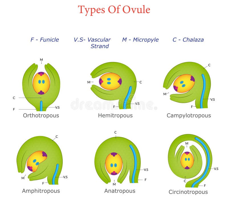
Free with trial An illustration depicting different types of ovules, including anatropous, orthotropous, campylotropous, and others, with labeled details. Integument vectors Six Types of Ovules Illustrated Classification and Structure. An illustration depicting different types of ovules, including anatropous, orthotropous. An illustration depicting different types of ovules, including anatropous, orthotropous, campylotropous, and others, with labeled details
Free with trial An illustration of a campylotropous ovule, showing its curved structure with labeled parts like nucellus, integuments, chalaza, micropyle, and funiculus. Integument vectors Campylotropous Ovule Diagram Displaying Curved Structure with Labeled Parts. An illustration of a campylotropous ovule, showing its curved structure with labeled. An illustration of a campylotropous ovule, showing its curved structure with labeled parts like nucellus, integuments, chalaza, micropyle, and funiculus
Free with trial Mix icon for Membrane, integument and waterproof. Mix icon for Membrane, integument, based, textile, breathable, waterproof, layered and material. Integument vectors Mix icon for Membrane, integument and waterproof. Mix icon for Membrane, integument, based, textile, breathable, waterproof, layered and material
Free with trial Black line icon for Membrane, integument and waterproff. Black line icon for Membrane, integument, based, textile, breathable, waterproof and layered. Integument vectors Black line icon for Membrane, integument and waterproff. Black line icon for Membrane, integument, based, textile, breathable, waterproof and layered
Free with trial Black solid icon for Membrane, integument and waterproof. Black solid icon for Membrane, integument, based, textile, breathable and waterproof. Integument vectors Black solid icon for Membrane, integument and waterproof. Black solid icon for Membrane, integument, based, textile, breathable and waterproof
 Sign in