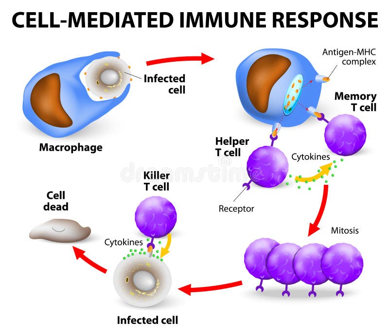
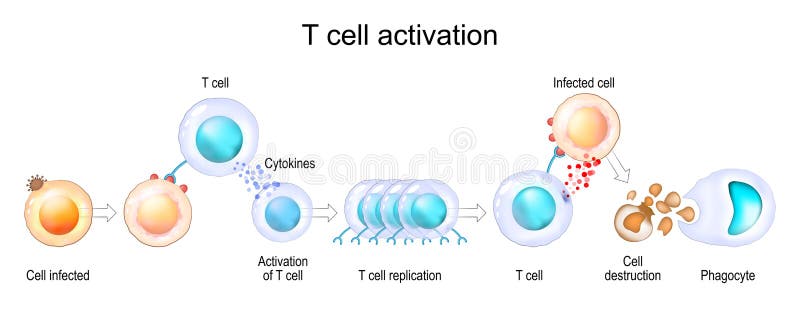
Free with trial Cell-mediated immunity. T lymphocytes do not secrete antibodies. this response incorporates activated macrophages, natural killer cells, antigen-specific cytotoxic T-lymphocytes as well as release of cytokines. Mediated vectors Cell-mediated immune response. Cell-mediated immunity. T lymphocytes do not secrete antibodies. this response incorporates activated macrophages, natural killer. Cell-mediated immunity. T lymphocytes do not secrete antibodies. this response incorporates activated macrophages, natural killer cells, antigen-specific cytotoxic T-lymphocytes as well as release of cytokines.
Free with trial Cell-mediated immunity. T lymphocytes do not secrete antibodies. this response incorporates activated macrophages, natural killer cells, antigen-specific cytotoxic T-lymphocytes as well as release of cytokines. Mediated vectors Cell-mediated immunity. T lymphocytes do not secrete antibodies. this response incorporates activated macrophages, natural killer cells, antigen-specific
Free with trial Cytokines - proteins that are important in cell signaling. They are released by cells and affect the behavior of other cells. Cytokines include interferons, interleukins, lymphokines and other. Cytokines are produced by a broad range of cells, including immune cells: macrophages, B lymphocytes, T lymphocytes and mast cells. Mediated vectors Properties of Cytokines. Cytokines - proteins that are important in cell signaling. They are released by cells and affect the behavior of other cells. Cytokines. Cytokines - proteins that are important in cell signaling. They are released by cells and affect the behavior of other cells. Cytokines include interferons, interleukins, lymphokines and other. Cytokines are produced by a broad range of cells, including immune cells: macrophages, B lymphocytes, T lymphocytes and mast cells.
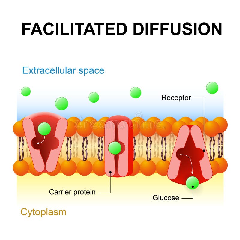
Free with trial Facilitated diffusion or passive-mediated transport. Facilitated diffusion or facilitated transport or passive-mediated transport. Carrier protein. Mediated vectors Facilitated diffusion or passive-mediated transport. Facilitated diffusion or facilitated transport or passive-mediated transport. Carrier protein
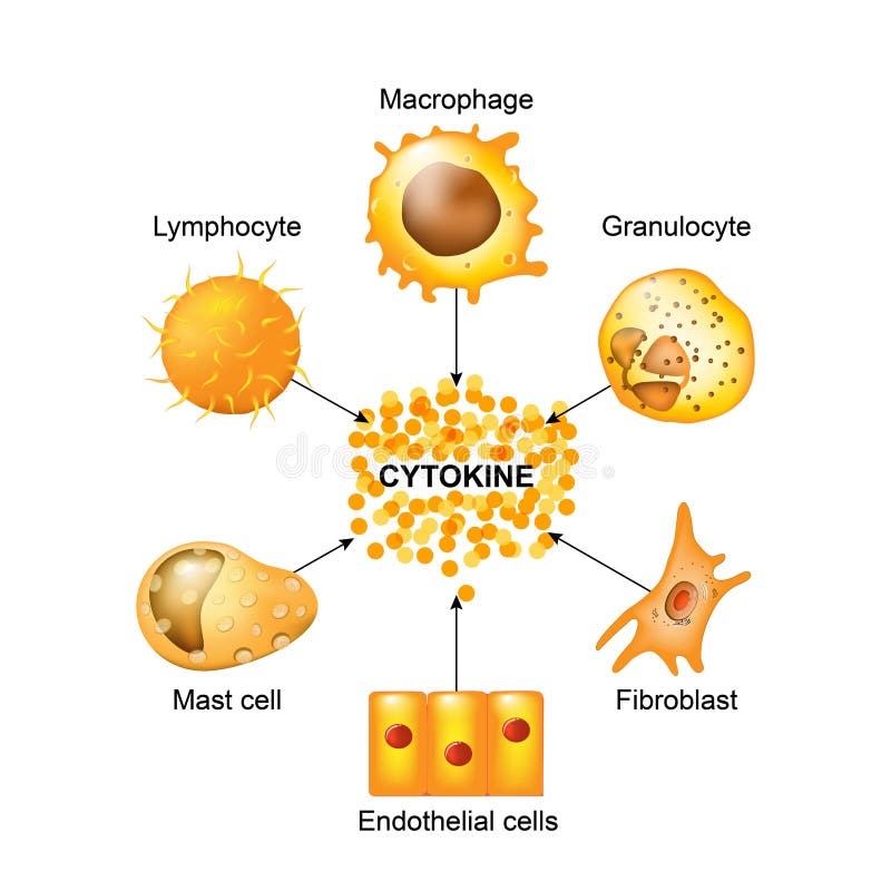
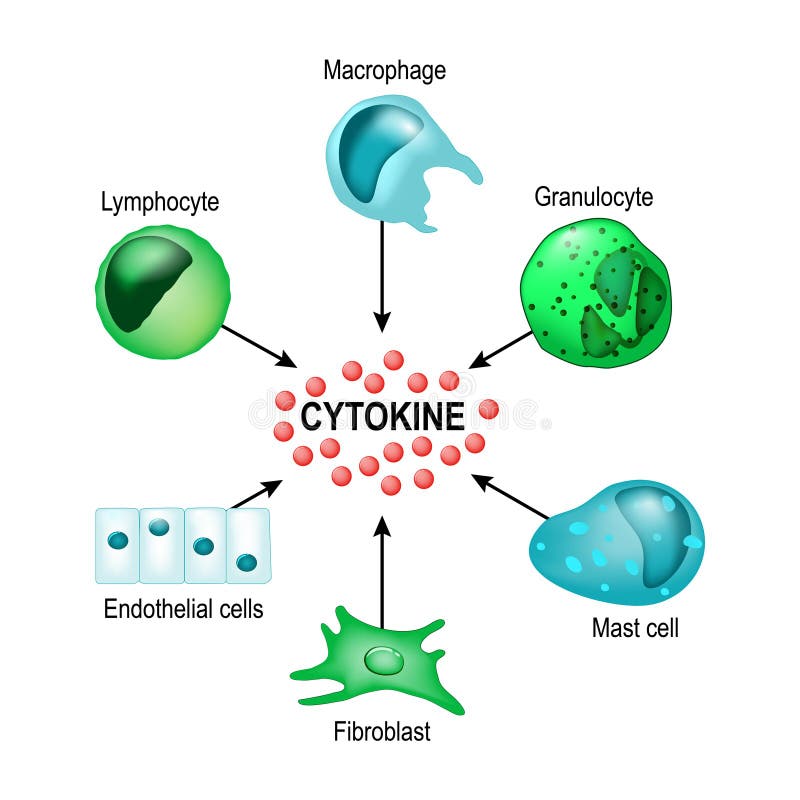
Free with trial Cytokines are produced by macrophages, lymphocytes, mast cells, endothelial cells and fibroblasts. Cytokines include chemokines, interferons, interleukins, lymphokines, and tumour necrosis factors, but not hormones or growth factors. Mediated vectors Cells that produce cytokines. Cytokines are produced by macrophages, lymphocytes, mast cells, endothelial cells and fibroblasts. Cytokines include chemokines. Cytokines are produced by macrophages, lymphocytes, mast cells, endothelial cells and fibroblasts. Cytokines include chemokines, interferons, interleukins, lymphokines, and tumour necrosis factors, but not hormones or growth factors
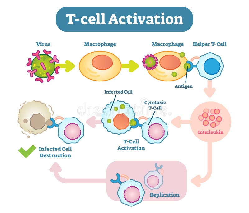
Free with trial A T cell, or T lymphocyte, is a type of lymphocyte a subtype of white blood cell that plays a central role in cell-mediated immunity. Mediated vectors T-Cell activation diagram, vector scheme illustration. A T cell, or T lymphocyte, is a type of lymphocyte a subtype of white blood cell that plays a central. A T cell, or T lymphocyte, is a type of lymphocyte a subtype of white blood cell that plays a central role in cell-mediated immunity.
Free with trial The different types of endocytosis: receptor-mediated endocytosis, pinocytosis cell drinking and phagocytosis cell eating. vesicle, coated vesicle, and phagosome. vector illustration for medical, educational and science use. Mediated vectors The different types of endocytosis. Receptor-mediated endocytosis, pinocytosis cell drinking and phagocytosis cell eating. vesicle, coated vesicle, and. The different types of endocytosis: receptor-mediated endocytosis, pinocytosis cell drinking and phagocytosis cell eating. vesicle, coated vesicle, and phagosome. vector illustration for medical, educational and science use
Free with trial Humoral immunity. antibody-mediated immunity. Viruse, Lymphocyte, antibody and antigen. Vector diagram for educational, biological, and science use. Mediated vectors Humoral immunity. antibody-mediated immunity. Viruse, Lymphocyte, antibody and antigen. Vector diagram for educational, biological. And science use
Free with trial Lymphocyte. Structure cell. Lymphocyte - type of white blood cell. Mediated vectors Lymphocyte. Structure cell. - type of white blood cell. Lymphocyte. Structure cell. Lymphocyte - type of white blood cell
Free with trial Properties of cytokines. signal transduction between cells. endocrine, paracrine and autocrine secretion. Mediated vectors Properties of cytokines. Signal transduction between cells. endocrine, paracrine and autocrine secretion
Free with trial Cytokines are produced by macrophages, lymphocytes, mast cells, endothelial cells and fibroblasts. Cytokines include chemokines, interferons, interleukins, lymphokines, and tumour necrosis factors, but not hormones or growth factors. Mediated vectors Cytokine. Vector concept. Cytokines are produced by macrophages, lymphocytes, mast cells, endothelial cells and fibroblasts. Cytokines include chemokines. Cytokines are produced by macrophages, lymphocytes, mast cells, endothelial cells and fibroblasts. Cytokines include chemokines, interferons, interleukins, lymphokines, and tumour necrosis factors, but not hormones or growth factors
Free with trial Cytokines. Are released by cells and affect the behavior of other cells. Vector concept. Mediated vectors Cytokines. Are released by cells and affect the behavior of other cells. Vector concept
Free with trial Types of T-cell. Lymphoid cell. T lymphocytes: Naive Regulatory Memory helper and T-killer or Cytotoxic T cells. Vector illustration. Mediated vectors Types of T-cell. T lymphocyte. Types of T-cell. Lymphoid cell. T lymphocytes: Naive Regulatory Memory helper and T-killer or Cytotoxic T cells. Vector. Types of T-cell. Lymphoid cell. T lymphocytes: Naive Regulatory Memory helper and T-killer or Cytotoxic T cells. Vector illustration
Free with trial Growth hormone or somatotropin or somatropin. Physiologic Effects of Growth Hormone. Direct and indirect effects. Mediated vectors Growth hormone or somatotropin or somatropin. Physiologic Effects of Growth Hormone. Direct and indirect effects
Free with trial Cytokines and Cytokine receptor. signal transduction between cells. endocrine, paracrine and autocrine secretion. Mediated vectors Cytokine receptor. signal transduction between cells. Cytokines and Cytokine receptor. signal transduction between cells. endocrine, paracrine and autocrine. Cytokines and Cytokine receptor. signal transduction between cells. endocrine, paracrine and autocrine secretion
Free with trial Cell signaling. Signalling mechanism in cells: intracrine, autocrine and endocrine signals. Mediated vectors Cell signaling. intracrine, autocrine and endocrine signals. Cell signaling. Signalling mechanism in cells: intracrine, autocrine and endocrine signals. Cell signaling. Signalling mechanism in cells: intracrine, autocrine and endocrine signals.
Free with trial T cell Activation. T lymphocyte, is a white blood cell. cell-mediated immunity. Mediated vectors T cell Activation. T lymphocyte, is a white blood cell. cell-mediated immunity
Free with trial Endocytosis process with closeup cell side view in anatomical outline diagram. Phagocytosis, pinocytosis or receptor mediated stages explanation vector illustration. Labeled intracellular invagination. Mediated vectors Endocytosis process with closeup cell side view in anatomical outline diagram. Phagocytosis, pinocytosis or receptor mediated stages explanation vector
Free with trial Immune response. humoral immunity. antibody-mediated immunity. activation of macrophage, B-cell and Plasma cell. Antibodies bind to pathogen. Vector poster for education. Mediated vectors Immune response. humoral immunity. Antibody-mediated immunity. activation of macrophage, B-cell and Plasma cell. Antibodies bind to pathogen. Vector poster for
Free with trial Cytokines - proteins that are important in cell signaling. They are released by cells and affect the behavior of other cells. Cytokines include interferons, interleukins, lymphokines and other. Cytokines are produced by a broad range of cells, including immune cells: macrophages, B lymphocytes, T lymphocytes and mast cells. Mediated vectors Cytokines include interferons, interleukins, lymphokines. Cytokines - proteins that are important in cell signaling. They are released by cells and affect the. Cytokines - proteins that are important in cell signaling. They are released by cells and affect the behavior of other cells. Cytokines include interferons, interleukins, lymphokines and other. Cytokines are produced by a broad range of cells, including immune cells: macrophages, B lymphocytes, T lymphocytes and mast cells.
Free with trial Cytokines - proteins that are important in cell signaling. They are released by cells and affect the behavior of other cells. Cytokines include interferons, interleukins, lymphokines and other. Cytokines are produced by a broad range of cells, including immune cells: macrophages, B lymphocytes, T lymphocytes and mast cells. Mediated vectors Cytokines. Proteins that are important in cell signaling. They are released by cells and affect the behavior of other cells. include interferons. Cytokines - proteins that are important in cell signaling. They are released by cells and affect the behavior of other cells. Cytokines include interferons, interleukins, lymphokines and other. Cytokines are produced by a broad range of cells, including immune cells: macrophages, B lymphocytes, T lymphocytes and mast cells.
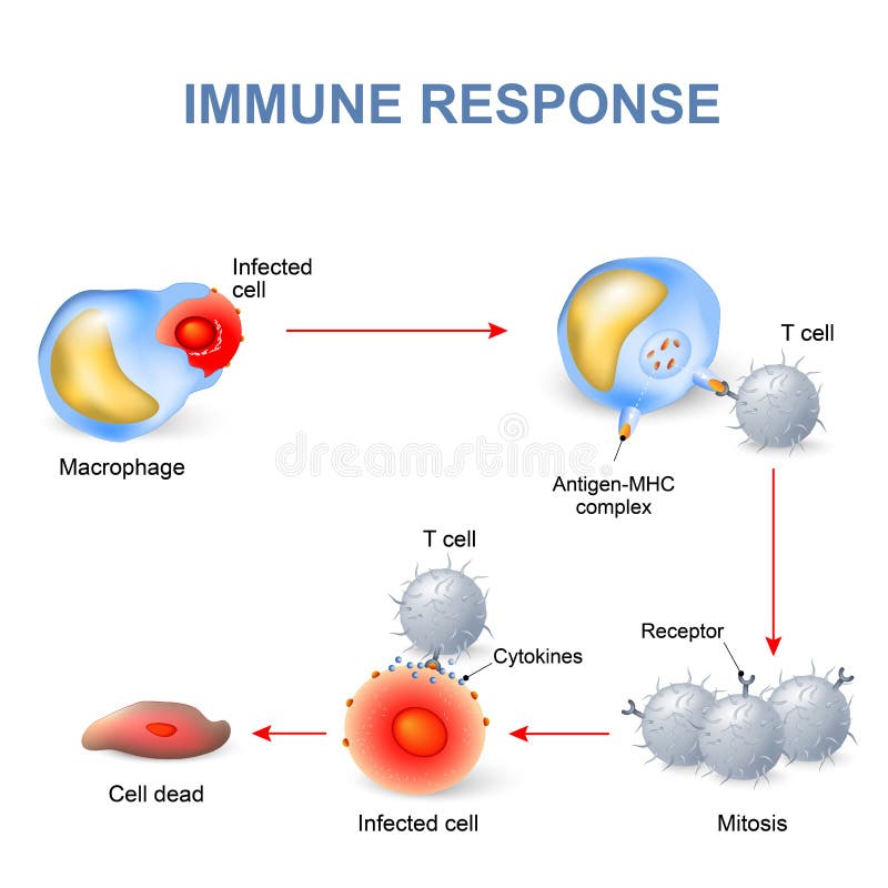
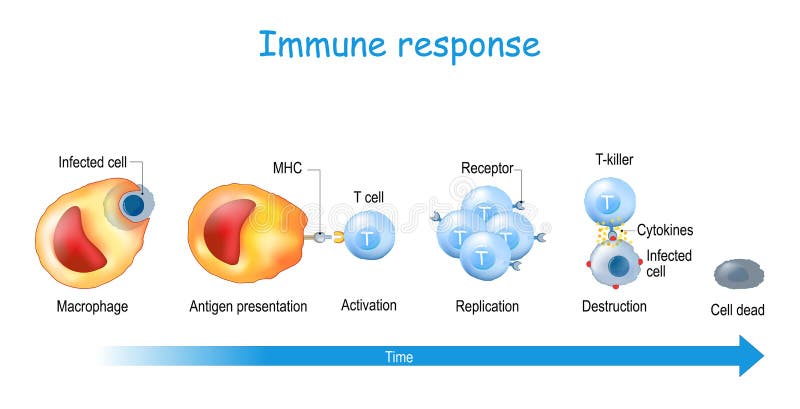
Free with trial Immune response and Antigen presentation. T-cell Activation. T lymphocyte, is a white blood cell. cell-mediated immunity. Mediated vectors Immune response and Antigen presentation. T-cell Activation. T lymphocyte, is a white blood cell. cell-mediated immunity
Free with trial Sore or white tongue. comparison of healthy tongue and oral disease: Lichen planus, Leukoplakia, Geographic tongue. Vector poster for medical use. Mediated vectors Sore or white tongue. comparison of healthy tongue and oral disease. Lichen planus, Leukoplakia, Geographic tongue. Vector poster for medical use
Free with trial Activation of leukocytes. T-cell encounters its cognate antigen on the surface of an infected cell. T-cells direct and regulate immune responses and attack infected or cancerous cells. Cell-mediated immunity. The Adaptive and Innate immune system. vector poster. Mediated vectors Activation of T cell leukocytes. Immune response. Activation of leukocytes. T-cell encounters its cognate antigen on the surface of an infected cell. T-cells. Activation of leukocytes. T-cell encounters its cognate antigen on the surface of an infected cell. T-cells direct and regulate immune responses and attack infected or cancerous cells. Cell-mediated immunity. The Adaptive and Innate immune system. vector poster
Free with trial Immune response and Antigen presentation. Cell-mediated immunity is an immune response that does not involve antibodies. T-cell Activation. Vector poster. Mediated vectors Immune response. Cell-mediated immunity. Immune response and Antigen presentation. Cell-mediated immunity is an immune response that does not involve antibodies. Immune response and Antigen presentation. Cell-mediated immunity is an immune response that does not involve antibodies. T-cell Activation. Vector poster
Free with trial Immune response. Cell-mediated immunity. activation of phagocytes, antigen-specific cytotoxic T-lymphocytes, and the release of cytokines in response to an antigen. Vector poster for educatio. Mediated vectors Immune response. Cell-mediated immunity. Activation of phagocytes, antigen-specific cytotoxic T-lymphocytes, and the release of cytokines in response to an
Free with trial Enterprise resource planning ERP integrated management. Of core business processes, mediated by software and technology, data warehouse, human resources vector. Mediated vectors Enterprise resource planning ERP integrated management. Of core business processes, mediated by software and technology, data warehouse, human resources vector
Free with trial Illustration of biology, Humoral immunity is also referred to as antibody mediated immunity, Humoral immunity is the aspect of immunity that is mediated by macromolecules, cellular immune elements. Mediated vectors Illustration of biology, Humoral immunity is also referred to as antibody mediated immunity,. Humoral immunity is the aspect of immunity that is mediated by. illustration of biology, Humoral immunity is also referred to as antibody mediated immunity, Humoral immunity is the aspect of immunity that is mediated by macromolecules, cellular immune elements
Free with trial AR developer. Augmented reality software development. Computer-mediated reality, digital media technology for adverisment and education. Flat vector illustration. Mediated vectors AR developer. Augmented reality software development. Computer-mediated. Reality, digital media technology for adverisment and education. Flat vector
Free with trial Vasovagal syncope explained, triggers overactivate the vagus nerve, causing bradycardia and hypotension that lead to fainting, brain, heart, and pressure gauge illustrated. Outline diagram. Mediated vectors Vasovagal syncope explained, triggers overactivate the vagus nerve, causing bradycardia ... Vasovagal syncope explained, triggers overactivate the vagus nerve. Vasovagal syncope explained, triggers overactivate the vagus nerve, causing bradycardia and hypotension that lead to fainting, brain, heart, and pressure gauge illustrated. Outline diagram
Free with trial Allergy. First contact with the allergen and Re-exposure to the allergen. Mast cell structure. Role of histamine, allergens, immunoglobulin IgE in Allergic responses. Detailed vector poster. Mediated vectors Allergy mechanism and Allergic responses. Allergy. First contact with the allergen and Re-exposure to the allergen. Mast cell structure. Role of histamine. Allergy. First contact with the allergen and Re-exposure to the allergen. Mast cell structure. Role of histamine, allergens, immunoglobulin IgE in Allergic responses. Detailed vector poster
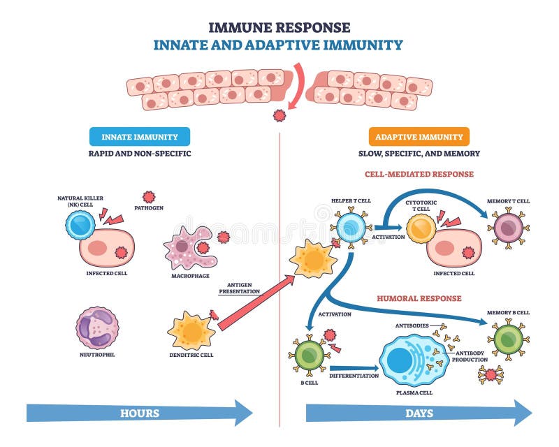
Free with trial Immune response brief flow shows innate to adaptive stages with T cells, B cells, and antibodies defeating pathogens, cells interact over time from exposure to memory. Outline diagram. Mediated vectors Immune response brief flow shows innate to adaptive stages with ... Immune response brief flow shows innate to adaptive stages with T cells, B cells, and. Immune response brief flow shows innate to adaptive stages with T cells, B cells, and antibodies defeating pathogens, cells interact over time from exposure to memory. Outline diagram
Free with trial Insulin function. INS. Fat accumulation, Ribosomal protein synthesis, Energy storage in liver, Blood sugar regulation, Glucose uptake. Hormone produced by beta cells of the pancreatic islets. Detailed vector poster. Mediated vectors Insulin role. Metabolic and Anabolic Functions of INS Pancreatic hormone. Insulin function. INS. Fat accumulation, Ribosomal protein synthesis, Energy storage in. Insulin function. INS. Fat accumulation, Ribosomal protein synthesis, Energy storage in liver, Blood sugar regulation, Glucose uptake. Hormone produced by beta cells of the pancreatic islets. Detailed vector poster
Free with trial GLP-1 mechanism of action. Glucagon-like peptide in pancreatic cell. GLP-1R mediated insulin release. Mediated vectors GLP-1 mechanism of action. Glucagon-like peptide in pancreatic cell. GLP-1R mediated insulin release
Free with trial The patients skin inflammation from the light. Illustration. Because of loss of tolerance to self leads to immune system mediated damage to self The body cannot produce normal immunity. Mediated vectors Systemic Lupus Erythematosus SLE. The patients skin inflammation from the light. Illustration. The patients skin inflammation from the light. Illustration. The patients skin inflammation from the light. Illustration. Because of loss of tolerance to self leads to immune system mediated damage to self The body cannot produce normal immunity.
Free with trial Passive Transport. Simple diffusion through the lipid bilayer and Facilitated Diffusion through a specific or non specific transporter. Vector illustration. Mediated vectors Passive Transport. Simple diffusion and facilitated diffusion. Passive Transport. Simple diffusion through the lipid bilayer and Facilitated Diffusion through a. Passive Transport. Simple diffusion through the lipid bilayer and Facilitated Diffusion through a specific or non specific transporter. Vector illustration.
Free with trial Augmented reality apps and games computer entertainment technology isometric ar poster with different levels experience vector illustration. Mediated vectors Augmented Reality AR Games Isometric Poster. Augmented reality apps and games computer entertainment technology isometric ar poster with different levels. Augmented reality apps and games computer entertainment technology isometric ar poster with different levels experience vector illustration
Free with trial Process of engulfing particles by eukaryotic cells. Phagocytosis, pinocytosis, and receptor-mediated endocytosis. Vector illustration. Didactic illustration. Mediated vectors Illustration of types of endocytosis. Process of engulfing particles by eukaryotic cells. Phagocytosis, pinocytosis, and receptor-mediated endocytosis. Vector. Process of engulfing particles by eukaryotic cells. Phagocytosis, pinocytosis, and receptor-mediated endocytosis. Vector illustration. Didactic illustration.
Free with trial Gray-orange Immunoglobulin Round Icon. Gray-orange symbolic immunoglobulin molecule round web icon. Immune cell emaciated, exhausted and tired of interferon. Mediated vectors Gray-orange Immunoglobulin Round Icon. Gray-orange symbolic immunoglobulin molecule round web icon. Immune cell emaciated, exhausted and tired of interferon
Free with trial Man in augmented reality smart glasses with computer vision learning foreign language home isometric poster vector illustration. Mediated vectors Augmented Reality Glases Symbols Isometric oster. Man in augmented reality smart glasses with computer vision learning foreign language home isometric poster. Man in augmented reality smart glasses with computer vision learning foreign language home isometric poster vector illustration
Free with trial The pathogenesis of acanthosis nigricans vector scheme. The pathogenesis of acanthosis nigricans scheme, vector medical illustration. Mediated vectors The pathogenesis of acanthosis nigricans vector scheme. The pathogenesis of acanthosis nigricans scheme, vector medical illustration
Free with trial Eosinophil. Immune blood cell. Chemokines. Vector illustration on isolated background. Mediated vectors Eosinophil. Immune blood cell. Chemokines. Vector illustration on isolated background.
Free with trial Proton quarks 3D scheme. The quark content of proton in 3D decoration. Strong forces between quarks are mediated by gluons. Vector illustration. Mediated vectors Proton quarks 3D scheme. The quark content of proton in 3D decoration. Strong forces between quarks are mediated by gluons. Vector illustration
Free with trial Proton quarks schematical icon. The quark content of proton in flat decoration. Strong forces between quarks are mediated by gluons. Vector illustration. Mediated vectors Proton quarks schematical icon. The quark content of proton in flat decoration. Strong forces between quarks are mediated by gluons. Vector illustration
Free with trial Cell-Mediated Immunity from antigen presentation to cytotoxic response and Memory T Cell formation. Adaptive immunity. Vector illustration. Medical poster. Schematic diagram. Mediated vectors Adaptive immunity. Cell-Mediated Immunity. Cell-Mediated Immunity from antigen presentation to cytotoxic response and Memory T Cell formation. Adaptive immunity. Cell-Mediated Immunity from antigen presentation to cytotoxic response and Memory T Cell formation. Adaptive immunity. Vector illustration. Medical poster. Schematic diagram
Free with trial Humoral Immunity process diagram medical poster, Immune response. antibody-mediated immunity, labeled lymphocyte, antibodies, virus, bacteria, biology education. Mediated vectors Humoral Immunity process diagram medical poster, Immune response. antibody-mediated immunity, labeled lymphocyte, antibodies,. Virus, bacteria, biology
Free with trial Endocytosis process diagram with side view, labeled Phagocytosis, pinocytosis or receptor mediated stages, intracellular invagination, vacuole, coated vesicle, pseudopodia, plasma membrane. Mediated vectors Endocytosis process diagram with side view, labeled Phagocytosis, pinocytosis or receptor mediated stages, intracellular. Invagination, vacuole, coated vesicle. Endocytosis process diagram with side view, labeled Phagocytosis, pinocytosis or receptor mediated stages, intracellular invagination, vacuole, coated vesicle, pseudopodia, plasma membrane
Free with trial Cell mediated immunity diagram process, Immune response. activation of phagocytes, labeled macrophage, pathogen, t-lymphocyte, release cytokines in response to antigen, cytotoxic. Biology education. Mediated vectors Cell mediated immunity diagram process, Immune response. activation of phagocytes, labeled macrophage, pathogen, t-lymphocyte,. Release cytokines in response to. Cell mediated immunity diagram process, Immune response. activation of phagocytes, labeled macrophage, pathogen, t-lymphocyte, release cytokines in response to antigen, cytotoxic. Biology education
Free with trial Bulk transport process diagram, Endocytosis and exocytosis, eukaryotes, Phagocytosis, pinocytosis or receptor mediated stages, intracellular invagination, vacuole, coated vesicle, pseudopodia, plasma. Mediated vectors Bulk transport process diagram, Endocytosis and exocytosis, eukaryotes, Phagocytosis, pinocytosis or receptor mediated stages,. Intracellular invagination. Bulk transport process diagram, Endocytosis and exocytosis, eukaryotes, Phagocytosis, pinocytosis or receptor mediated stages, intracellular invagination, vacuole, coated vesicle, pseudopodia, plasma
Free with trial Celiac disease immune-mediated damage of the small intestinal mucosa characterized by villous atrophy and impaired nutrient absorption. Mediated vectors Celiac disease immune-mediated damage of the small intestinal mucosa characterized by villous atrophy and impaired nutrient. Absorption
Free with trial Kidney surrounded by flying antibodies Antibody-mediated transplant rejection, autoimmune kidney disease, immunotherapy, monoclonal antibody treatment, kidney-related biologic medicine concept Vector. Mediated vectors Kidney surrounded by flying antibodies. Antibody-mediated transplant rejection, autoimmune kidney disease, immunotherapy. Kidney surrounded by flying antibodies. Kidney surrounded by flying antibodies Antibody-mediated transplant rejection, autoimmune kidney disease, immunotherapy, monoclonal antibody treatment, kidney-related biologic medicine concept Vector.
Free with trial An icon showing two figures maintaining distance, with a heart and gears between them, symbolizing social connection through technology. This represents modern relationships, remote work, and digitally mediated interactions. Ideal for social media, community, and tech topics, vector design Generative AI. Mediated vectors Social Distancing, Love in Tech, Heart Connection, vector design Generative AI. An icon showing two figures maintaining distance, with a heart and gears between. An icon showing two figures maintaining distance, with a heart and gears between them, symbolizing social connection through technology. This represents modern relationships, remote work, and digitally mediated interactions. Ideal for social media, community, and tech topics, vector design Generative AI
Free with trial Illustration of biology, Receptor mediated endocytosis is a process by which cells absorb metabolites, hormones, proteins and in some cases viruses by the inward budding of the plasma membrane. Mediated vectors Illustration of biology, Receptor mediated endocytosis is a process by which cells absorb metabolites. Hormones, proteins and in some cases viruses by the. illustration of biology, Receptor mediated endocytosis is a process by which cells absorb metabolites, hormones, proteins and in some cases viruses by the inward budding of the plasma membrane
Free with trial Cell endocytosis and exocytosis, infographic shows how the cell membrane forms vesicles to import and export cargo, key objects are cell membrane, vesicles, and Golgi apparatus. Mediated vectors Cell endocytosis and exocytosis, infographic shows how the cell membrane ... Cell endocytosis and exocytosis, infographic shows how the cell membrane forms. Cell endocytosis and exocytosis, infographic shows how the cell membrane forms vesicles to import and export cargo, key objects are cell membrane, vesicles, and Golgi apparatus.
Free with trial Macrophage polarization process. Functions of M1 and M2 macrophages. immune response. Vector illustration. Medical poster. Schematic diagram. Mediated vectors Macrophage polarization process. Functions of M1 and M2 macrophages. immune response. Vector illustration. Medical poster. Schematic diagram
Free with trial T cell functions outline depicts helper CD4, cytotoxic CD8, and regulatory cells guiding activation, killing infected cells, and suppression with arrows and labels. Outline diagram. Mediated vectors T cell functions outline depicts helper CD4, cytotoxic CD8, and ... T cell functions outline depicts helper CD4, cytotoxic CD8, and regulatory cells guiding. T cell functions outline depicts helper CD4, cytotoxic CD8, and regulatory cells guiding activation, killing infected cells, and suppression with arrows and labels. Outline diagram
Free with trial Immune cell lineage brief diagram maps stem-to-progenitor branching, showing hematopoietic stem cell splitting into myeloid and lymphoid paths, key T cell, B cell, macrophage. Outline diagram. Mediated vectors Immune cell lineage brief diagram maps stem-to-progenitor branching. Showing hematopoietic stem cell splitting into myeloid and lymphoid paths, key T cell, B. Immune cell lineage brief diagram maps stem-to-progenitor branching, showing hematopoietic stem cell splitting into myeloid and lymphoid paths, key T cell, B cell, macrophage. Outline diagram
Free with trial Types of immunity brief diagram comparing innate and adaptive pathways into active and passive, shield, lock, syringe highlight defenses and vaccination concept. Outline diagram. Mediated vectors Types of immunity brief diagram comparing innate and adaptive pathways ... Types of immunity brief diagram comparing innate and adaptive pathways into active. Types of immunity brief diagram comparing innate and adaptive pathways into active and passive, shield, lock, syringe highlight defenses and vaccination concept. Outline diagram
Free with trial Immunodeficiency disorders outline showing innate vs adaptive defects, key icons, B-cell, T-cell, phagocyte. Explains roles and causes across categories. Outline diagram. Mediated vectors Immunodeficiency disorders outline showing innate vs adaptive defects, key icons, ... Immunodeficiency disorders outline showing innate vs adaptive defects, key. Immunodeficiency disorders outline showing innate vs adaptive defects, key icons, B-cell, T-cell, phagocyte. Explains roles and causes across categories. Outline diagram
Free with trial Hormone receptors are illustrated with membrane channels, DNA response elements, and signaling molecules, showing cellular communication. Outline style collection. Mediated vectors Hormone receptors are illustrated with membrane channels, DNA response elements, ... Hormone receptors are illustrated with membrane channels, DNA response. Hormone receptors are illustrated with membrane channels, DNA response elements, and signaling molecules, showing cellular communication. Outline style collection
Free with trial Cytokines. Cell signaling from Cytokine Producing cell to Target Cell. Interferons, interleukins, lymphokines. Adaptive and Innate immunity. Chronic inflammation. Detailed Vector poster. Isometric Flat illustration. Schematic diagram. Mediated vectors Cytokine Producing cell and Target Cell. Interferons, interleukins, lymphokines. Cytokines. Cell signaling from Cytokine Producing cell to Target Cell. Cytokines. Cell signaling from Cytokine Producing cell to Target Cell. Interferons, interleukins, lymphokines. Adaptive and Innate immunity. Chronic inflammation. Detailed Vector poster. Isometric Flat illustration. Schematic diagram
Free with trial Medical showing immune recognition of breast cancer cells, checkpoint-mediated suppression, and restored T-cell activity after checkpoint blockade therapy. Mediated vectors Immune therapy blocking cancer cell escape Vector. Medical showing immune recognition of breast cancer cells, checkpoint-mediated suppression, and restored T. Medical showing immune recognition of breast cancer cells, checkpoint-mediated suppression, and restored T-cell activity after checkpoint blockade therapy
Free with trial Centralized Two Way Communication Icon Set. Vol 34442. Mediated vectors Centralized Two Way Communication Icon Set. Vol 34442
Free with trial T-cell Activation and Response. Adaptive immunity. Vector illustration. Medical poster. Schematic diagram. Mediated vectors T-cell Activation and Response. Adaptive immunity. Vector illustration. Medical poster. Schematic diagram
Free with trial The ubiquitin-proteasome pathway illustrates protein ubiquitination and degradation through E1, E2, and E3 enzymes, followed by proteasome-mediated breakdown of targeted proteins—a clean scientific medical infographic for molecular biology, biochemistry, biotechnology, and life science education. Mediated vectors The Ubiquitin Proteasome Pathway Scientific Design Vector Illustration. The ubiquitin-proteasome pathway illustrates protein ubiquitination and degradation. The ubiquitin-proteasome pathway illustrates protein ubiquitination and degradation through E1, E2, and E3 enzymes, followed by proteasome-mediated breakdown of targeted proteins—a clean scientific medical infographic for molecular biology, biochemistry, biotechnology, and life science education.
Free with trial Innate and adaptive immune response visual showing pathogen defense pathways, key elements, macrophage, T cell activation, antibody production. Outline diagram. Mediated vectors Innate and adaptive immune response visual showing pathogen defense pathways, ... Innate and adaptive immune response visual showing pathogen defense pathways. Innate and adaptive immune response visual showing pathogen defense pathways, key elements, macrophage, T cell activation, antibody production. Outline diagram
Free with trial T lymphocytes diagram shows helper, cytotoxic, and regulatory T cells coordinating immunity, key objects, helper T cell, cytotoxic T cell, infected cell. Outline diagram. Mediated vectors T lymphocytes diagram shows helper, cytotoxic, and regulatory T cells ... T lymphocytes diagram shows helper, cytotoxic, and regulatory T cells coordinating. T lymphocytes diagram shows helper, cytotoxic, and regulatory T cells coordinating immunity, key objects, helper T cell, cytotoxic T cell, infected cell. Outline diagram
Free with trial The ubiquitin proteasome pathway illustrating protein ubiquitination and degradation through E1, E2, and E3 enzymes, followed by proteasome-mediated breakdown of targeted proteins. Clean scientific medical infographic for molecular biology, biochemistry, biotechnology, and life science education. Mediated vectors The Ubiquitin Proteasome Pathway Biological Process Design Vector Illustration. The ubiquitin proteasome pathway illustrating protein ubiquitination and. The ubiquitin proteasome pathway illustrating protein ubiquitination and degradation through E1, E2, and E3 enzymes, followed by proteasome-mediated breakdown of targeted proteins. Clean scientific medical infographic for molecular biology, biochemistry, biotechnology, and life science education.
Free with trial Educational vector illustration explaining the ubiquitination and deubiquitination pathway. The diagram shows ubiquitin activation (E1), conjugation (E2), ligation (E3), polyubiquitination of target proteins, proteasome-mediated protein degradation, and deubiquitinating enzymes (DUBs). Ideal for molecular biology, biochemistry, cell biology, proteomics, biotechnology, medical education, textbooks, scientific presentations, and research publications. Mediated vectors Ubiquitination and Deubiquitination Pathway Protein Degradation and Proteasome Process Vector Illustration. Educational vector illustration explaining the. Educational vector illustration explaining the ubiquitination and deubiquitination pathway. The diagram shows ubiquitin activation (E1), conjugation (E2), ligation (E3), polyubiquitination of target proteins, proteasome-mediated protein degradation, and deubiquitinating enzymes (DUBs). Ideal for molecular biology, biochemistry, cell biology, proteomics, biotechnology, medical education, textbooks, scientific presentations, and research publications.
Free with trial Cytokines. Proteins for cell signaling. Cytokines are produced by macrophage, lymphocyte, mast cell, endothelial cells and fibroblast. Cytokine storm. Vector illustration. Mediated vectors Cytokines are produced by macrophage, lymphocyte, mast cell, endothelial cells and fibroblast. Cytokines. Proteins for cell signaling. Cytokines are produced by. Cytokines. Proteins for cell signaling. Cytokines are produced by macrophage, lymphocyte, mast cell, endothelial cells and fibroblast. Cytokine storm. Vector illustration
Free with trial Cytokines are produced by macrophage, lymphocyte, mast cell, endothelial cells, fibroblast and Granulocyte. Cytokines include chemokines, interferons, interleukins, lymphokines, and tumour necrosis factors, but not hormones or growth factors. Isometric Flat vector illustration. Mediated vectors Cytokines that produced by macrophage, lymphocyte, mast cell, endothelial cells, fibroblast and Granulocyte. Cytokines are produced by macrophage, lymphocyte. Cytokines are produced by macrophage, lymphocyte, mast cell, endothelial cells, fibroblast and Granulocyte. Cytokines include chemokines, interferons, interleukins, lymphokines, and tumour necrosis factors, but not hormones or growth factors. Isometric Flat vector illustration
Free with trial Centralized Two Way Communication Icon Set. Vol 34442. Mediated vectors Centralized Two Way Communication Icon Set. Vol 34442
Free with trial Medical vector illustration of multiple sclerosis depicting immune-mediated demyelination of neurons in the brain and spinal cord, leading to disrupted nerve signals and progressive neurological symptoms. Mediated vectors Multiple sclerosis vector illustration. Medical vector illustration of multiple sclerosis depicting immune-mediated demyelination of neurons in the brain and. Medical vector illustration of multiple sclerosis depicting immune-mediated demyelination of neurons in the brain and spinal cord, leading to disrupted nerve signals and progressive neurological symptoms.
Free with trial Educational vector illustration explaining the adaptive immune response in coeliac disease. The diagram shows gluten digestion, protease activity, TG2-mediated deamidation, antigen presentation, activation of CD4+ T cells, B cells, plasma cells, cytokine release, and production of anti-gluten and anti-TG2 antibodies. Ideal for immunology, gastroenterology, pathology, medical education, biology textbooks, scientific presentations, and healthcare research. Mediated vectors Adaptive Immune Response in Coeliac Disease Gluten-Induced Autoimmune Pathway Vector Illustration. Educational vector illustration explaining the adaptive. Educational vector illustration explaining the adaptive immune response in coeliac disease. The diagram shows gluten digestion, protease activity, TG2-mediated deamidation, antigen presentation, activation of CD4+ T cells, B cells, plasma cells, cytokine release, and production of anti-gluten and anti-TG2 antibodies. Ideal for immunology, gastroenterology, pathology, medical education, biology textbooks, scientific presentations, and healthcare research.
Free with trial Black and white icon shows a person and a video camera connected. The icon represents video conferencing, virtual meetings, or video calls. Represents technology-mediated communication. Mediated vectors Video Call Icon Representing Communication and Virtual Connection, Simple Black Design. Black and white icon shows a person and a video camera connected. The. Black and white icon shows a person and a video camera connected. The icon represents video conferencing, virtual meetings, or video calls. Represents technology-mediated communication.
Free with trial Educational vector illustration explaining the ubiquitination and deubiquitination pathway. The diagram shows ubiquitin activation (E1), conjugation (E2), ligation (E3), polyubiquitination of target proteins, proteasome-mediated protein degradation, and deubiquitinating enzymes (DUBs). Ideal for molecular biology, biochemistry, cell biology, proteomics, biotechnology, medical education, textbooks, scientific presentations, and research publications. Mediated vectors Ubiquitination and Deubiquitination Pathway Protein Degradation and Proteasome Process Vector Illustration. Educational vector illustration explaining the. Educational vector illustration explaining the ubiquitination and deubiquitination pathway. The diagram shows ubiquitin activation (E1), conjugation (E2), ligation (E3), polyubiquitination of target proteins, proteasome-mediated protein degradation, and deubiquitinating enzymes (DUBs). Ideal for molecular biology, biochemistry, cell biology, proteomics, biotechnology, medical education, textbooks, scientific presentations, and research publications.
Free with trial This vector illustration explains the T-cell activation pathway in the adaptive immune system. It demonstrates antigen presentation by antigen-presenting cells through MHC class II, activation of helper T cells, cytokine release, differentiation of CD8 cytotoxic T cells, recognition of infected cells via MHC class I, and the release of perforin and granzyme leading to apoptosis. Ideal for medical education, immunology textbooks, healthcare presentations, scientific publications, biology courses, and educational infographics. Mediated vectors T-Cell Activation Pathway Diagram Adaptive Immune Response and Cytotoxic T Lymphocyte Vector Illustration. This vector illustration explains the T-cell. This vector illustration explains the T-cell activation pathway in the adaptive immune system. It demonstrates antigen presentation by antigen-presenting cells through MHC class II, activation of helper T cells, cytokine release, differentiation of CD8 cytotoxic T cells, recognition of infected cells via MHC class I, and the release of perforin and granzyme leading to apoptosis. Ideal for medical education, immunology textbooks, healthcare presentations, scientific publications, biology courses, and educational infographics.
Free with trial This vector illustration explains the T-cell activation pathway in the adaptive immune system. It demonstrates antigen presentation by antigen-presenting cells through MHC class II, activation of helper T cells, cytokine release, differentiation of CD8 cytotoxic T cells, recognition of infected cells via MHC class I, and the release of perforin and granzyme leading to apoptosis. Ideal for medical education, immunology textbooks, healthcare presentations, scientific publications, biology courses, and educational infographics. Mediated vectors T-Cell Activation Pathway Diagram Adaptive Immune Response and Cytotoxic T Lymphocyte Vector Illustration. This vector illustration explains the T-cell. This vector illustration explains the T-cell activation pathway in the adaptive immune system. It demonstrates antigen presentation by antigen-presenting cells through MHC class II, activation of helper T cells, cytokine release, differentiation of CD8 cytotoxic T cells, recognition of infected cells via MHC class I, and the release of perforin and granzyme leading to apoptosis. Ideal for medical education, immunology textbooks, healthcare presentations, scientific publications, biology courses, and educational infographics.
Free with trial This vector illustration explains the T-cell activation pathway in the adaptive immune system. It demonstrates antigen presentation by antigen-presenting cells through MHC class II, activation of helper T cells, cytokine release, differentiation of CD8 cytotoxic T cells, recognition of infected cells via MHC class I, and the release of perforin and granzyme leading to apoptosis. Ideal for medical education, immunology textbooks, healthcare presentations, scientific publications, biology courses, and educational infographics. Mediated vectors T-Cell Activation Pathway Diagram Adaptive Immune Response and Cytotoxic T Lymphocyte Vector Illustration. This vector illustration explains the T-cell. This vector illustration explains the T-cell activation pathway in the adaptive immune system. It demonstrates antigen presentation by antigen-presenting cells through MHC class II, activation of helper T cells, cytokine release, differentiation of CD8 cytotoxic T cells, recognition of infected cells via MHC class I, and the release of perforin and granzyme leading to apoptosis. Ideal for medical education, immunology textbooks, healthcare presentations, scientific publications, biology courses, and educational infographics.
Free with trial This vector illustration explains the T-cell activation pathway in the adaptive immune system. It demonstrates antigen presentation by antigen-presenting cells through MHC class II, activation of helper T cells, cytokine release, differentiation of CD8 cytotoxic T cells, recognition of infected cells via MHC class I, and the release of perforin and granzyme leading to apoptosis. Ideal for medical education, immunology textbooks, healthcare presentations, scientific publications, biology courses, and educational infographics. Mediated vectors T-Cell Activation Pathway Diagram Adaptive Immune Response and Cytotoxic T Lymphocyte Vector Illustration. This vector illustration explains the T-cell. This vector illustration explains the T-cell activation pathway in the adaptive immune system. It demonstrates antigen presentation by antigen-presenting cells through MHC class II, activation of helper T cells, cytokine release, differentiation of CD8 cytotoxic T cells, recognition of infected cells via MHC class I, and the release of perforin and granzyme leading to apoptosis. Ideal for medical education, immunology textbooks, healthcare presentations, scientific publications, biology courses, and educational infographics.
 Sign in