Free with trial Bunion in foot. Deformity of the big toe joint usually due to tight fitting shoes, eps10. Metatarsal bones vectors Bunion in foot. Deformity of the big toe joint usually due to tight fitting shoes, eps10
Free with trial Turf toe. Sprain or tears of ligaments of the big toe joint, a common sport injury, eps10. Metatarsal bones vectors Turf toe. Sprain or tears of ligaments of the big toe joint, a common sport injury, eps10
Free with trial Pott fracture. A common sport injury of the ankle joint, eps8. Metatarsal bones vectors Pott fracture. A common sport injury of the ankle joint, eps8
Free with trial Vector diagram of forefoot pain, metatarsalgia symptom, tenderness in the balls of metatarsal bones of the foot. Used: Gradient, transparency, blend mode. Metatarsal bones vectors Metatarsalgia. Vector diagram of forefoot pain, metatarsalgia symptom, tenderness in the balls of metatarsal bones of the foot. Used: Gradient, transparency. Vector diagram of forefoot pain, metatarsalgia symptom, tenderness in the balls of metatarsal bones of the foot. Used: Gradient, transparency, blend mode.
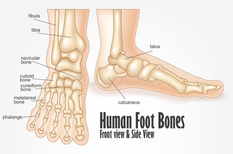
Free with trial Human foot bones front and side view anatomy. Illustration Of Human foot bones front and side view anatomy. Metatarsal bones vectors Human foot bones front and side view anatomy. Illustration Of Human foot bones front and side view anatomy
Free with trial Broken toe. Common sites of toe bone fractures, eps10. Metatarsal bones vectors Broken toe. Common sites of toe bone fractures, eps10
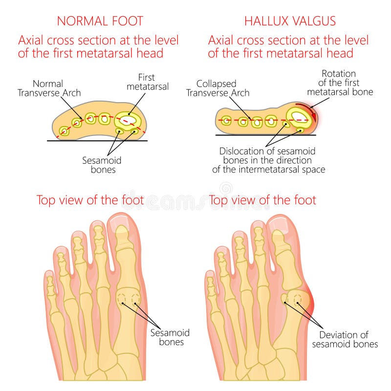
Free with trial Vector illustration of a healthy human forefoot and a foot with hallux valgus, dislocation of sesamoid bones. Top view and cross section of of the foot. For advertising, medical publications. EPS 10. Metatarsal bones vectors Normal foot and Hallux valgus with rotation of the first mematarsal bone. Vector illustration of a healthy human forefoot and a foot with hallux valgus. Vector illustration of a healthy human forefoot and a foot with hallux valgus, dislocation of sesamoid bones. Top view and cross section of of the foot. For advertising, medical publications. EPS 10.
Free with trial Deviation of the first and the fifth metatarsals, transverse flatfoot. Used: gradient, transparency, blend mode. Metatarsal bones vectors Deviation of the first and the fifth metatarsals transverse flat. Deviation of the first and the fifth metatarsals, transverse flatfoot. Used: gradient. Deviation of the first and the fifth metatarsals, transverse flatfoot. Used: gradient, transparency, blend mode.
Free with trial Illustration of the normal foot and hallux valgus with indicating of the first metatarsophalangeal and the first-second intermetatarsal angles. Used: gradient, transparency. Blend mode. Metatarsal bones vectors Normal foot and valgus deviation of the first toe with indicating of the deflection angles. Illustration of the normal foot and hallux valgus with indicating of. Illustration of the normal foot and hallux valgus with indicating of the first metatarsophalangeal and the first-second intermetatarsal angles. Used: gradient, transparency. Blend mode.
Free with trial Bones of foot. Human Anatomy. The diagram shows the placement and names of all bones of foot. Metatarsal bones vectors Bones of foot. Human Anatomy. The diagram shows the placement and names of all
Free with trial Bones of the foot and ankle joint medical vector illustration isolated on white background. Eps 10 infographic. Metatarsal bones vectors Bones of the foot and ankle joint medical vector illustration isolated on white background. Eps 10 infographic
Free with trial Human Anatomy Leg Bones with Detail. Vector Illustration. Metatarsal bones vectors Human Anatomy Leg Bones with Detail. Vector Illustration
Free with trial Diagnosis of the tailor's bunion with angles of deviation. Metatarsal bones vectors Diagnosis of the tailor's bunion. With angles of deviation
Free with trial Deviation of the first and the fifth metatarsals transverse flat. Foot with orthopedic insole. Used: gradient, transparency, blend mode. Metatarsal bones vectors Deviation of the first and the fifth metatarsals transverse flat. Foot with orthopedic insole. Used: gradient, transparency, blend mode
Free with trial Stress fracture. Marching fracture of the foot. Fracture of the metatarsal bones in the foot. Anatomical structure of the foot. Skeleton. Broken bones. Vector illustration on isolated background. Metatarsal bones vectors Stress fracture. Marching fracture of the foot. Fracture of the metatarsal bones in the foot. Anatomical structure of. The foot. Skeleton. Broken bones. Vector. Stress fracture. Marching fracture of the foot. Fracture of the metatarsal bones in the foot. Anatomical structure of the foot. Skeleton. Broken bones. Vector illustration on isolated background
Free with trial Fracture of the metatarsal bones in the foot. Anatomical structure of the foot. Skeleton. Broken bones. Vector illustration on isolated background. Metatarsal bones vectors Fracture of the metatarsal bones in the foot. Anatomical structure of the foot. Skeleton. Broken bones. Vector. Illustration on isolated background
Free with trial Foot bones anatomy. Structure and names. Metatarsal bones location. Medical scheme. Vector illustration in realistic style isolated on green background. Educational poster. Medicine concept. Metatarsal bones vectors Foot bones anatomy. Structure and names. Metatarsal bones location. Medical scheme. Vector illustration in realistic style isolated on green background
Free with trial In the dorsal (posterior) view of the bones of the feet, the tarsal bones are visible, including the talus, calcaneus, navicular, cuboid, and the three cuneiform bones. The metatarsal bones, numbering from one to five, can also be observed, extending towards the toes. Additionally, the phalanges, consisting of proximal, middle, and distal phalanges, complete the skeletal structure of the feet in the dorsal view. Metatarsal bones vectors Bones of feet, dorsal (posterior) view. In the dorsal (posterior) view of the bones of the feet, the tarsal bones are visible, including the talus, calcaneus. In the dorsal (posterior) view of the bones of the feet, the tarsal bones are visible, including the talus, calcaneus, navicular, cuboid, and the three cuneiform bones. The metatarsal bones, numbering from one to five, can also be observed, extending towards the toes. Additionally, the phalanges, consisting of proximal, middle, and distal phalanges, complete the skeletal structure of the feet in the dorsal view.
Free with trial Bones of foot. Human Anatomy. The diagram shows the placement and names of all bones of foot. Metatarsal bones vectors Bones of foot. Human Anatomy. The diagram shows the placement and names of all
Free with trial Ankle, foot bones. Vector illustration of the ankle bones of the foot. Metatarsal bones vectors Ankle, foot bones. Vector illustration of the ankle bones of the foot
Free with trial Anatomy Bones of the Feet. Superior view. Orthotics for foot Superior view. Metatarsal bones vectors Anatomy Bones of the Feet. Superior view. Anatomy Bones of the Feet. Orthotics for foot Superior view
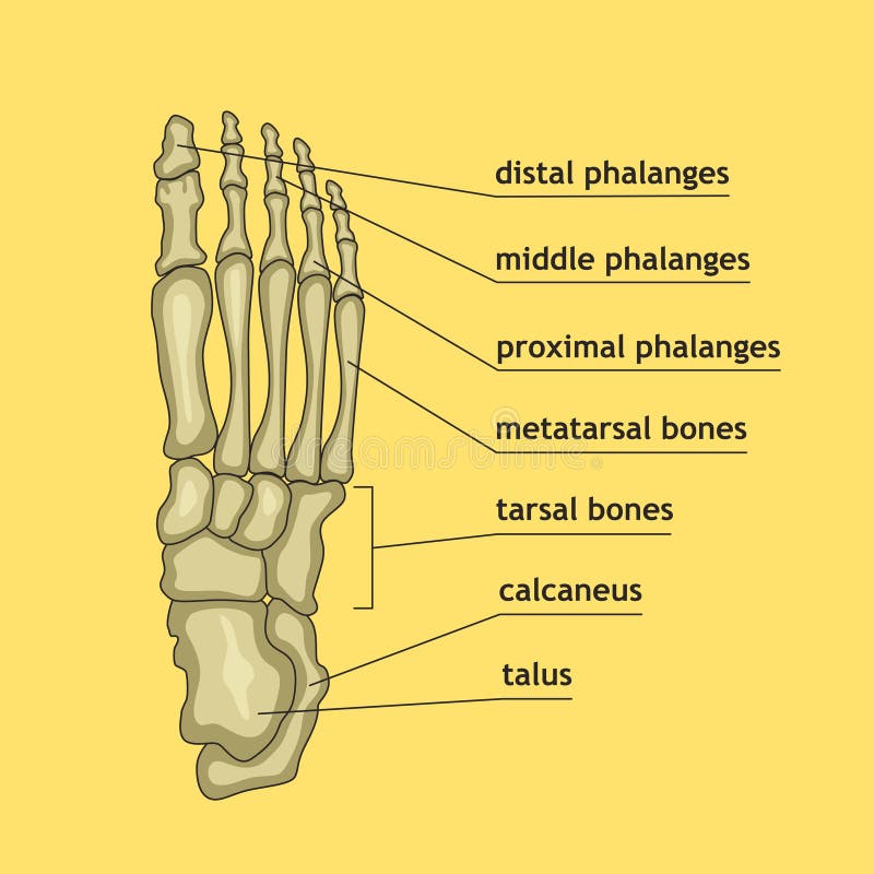
Free with trial Bones of the Foot. Tarsals or tarsus, Metatarsals, Phalanges. Metatarsal bones vectors Bones of the Foot .Tarsals or tarsus, Metatarsals, Phalanges. Bones of the Foot.Tarsals or tarsus, Metatarsals, Phalanges Anatomy
Free with trial Foot bones anatomy. Human skeleton. Horizontal medical poster. Medical education. Editable vector illustration isolated on light background. Graphic design for any purposes. Metatarsal bones vectors Foot bones anatomy. Human skeleton. Horizontal medical poster. Medical education. Editable vector illustration isolated on light background. Graphic design for
Free with trial Foot bones anatomy. Structure and names. Cuboid bone and cuneifroms location. Medical scheme. Vector illustration in realistic style isolated on green background. Educational poster. Medicine concept. Metatarsal bones vectors Foot bones anatomy. Structure and names. Cuboid bone and cuneifroms location. Medical scheme. Vector illustration in realistic style isolated on green
Free with trial Foot bones anatomy. Structure and names. Talus bone location. Medical scheme. Vector illustration in realistic style isolated on green background. Educational poster. Medicine science concept. Metatarsal bones vectors Foot bones anatomy. Structure and names. Talus bone location. Medical scheme. Vector illustration in realistic style isolated on green background. Educational
Free with trial Foot bones anatomy. Structure and names. Distal phalanges location. Medical scheme. Vector illustration in realistic style isolated on green background. Educational poster. Medicine science concept. Metatarsal bones vectors Foot bones anatomy. Structure and names. Distal phalanges location. Medical scheme. Vector illustration in realistic style isolated on green background
Free with trial Foot bones anatomy. Human skeleton. Horizontal medical poster. Medical education. Editable vector illustration isolated on darkt background. Graphic design for any purposes. Metatarsal bones vectors Foot Bones Anatomy. Human skeleton. Horizontal medical poster. Medical education. Editable vector illustration isolated on darkt background. Graphic design for
Free with trial Foot bones anatomy. Structure and names. Middle phalanges location. Medical scheme. Vector illustration in realistic style isolated on green background. Educational poster. Medicine science concept. Metatarsal bones vectors Foot bones anatomy. Structure and names. Middle phalanges location. Medical scheme. Vector illustration in realistic style isolated on green background
Free with trial Foot bones anatomy. Human skeleton. Horizontal medical poster. Medical education. Editable vector illustration isolated on light background. Graphic design for any purposes. Metatarsal bones vectors Foot bones anatomy. Human skeleton. Horizontal medical poster. Medical education. Editable vector illustration isolated on light background. Graphic design for
Free with trial Foot bones anatomy. Structure and names. Navicular bone location. Medical scheme. Vector illustration in realistic style isolated on green background. Educational poster. Medicine science concept. Metatarsal bones vectors Foot bones anatomy. Structure and names. Navicular bone location. Medical scheme. Vector illustration in realistic style isolated on green background
Free with trial Foot bones anatomy. Structure and names. Proximal phalanges location. Medical scheme. Vector illustration in realistic style isolated on green background. Educational poster. Medicine science concept. Metatarsal bones vectors Foot bones anatomy. Structure and names. Proximal phalanges location. Medical scheme. Vector illustration in realistic style isolated on green background
Free with trial Medical illustration of Abductor Hallucis muscle foot, oblique side view of the foot. The metatarsal bones and muscles of the feet. Line drawings for medicine and sports science or education learning. Metatarsal bones vectors Medical illustration of Abductor Hallucis muscle foot, oblique side view of the foot. The metatarsal bones and muscles of the feet. Line drawings for medicine
Free with trial Medical orthopedic watercolor background. Treatment for orthopedics traumatology of foot and ankle bones and joints injury. Medical presentation, hospital. Vector illustration. Metatarsal bones vectors Medical orthopedic watercolor background. Treatment for orthopedics traumatology of foot and ankle bones and joints injury. Medical presentation, hospital
Free with trial Bones of the lower extremity. Metatarsal bone and phalanges of the third toe of the right side. Detailed medical illustration. Isolated on a white background. Bright and clean design. Metatarsal bones vectors Bones of the foot. Bones of the lower extremity. Metatarsal bone and phalanges of the third toe of the right side. Detailed medical illustration. Isolated on a. Bones of the lower extremity. Metatarsal bone and phalanges of the third toe of the right side. Detailed medical illustration. Isolated on a white background. Bright and clean design.
Free with trial Vector illustration of foot bones with explanation. Skeleton part for medical or health care design. Anatomy guide skeletal human foot. Metatarsal bones vectors Foot bones with explanation. Vector illustration of foot bones with explanation. Skeleton part for medical or health care design. Anatomy guide skeletal human. Vector illustration of foot bones with explanation. Skeleton part for medical or health care design. Anatomy guide skeletal human foot.
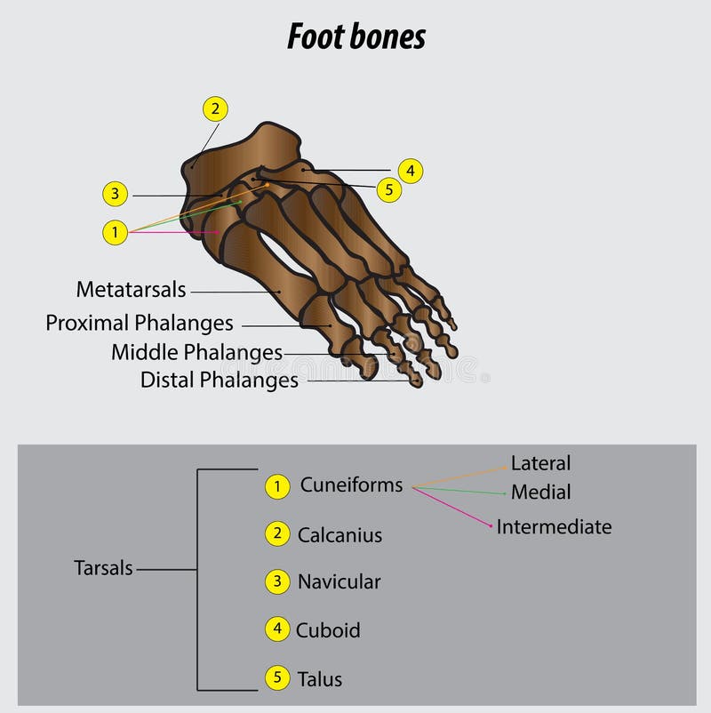
Free with trial Foot bones vector illustration with labels. Medical diagram with tibia, fibula, malleous, talus and navicular. Educational scheme with labeled cuneiforms, cuboid, lateral, calcanius and phalanges. Metatarsal bones vectors Vector illustration with foot bones. Medical diagram and educational scheme with tibia, fibula, malleous, talus and navicular. Foot bones vector illustration. Foot bones vector illustration with labels. Medical diagram with tibia, fibula, malleous, talus and navicular. Educational scheme with labeled cuneiforms, cuboid, lateral, calcanius and phalanges.
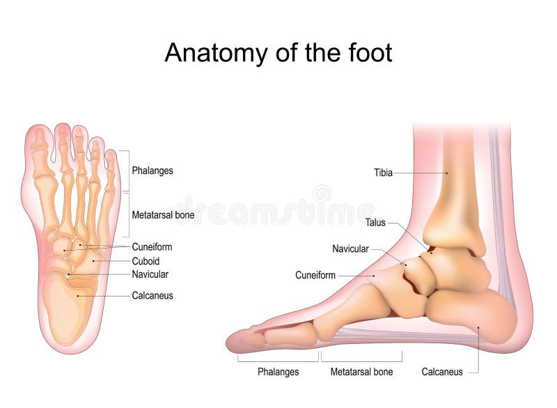
Free with trial Foot anatomy. Human foot with the name and description of all bones and sites. top view and side view. Arches of the feet. skeleton anatomy. Vector illustration. Metatarsal bones vectors Foot anatomy. Human foot with the name and description of all bones and sites. Top view and side view. Arches of the feet. skeleton anatomy. Vector
Free with trial Detailed medical illustration showing the step-by-step removal of Morton s neuroma, a benign nerve tumor located between the metatarsal bones of the foot. Step-by-Step Diagram of Peripheral Nerve Tumor Surgery in Foot. Metatarsal bones vectors Detailed medical illustration showing the step-by-step removal of Morton s neuroma, a benign nerve tumor located between the. Metatarsal bones of the foot. Step. Detailed medical illustration showing the step-by-step removal of Morton s neuroma, a benign nerve tumor located between the metatarsal bones of the foot. Step-by-Step Diagram of Peripheral Nerve Tumor Surgery in Foot
Free with trial Hammer toe medical problem anatomy with foot phalanges deformation outline diagram. Labeled educational scheme with bent metatarsal, proximal, middle and distal bones deformity vector illustration. Metatarsal bones vectors Hammer toe medical problem as foot phalanges deformation outline diagram. Hammer toe medical problem anatomy with foot phalanges deformation outline diagram. Hammer toe medical problem anatomy with foot phalanges deformation outline diagram. Labeled educational scheme with bent metatarsal, proximal, middle and distal bones deformity vector illustration.
Free with trial Front view foot bones. Hand drawn illustration of the foot bones on white, artistic anatomy graphic study. Metatarsal bones vectors Front view foot bones. Hand drawn illustration of the foot bones on white, artistic anatomy graphic study
Free with trial Human leg bones anatomy. Hip, knee, pelvis,femur, foot, toe, joint symbol. Vector flat concept illustration. Isolated on white background. Medical, educational and science bannern. Metatarsal bones vectors Human leg bones anatomy. Hip, knee, pelvis,femur, foot, toe, joint symbol. Vector flat concept illustration. Isolated on white background. Medical, educational
Free with trial Anatomical drawing of the foot. Foot anatomy, a list of all the bones of the foot. Metatarsal bones vectors Anatomical drawing of the foot. Foot anatomy, a list of all the bones of the foot
Free with trial Foot bone anatomy concept. Descriptions of the human feet bones and joints. Talus, ligaments, calcaneus, navicular anatomy. Skeletal medical anatomical poster. Ankle X ray isolated vector illustration. Metatarsal bones vectors Foot bones anatomy. Foot bone anatomy concept. Descriptions of the human feet bones and joints. Talus, ligaments, calcaneus, navicular anatomy. Skeletal medical. Foot bone anatomy concept. Descriptions of the human feet bones and joints. Talus, ligaments, calcaneus, navicular anatomy. Skeletal medical anatomical poster. Ankle X ray isolated vector illustration
Free with trial Skeleton Human leg with pelvis hip silhouette body bones - Thighs, femur, patella, tibia, fibula front view flat black color concept Vector illustration of anatomy isolated on white background. Metatarsal bones vectors Skeleton Human leg with pelvis hip silhouette body bones - Thighs, femur, patella, tibia, fibula front view flat black. Color concept Vector illustration of. Skeleton Human leg with pelvis hip silhouette body bones - Thighs, femur, patella, tibia, fibula front view flat black color concept Vector illustration of anatomy isolated on white background
Free with trial Skeleton Human foot silhouette body bones - front Anterior ventral view. Feet, ankle, tarsals metatarsals phalanges flat black color concept Vector illustration of anatomy isolated on white background. Metatarsal bones vectors Skeleton Human foot silhouette body bones - front Anterior ventral view. Feet, ankle, tarsals metatarsals phalanges. Flat black color concept Vector. Skeleton Human foot silhouette body bones - front Anterior ventral view. Feet, ankle, tarsals metatarsals phalanges flat black color concept Vector illustration of anatomy isolated on white background
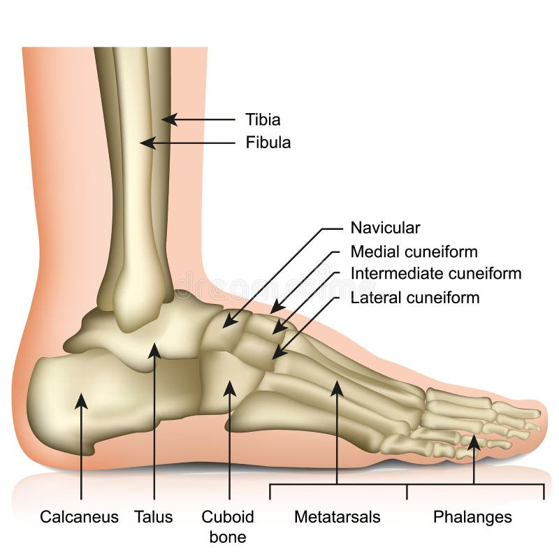
Free with trial Vector illustration of a human leg with denominations of the bones of the foot. Anatomy of lateral or side view of the foot. For advertising or medical publications. EPS 10. Metatarsal bones vectors Anatomy_bones of the human foot lateral view. Vector illustration of a human leg with denominations of the bones of the foot. Anatomy of lateral or side view of. Vector illustration of a human leg with denominations of the bones of the foot. Anatomy of lateral or side view of the foot. For advertising or medical publications. EPS 10.
Free with trial Vector illustration of a human leg with denominations of the bones of the foot. Anatomy of medial or side view of the foot. For advertising or medical publications. EPS 10. Metatarsal bones vectors Anatomy_bones of the human foot medial view. Vector illustration of a human leg with denominations of the bones of the foot. Anatomy of medial or side view of. Vector illustration of a human leg with denominations of the bones of the foot. Anatomy of medial or side view of the foot. For advertising or medical publications. EPS 10.
Free with trial Vector illustration of a human leg with denominations of the bones of the foot. Anatomy of dorsal and plantar views of the foot. For advertising or medical publications. EPS 10. Metatarsal bones vectors Anatomy_bones of the human foot dorsal and plantar view. Vector illustration of a human leg with denominations of the bones of the foot. Anatomy of dorsal and. Vector illustration of a human leg with denominations of the bones of the foot. Anatomy of dorsal and plantar views of the foot. For advertising or medical publications. EPS 10.
Free with trial Mortons neuroma. Foot pain, plantar fasciitis inflammation and ruptures strain. Bones realistic illustration. Bottom view. Anatomy of joints, human leg black and yellow transparente skeleton. For medical orthopedic advertising. Vector illustration stock vector. Metatarsal bones vectors Mortons neuroma. Foot pain strain bottom view. Realistic anatomy illustration. Mortons neuroma. Foot pain, plantar fasciitis inflammation and ruptures strain. Mortons neuroma. Foot pain, plantar fasciitis inflammation and ruptures strain. Bones realistic illustration. Bottom view. Anatomy of joints, human leg black and yellow transparente skeleton. For medical orthopedic advertising. Vector illustration stock vector.
Free with trial Vector drawing of human foot human foot anatomy drawing labeled. Drawing with layers reedit able. Metatarsal bones vectors Vector drawing of human foot human foot anatomy drawing labeled. Drawing with layers reedit able
Free with trial Arches of the feet vector. Foot skeleton anatomy. vector. illustration. Metatarsal bones vectors Arches of the feet vector. Foot skeleton anatomy. Vector. illustration
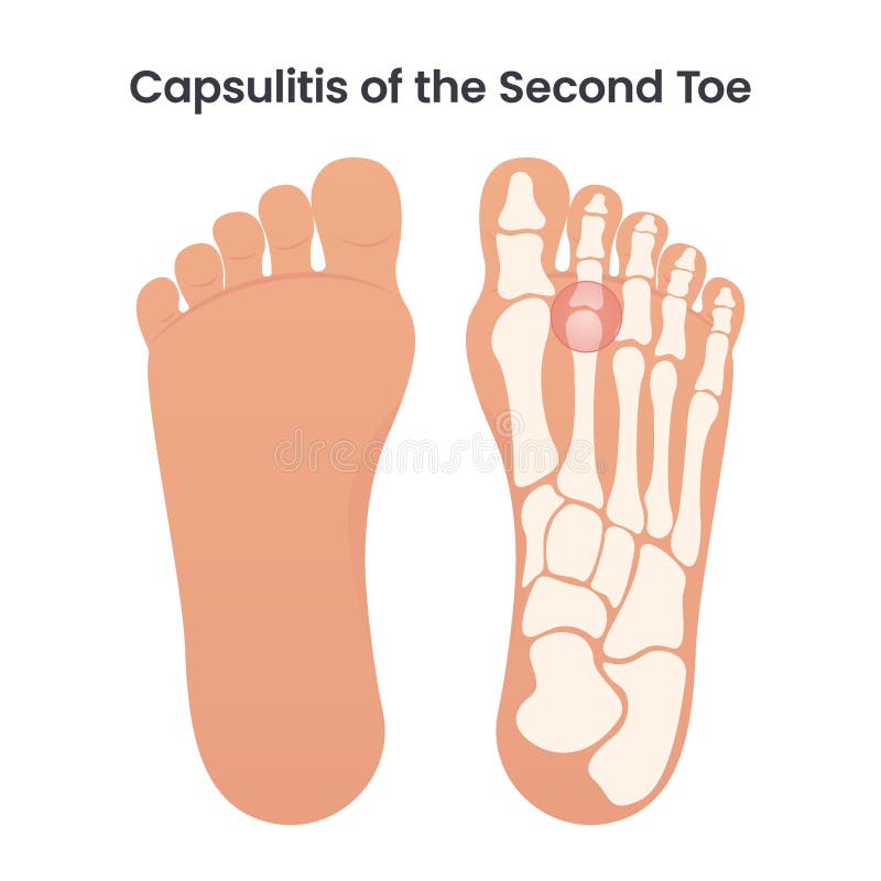
Free with trial Capsulitis of the second toe occurs in the ligaments surrounding the joint at the base of the second toe. These ligaments form a capsule, which helps the joint to function properly. Capsulitis is a condition in which these ligaments have become inflamed. Metatarsal bones vectors Capsulitis of the Second Bone educational medical vector illustration. Capsulitis of the second toe occurs in the ligaments surrounding the joint at the base of. Capsulitis of the second toe occurs in the ligaments surrounding the joint at the base of the second toe. These ligaments form a capsule, which helps the joint to function properly. Capsulitis is a condition in which these ligaments have become inflamed
Free with trial Illustration of biology and medical, Foot arches, metatarsal bones, the ligaments and tendons in the foot, the foot to support the weight of the body. Metatarsal bones vectors Illustration of biology and medical, Foot arches, metatarsal bones. The ligaments and tendons in the foot, the foot to support the weight of the body
Free with trial A black-and-white anatomical diagram presents eight labeled bones of the foot: the calcaneus, talus, navicular, cuboid, medial, intermediate and lateral cuneiforms, and first metatarsal. Each bone is shown separately as a simplified, outlined with shaded areas suggesting its shape and contours; the first metatarsal appears elongated, while the tarsal bones are more compact and irregular. The clean layout on a white background emphasizes identification and comparison, with bold labels beneath each drawing. Metatarsal bones vectors A black-and-white anatomical diagram presents eight labeled bones of the foot: the calcaneus, talus, navicular, cuboid, medial. Intermediate and lateral. A black-and-white anatomical diagram presents eight labeled bones of the foot: the calcaneus, talus, navicular, cuboid, medial, intermediate and lateral cuneiforms, and first metatarsal. Each bone is shown separately as a simplified, outlined with shaded areas suggesting its shape and contours; the first metatarsal appears elongated, while the tarsal bones are more compact and irregular. The clean layout on a white background emphasizes identification and comparison, with bold labels beneath each drawing.
Free with trial Human Ankle Joint Bones Skeleton Icons. Presented in outline and solid black styles for medical or anatomical representation. Metatarsal bones vectors Human Ankle Joint Bones Skeleton Icons. Presented in outline and solid black styles for medical or anatomical representation
Free with trial Human foot bones illustration. Detailed anatomical illustration of a human foot showcasing its bones and structure, ideal for medical or educational purposes. Metatarsal bones vectors Human foot bones illustration. Detailed anatomical illustration of a human foot showcasing its bones and structure, ideal for medical or educational purposes
Free with trial Detailed anatomical illustration of human feet bones in a vintage style, set against a dark background, showcasing skeletal structure and medical detail. Metatarsal bones vectors Anatomical Illustration of Human Feet Bones on Dark Background. Detailed anatomical illustration of human feet bones in a vintage style, set against a dark. Detailed anatomical illustration of human feet bones in a vintage style, set against a dark background, showcasing skeletal structure and medical detail
Free with trial This is a detailed illustration of a human foot skeleton, showcasing the intricate structure of the skeletal system. The image features a gray foot skeleton with a transparent background, allowing for clear visibility of the bones. The skeleton is composed of multiple bones, including the phalanges, metatarsal, tarsal, calcaneus, talus, cuboid, navicular, and cuneiform. This image is ideal for medical, scientific, and educational purposes, providing a comprehensive view of the human foot anatomy. Metatarsal bones vectors A detailed illustration of a human foot skeleton on transparent background. This is a detailed illustration of a human foot skeleton, showcasing the intricate. This is a detailed illustration of a human foot skeleton, showcasing the intricate structure of the skeletal system. The image features a gray foot skeleton with a transparent background, allowing for clear visibility of the bones. The skeleton is composed of multiple bones, including the phalanges, metatarsal, tarsal, calcaneus, talus, cuboid, navicular, and cuneiform. This image is ideal for medical, scientific, and educational purposes, providing a comprehensive view of the human foot anatomy.
Free with trial Illustration shows human foot and lower leg bones in simple line art style for medical education and anatomical study, clear and informative mood. Generative AI. Metatarsal bones vectors Human foot and lower leg bones anatomy line art medical vector illustration. Illustration shows human foot and lower leg bones in simple line art style for. Illustration shows human foot and lower leg bones in simple line art style for medical education and anatomical study, clear and informative mood. Generative AI
Free with trial Anatomical human foot skeleton showing the metatarsal bones in tea-stained bone in top angle view on a plain white background. Sharp detail and even light suit education and editorial design. Metatarsal bones vectors Human foot skeleton top angle view isolated on white background. Anatomical human foot skeleton showing the metatarsal bones in tea-stained bone in top angle. Anatomical human foot skeleton showing the metatarsal bones in tea-stained bone in top angle view on a plain white background. Sharp detail and even light suit education and editorial design
Free with trial Anatomical human foot skeleton showing the metatarsal bones in tea-stained bone in three quarter view on a plain white background. Sharp detail and even light suit education and editorial design. Metatarsal bones vectors Human foot skeleton three quarter view isolated on white background. Anatomical human foot skeleton showing the metatarsal bones in tea-stained bone in three. Anatomical human foot skeleton showing the metatarsal bones in tea-stained bone in three quarter view on a plain white background. Sharp detail and even light suit education and editorial design
Free with trial Anatomical human foot skeleton showing the metatarsal bones in tea-stained bone in side view on a plain white background. A clean studio photo with a crisp outline ready for design and print. Metatarsal bones vectors Human foot skeleton side view isolated on white background studio photo. Anatomical human foot skeleton showing the metatarsal bones in tea-stained bone in side. Anatomical human foot skeleton showing the metatarsal bones in tea-stained bone in side view on a plain white background. A clean studio photo with a crisp outline ready for design and print
Free with trial Illustration shows human leg and foot skeleton with detailed bones in black and white line art style, clear anatomical focus, educational tone. Generative AI. Metatarsal bones vectors Human Leg and Foot Skeleton Anatomy Illustration with Detailed Bones Vector Graphic. Illustration shows human leg and foot skeleton with detailed bones in black. Illustration shows human leg and foot skeleton with detailed bones in black and white line art style, clear anatomical focus, educational tone. Generative AI
Free with trial Anatomical human foot skeleton showing the metatarsal bones in tea-stained bone in front view on a plain white background. A clean studio photo with a crisp outline ready for design and print. Metatarsal bones vectors Human foot skeleton front view isolated on white background studio photo. Anatomical human foot skeleton showing the metatarsal bones in tea-stained bone in. Anatomical human foot skeleton showing the metatarsal bones in tea-stained bone in front view on a plain white background. A clean studio photo with a crisp outline ready for design and print
Free with trial Illustration shows human foot bones with highlighted ankle joints in a clean medical style vector image. Educational and informative mood. Generative AI. Metatarsal bones vectors Human foot bones skeletal anatomy illustration with highlighted ankle joints in medical style vector. Illustration shows human foot bones with highlighted ankle. Illustration shows human foot bones with highlighted ankle joints in a clean medical style vector image. Educational and informative mood. Generative AI
Free with trial A clean side-view anatomical shows the bones and soft tissues of a human foot and lower ankle, with the heel at left and toes extending to the right. Cream-colored bones trace the ankle, arch, and five toes, while white tendons and ligaments cross the pink and red muscle structures, all outlined in dark ink. The uncluttered white background and warm colors make the layered anatomy easy to distinguish, giving the image a clear educational reference quality. Metatarsal bones vectors Human Foot Bones and Tendons Anatomy Illustration – Side View Skeletal Structure with Ankle, Heel and Ligaments. A clean side-view anatomical shows the bones. A clean side-view anatomical shows the bones and soft tissues of a human foot and lower ankle, with the heel at left and toes extending to the right. Cream-colored bones trace the ankle, arch, and five toes, while white tendons and ligaments cross the pink and red muscle structures, all outlined in dark ink. The uncluttered white background and warm colors make the layered anatomy easy to distinguish, giving the image a clear educational reference quality.
Free with trial This detailed anatomical shows the human foot and ankle skeletal structure, highlighting the complex arrangement of bones and joints in a clear side profile view. Metatarsal bones vectors Anatomical of the human foot and ankle skeletal structure showing bones and joints in a side profile view for medical. This detailed anatomical shows the human. This detailed anatomical shows the human foot and ankle skeletal structure, highlighting the complex arrangement of bones and joints in a clear side profile view
Free with trial A high-quality, detailed anatomical of the human foot skeleton, showcasing the intricate structure of bones including the calcaneus, talus, and phalanges. Metatarsal bones vectors Detailed anatomical diagram of human foot bones showing skeletal structure with labels for medical education isolated on white. A high-quality, detailed. A high-quality, detailed anatomical of the human foot skeleton, showcasing the intricate structure of bones including the calcaneus, talus, and phalanges
Free with trial Orthopedic insole supporting metatarsal bones and arch of the foot, helping to reduce pain and improve posture. Metatarsal bones vectors Orthopedic insole for foot maintaining correct position while walking. Orthopedic insole supporting metatarsal bones and arch of the foot, helping to reduce pain. Orthopedic insole supporting metatarsal bones and arch of the foot, helping to reduce pain and improve posture
Free with trial Healthy foot and a foot with Sesamoid boneS fracture. Vector illustration for medical publiatios. Metatarsal bones vectors Healthy foot and a foot with Sesamoid boneS fracture. Vector illustration for medical publiatios
Free with trial Plantar fascial fibromatosis. Plantar fibroma. Ledderhoses disease. Morbus Ledderhose. Skeleton of foot. Medial aspect of bones and plantar fascia with non-malignant thickening of connective tissue. Detailed Vector poster. Metatarsal bones vectors Plantar fascial fibromatosis. Plantar fibroma. Ledderhoses disease. Morbus Ledderhose. Skeleton of foot. Medial aspect of bones and plantar fascia with non
Free with trial Foot Anterior View X Ray Displaying Clear Fracture Line, Metatarsal Bones, Phalanges, And Joint Alignment For Accurate Clinical Evaluation. High Resolution Radiographic Detail Highlights Bone Integrity And Fracture Visualization. Suitable For Orthopedic Diagnosis, Radiology Education, And Medical Publications. Isolated On A Transparent Background For Seamless Integration Into Healthcare And Scientific Visuals. Metatarsal bones vectors Foot Anterior View X Ray Showing Fracture Isolated On A Transparent Background. Foot Anterior View X Ray Displaying Clear Fracture Line, Metatarsal Bones. Foot Anterior View X Ray Displaying Clear Fracture Line, Metatarsal Bones, Phalanges, And Joint Alignment For Accurate Clinical Evaluation. High Resolution Radiographic Detail Highlights Bone Integrity And Fracture Visualization. Suitable For Orthopedic Diagnosis, Radiology Education, And Medical Publications. Isolated On A Transparent Background For Seamless Integration Into Healthcare And Scientific Visuals.
Free with trial Detailed Anterior View Foot Fracture Radiograph Illustrating Bone Discontinuity, Metatarsal Structure, And Joint Alignment With Clear Radiographic Detail. This High Resolution Medical Image Supports Orthopedic Diagnosis, Radiology Education, And Clinical Evaluation. Suitable For Healthcare Publications And Scientific Materials. Metatarsal bones vectors Detailed Anterior View Foot Fracture Radiograph Isolated On A Transparent Background. Detailed Anterior View Foot Fracture Radiograph Illustrating Bone. Detailed Anterior View Foot Fracture Radiograph Illustrating Bone Discontinuity, Metatarsal Structure, And Joint Alignment With Clear Radiographic Detail. This High Resolution Medical Image Supports Orthopedic Diagnosis, Radiology Education, And Clinical Evaluation. Suitable For Healthcare Publications And Scientific Materials.
Free with trial Detailed Anatomical Illustration of Human Foot, Ankle, Bones, Muscles, and Achilles Tendon. Metatarsal bones vectors Detailed Anatomical Illustration of Human Foot, Ankle, Bones, Muscles, and Achilles Tendon
Free with trial Detailed illustration of a human foot skeleton showcasing bones and joints perfect for anatomical diagrams or medical education resources. Metatarsal bones vectors Human foot bones with transparent background. Detailed illustration of a human foot skeleton showcasing bones and joints perfect for anatomical diagrams or. Detailed illustration of a human foot skeleton showcasing bones and joints perfect for anatomical diagrams or medical education resources
Free with trial Detailed diagram of a human foot showcasing its skeletal structure and individual bones, perfect for educational materials or medical illustrations. This image has a transparent background. Metatarsal bones vectors Human foot bones diagram with transparent background. Detailed diagram of a human foot showcasing its skeletal structure and individual bones, perfect for. Detailed diagram of a human foot showcasing its skeletal structure and individual bones, perfect for educational materials or medical illustrations. This image has a transparent background
Free with trial A clear, line-art illustration depicting the skeletal structure of the human foot and ankle, showing the tibia, fibula, tarsals, metatarsals, and phalanges. Generated using AI. Metatarsal bones vectors Anatomical Illustration of the Human Foot and Ankle Bones. A clear, line-art illustration depicting the skeletal structure of the human foot and ankle, showing. A clear, line-art illustration depicting the skeletal structure of the human foot and ankle, showing the tibia, fibula, tarsals, metatarsals, and phalanges. Generated using AI.
Free with trial Thickening of a plantar digital nerve, usually between the 3rd–4th toes. Signs Symptoms: Burning forefoot pain, tingling, numbness, 'pebble in shoe' feeling. Triggers: Tight or high-heeled footwear, walking. Nature: Benign nerve compression condition. Metatarsal bones vectors Morton\'s Neuroma Foot Medical Flashcard. Thickening of a plantar digital nerve, usually between the 3rd–4th toes.Signs Symptoms: Burning forefoot pain. Thickening of a plantar digital nerve, usually between the 3rd–4th toes.Signs Symptoms: Burning forefoot pain, tingling, numbness, 'pebble in shoe' feeling.Triggers: Tight or high-heeled footwear, walking.Nature: Benign nerve compression condition.
Free with trial Anatomy of bone ankle toe talus, concept Vector illustration of anatomy isolated on white background. Metatarsal bones vectors Anatomy of bone ankle toe talus. Concept Vector illustration of anatomy isolated on white background
Free with trial High Resolution Foot Anterior View X Ray Depicting Clear Fracture Line, Bone Alignment, And Joint Structure With Precise Radiographic Detail. This Professional Medical Illustration Supports Orthopedic Diagnosis, Radiology Education, And Clinical Evaluation. Accurate Anatomical Representation Ensures Effective Use In Scientific Publications And Healthcare Training Materials. Metatarsal bones vectors High Resolution Foot Anterior View Fracture X Ray Isolated On A Transparent Background. High Resolution Foot Anterior View X Ray Depicting Clear Fracture Line. High Resolution Foot Anterior View X Ray Depicting Clear Fracture Line, Bone Alignment, And Joint Structure With Precise Radiographic Detail. This Professional Medical Illustration Supports Orthopedic Diagnosis, Radiology Education, And Clinical Evaluation. Accurate Anatomical Representation Ensures Effective Use In Scientific Publications And Healthcare Training Materials.
 Sign in