Free with trial Bunion in foot. Deformity of the big toe joint usually due to tight fitting shoes, eps10. Metatarsal vectors Bunion in foot. Deformity of the big toe joint usually due to tight fitting shoes, eps10
Free with trial The ankle joint. Lateral and front views of the ankle joint, eps8. Metatarsal vectors The ankle joint. Lateral and front views of the ankle joint, eps8
Free with trial Hallux valgus. Medical illustration of the symptoms of hallux valgus. Metatarsal vectors Hallux valgus. Medical illustration of the symptoms of hallux valgus
Free with trial Illustration (diagram) of the fifth metatarsal Fractures (types and zones classification) in the foot. Metatarsal vectors Fractures of the 5th metatarsal. Illustration (diagram) of the fifth metatarsal Fractures (types and zones classification) in the foot. Illustration (diagram) of the fifth metatarsal Fractures (types and zones classification) in the foot.
Free with trial Vector diagram of forefoot pain, metatarsalgia symptom, tenderness in the balls of metatarsal bones of the foot. Used: Gradient, transparency, blend mode. Metatarsal vectors Metatarsalgia. Vector diagram of forefoot pain, metatarsalgia symptom, tenderness in the balls of metatarsal bones of the foot. Used: Gradient, transparency. Vector diagram of forefoot pain, metatarsalgia symptom, tenderness in the balls of metatarsal bones of the foot. Used: Gradient, transparency, blend mode.
Free with trial Vector illustration of a healthy human foot and a foot with lisfranc injury with weight bearing and mechanism of injury, dislocation of the second metatarsal bone. Medial, top view of the foot. EPS 10. Metatarsal vectors Lisfranc Injury. Vector illustration of a healthy human foot and a foot with lisfranc injury with weight bearing and mechanism of injury, dislocation of the. Vector illustration of a healthy human foot and a foot with lisfranc injury with weight bearing and mechanism of injury, dislocation of the second metatarsal bone. Medial, top view of the foot. EPS 10
Free with trial Turf toe. Sprain or tears of ligaments of the big toe joint, a common sport injury, eps10. Metatarsal vectors Turf toe. Sprain or tears of ligaments of the big toe joint, a common sport injury, eps10
Free with trial Bunion. Human anatomy. Skeleton. Healthy foot and foot with Bunion. Metatarsal vectors Bunion. Human anatomy. Skeleton. Healthy foot and foot with Bunion. Human anatomy. Skeleton
Free with trial Pott fracture. A common sport injury of the ankle joint, eps8. Metatarsal vectors Pott fracture. A common sport injury of the ankle joint, eps8
Free with trial Plantar fasciitis. Illustration of Plantar fasciitis. Bottom and side view of skeletal ankle. Used: gradient, transparency, Blend mode. Metatarsal vectors Plantar fasciitis. Illustration of Plantar fasciitis. Bottom and side view of skeletal ankle. Used: gradient, transparency, Blend mode. Illustration of Plantar fasciitis. Bottom and side view of skeletal ankle. Used: gradient, transparency, Blend mode.
Free with trial Gout is a form of inflammatory arthritis. Characterized by elevated levels of uric acid in the blood. This uric acid crystallizes and the crystals deposit in joints, tendons, and surrounding tissues. Vector illustration for medical use. Metatarsal vectors Gout is a form of inflammatory arthritis. Characterized by elevated levels of uric acid in the blood. This uric acid crystallizes and the crystals deposit in
Free with trial Broken toe. Common sites of toe bone fractures, eps10. Metatarsal vectors Broken toe. Common sites of toe bone fractures, eps10
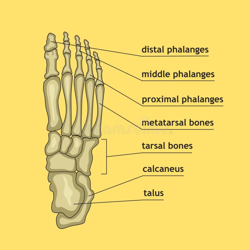
Free with trial Anatomy of a skeleton foot. Vector illustration of anatomy of the skeleton of the foot. Metatarsal vectors Anatomy of a skeleton foot. Vector illustration of anatomy of the skeleton of the foot
Free with trial Illustration (diagram) of normal function, limited motion and rigid deformity (with degenerative arthritis) of the first metatarsophalangeal joint of the foot. Used: Gradient, blend, transparecy, blend mode. Metatarsal vectors Hallux rigidus. Illustration (diagram) of normal function, limited motion and rigid deformity (with degenerative arthritis) of the first metatarsophalangeal. Illustration (diagram) of normal function, limited motion and rigid deformity (with degenerative arthritis) of the first metatarsophalangeal joint of the foot. Used: Gradient, blend, transparecy, blend mode.
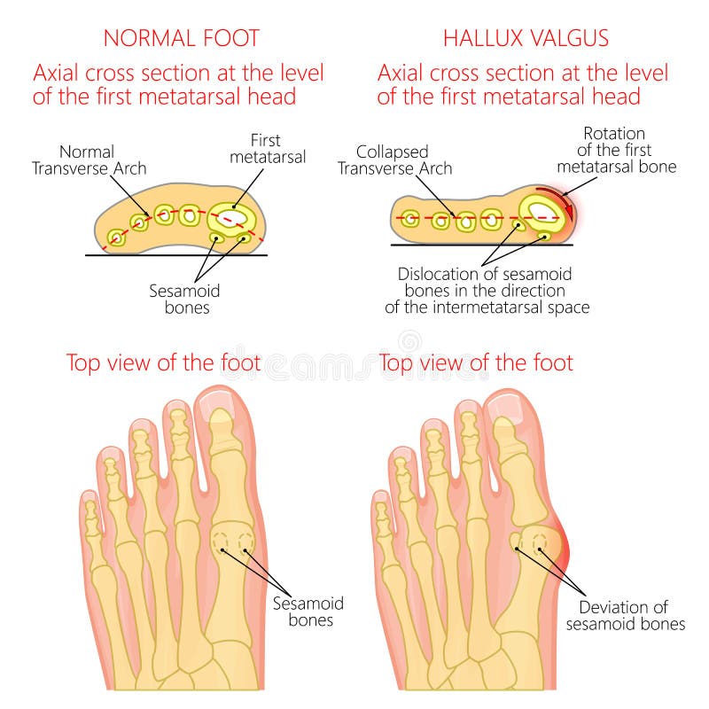
Free with trial Vector illustration of a healthy human forefoot and a foot with hallux valgus, dislocation of sesamoid bones. Top view and cross section of of the foot. For advertising, medical publications. EPS 10. Metatarsal vectors Normal foot and Hallux valgus with rotation of the first mematarsal bone. Vector illustration of a healthy human forefoot and a foot with hallux valgus. Vector illustration of a healthy human forefoot and a foot with hallux valgus, dislocation of sesamoid bones. Top view and cross section of of the foot. For advertising, medical publications. EPS 10.
Free with trial Different views of a human foot front, back, side, lateral, medial, dorsal and plantar isolated on white background. Vector illustration for medical health care use. EPS 10. Metatarsal vectors Different views of a human foot on white background_Anatomy. Different views of a human foot front, back, side, lateral, medial, dorsal and plantar isolated on. Different views of a human foot front, back, side, lateral, medial, dorsal and plantar isolated on white background. Vector illustration for medical health care use. EPS 10.
Free with trial Deviation of the first and the fifth metatarsals, transverse flatfoot. Used: gradient, transparency, blend mode. Metatarsal vectors Deviation of the first and the fifth metatarsals transverse flat. Deviation of the first and the fifth metatarsals, transverse flatfoot. Used: gradient. Deviation of the first and the fifth metatarsals, transverse flatfoot. Used: gradient, transparency, blend mode.
Free with trial Illustration of the normal foot and hallux valgus with indicating of the first metatarsophalangeal and the first-second intermetatarsal angles. Used: gradient, transparency. Blend mode. Metatarsal vectors Normal foot and valgus deviation of the first toe with indicating of the deflection angles. Illustration of the normal foot and hallux valgus with indicating of. Illustration of the normal foot and hallux valgus with indicating of the first metatarsophalangeal and the first-second intermetatarsal angles. Used: gradient, transparency. Blend mode.
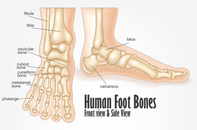
Free with trial Human foot bones front and side view anatomy. Illustration Of Human foot bones front and side view anatomy. Metatarsal vectors Human foot bones front and side view anatomy. Illustration Of Human foot bones front and side view anatomy
Free with trial Illustration (Diagram) of Metatarsal Stress fractures in the foot. Metatarsal vectors Metatarsal Stress fractures. Illustration (Diagram) of Metatarsal Stress fractures in the foot. Illustration (Diagram) of Metatarsal Stress fractures in the foot.
Free with trial Peroneus brevis leg muscle with longus and tertius muscular part location outline diagram. Labeled educational foot skeletal structure from later view vector illustration. Tibia and metatarsal bone. Metatarsal vectors Peroneus brevis leg muscle with longus and tertius location outline diagram. Peroneus brevis leg muscle with longus and tertius muscular part location outline. Peroneus brevis leg muscle with longus and tertius muscular part location outline diagram. Labeled educational foot skeletal structure from later view vector illustration. Tibia and metatarsal bone.
Free with trial Bones of foot. Human Anatomy. The diagram shows the placement and names of all bones of foot. Metatarsal vectors Bones of foot. Human Anatomy. The diagram shows the placement and names of all
Free with trial Peroneus tertius leg muscle with longus and brevis location outline diagram. Labeled educational anatomical scheme with human foot skeletal and muscular system from lateral view vector illustration. Metatarsal vectors Peroneus tertius leg muscle with longus and brevis location outline diagram. Labeled educational anatomical scheme with human foot skeletal and muscular system
Free with trial Vector illustration of peroneus longus and peroneus brevis muscle. Lateral view of human leg and ankle and bottom or plantar view of the foot. EPS 10. Metatarsal vectors Peroneus longus muscle. Vector illustration of peroneus longus and peroneus brevis muscle. Lateral view of human leg and ankle and bottom or plantar view of the. Vector illustration of peroneus longus and peroneus brevis muscle. Lateral view of human leg and ankle and bottom or plantar view of the foot. EPS 10.
Free with trial Illustration (diagram) of toe fracture types. Metatarsal vectors Toe fractures. Illustration (diagram) of toe fracture types. Illustration (diagram) of toe fracture types.
Free with trial Illustration of Skeletal ankle (side view) with Retrocalcaneal and Subcutaneous calcaneal bursitis. Used: Gradient, blend, transparency, blend mode. Metatarsal vectors Achilles bursitis. Illustration of Skeletal ankle (side view) with Retrocalcaneal and Subcutaneous calcaneal bursitis. Used: Gradient, blend, transparency, blend. Illustration of Skeletal ankle (side view) with Retrocalcaneal and Subcutaneous calcaneal bursitis. Used: Gradient, blend, transparency, blend mode.
Free with trial Vector illustration diagram. Turf toe. Sport trauma. Mechanism of rupture of plantar plate of the first toe of the foot. External and skeletal views of an ankle. Used: blend. Metatarsal vectors Turf toe. Rupture of the plantar plate of the first toe of the f. Vector illustration diagram.Turf toe. Sport trauma. Mechanism of rupture of plantar plate of. Vector illustration diagram.Turf toe. Sport trauma. Mechanism of rupture of plantar plate of the first toe of the foot. External and skeletal views of an ankle. Used: blend.
Free with trial Diagnosis of the tailor's bunion with angles of deviation. Metatarsal vectors Diagnosis of the tailor's bunion. With angles of deviation
Free with trial Bunionectomy is a procedure to correct of pathologies and deformity of the joint connecting the big toe to the foot. Before and after of Surgery. Healthy foot and foot with Bunion. Human anatomy. Skeleton. Metatarsal vectors Bunionectomy. surgery to correct pathologies of the foot. Bunionectomy is a procedure to correct of pathologies and deformity of the joint connecting the big. Bunionectomy is a procedure to correct of pathologies and deformity of the joint connecting the big toe to the foot. Before and after of Surgery. Healthy foot and foot with Bunion. Human anatomy. Skeleton
Free with trial Bones of the foot and ankle joint medical vector illustration isolated on white background. Eps 10 infographic. Metatarsal vectors Bones of the foot and ankle joint medical vector illustration isolated on white background. Eps 10 infographic
Free with trial Illustration of the normal foot and hallux varus. Used: gradient, transparency, Blend mode. Metatarsal vectors Normal foot and Hallux varus. Illustration of the normal foot and hallux varus. Used: gradient, transparency, Blend mode. Illustration of the normal foot and hallux varus. Used: gradient, transparency, Blend mode.
Free with trial Vector illustration of Peroneal Tendon Injuries. Peroneal tendonitis. Inflammation of peroneal tendons. Lateral ankle injury. Metatarsal vectors Peroneal Tendon Injuries_Peroneal tendonitis. Vector illustration of Peroneal Tendon Injuries. Peroneal tendonitis. Inflammation of peroneal tendons. Lateral. Vector illustration of Peroneal Tendon Injuries. Peroneal tendonitis. Inflammation of peroneal tendons. Lateral ankle injury.
Free with trial Different views of a human foot front, back, side, lateral, medial, dorsal and plantar isolated on white background. Vector illustration for medical health care use. EPS 10. Metatarsal vectors Different views of a human foot on white background_Skeleton. Different views of a human foot front, back, side, lateral, medial, dorsal and plantar isolated on. Different views of a human foot front, back, side, lateral, medial, dorsal and plantar isolated on white background. Vector illustration for medical health care use. EPS 10.
Free with trial Vector illustration diagram. Hammer toe. Plantar plate injury. Mechanism of rupture of plantar plate of the second toe of the foot. External and skeletal views of an ankle. Metatarsal vectors Plantar plate tear. Hammer toe. Vector illustration diagram. Hammer toe. Plantar plate injury. Mechanism of rupture of plantar plate of the second toe of the. Vector illustration diagram. Hammer toe. Plantar plate injury. Mechanism of rupture of plantar plate of the second toe of the foot. External and skeletal views of an ankle.
Free with trial Vector illustration diagram. Deviation of the toe. Mechanism of rupture of plantar plate of the seond toe of the foot. External and skeletal views of an ankle. Metatarsal vectors Plantar plate tear. Deviation of the toe. Vector illustration diagram. Deviation of the toe. Mechanism of rupture of plantar plate of the seond toe of the foot. Vector illustration diagram. Deviation of the toe. Mechanism of rupture of plantar plate of the seond toe of the foot. External and skeletal views of an ankle.
Free with trial Bunions vector illustration. Labeled feet bone disorder explanation scheme. Painful toe joint condition. Orthopedics inflammation problem with deformity. Medical barefoot xray infographic diagram. Metatarsal vectors Bunions vector illustration. Labeled feet bone disorder explanation scheme. Painful toe joint condition. Orthopedics inflammation problem with deformity
Free with trial Genu varum, genu valgum - a physical legs deformity, bowing of the leg in relation to the thigh. Metatarsal vectors Genu varum, genu valgum - a physical legs deformity. Bowing of the leg in relation to the thigh
Free with trial Abnormal of foot shape from deformity joint connecting the big toe. Problem of foot form wearing high heel make. Metatarsal vectors Abnormal of foot shape from deformity joint toe. Abnormal of foot shape from deformity joint connecting the big toe. Problem of foot form wearing high heel make. Abnormal of foot shape from deformity joint connecting the big toe. Problem of foot form wearing high heel make.
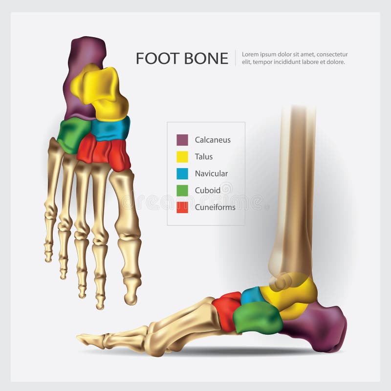
Free with trial Human Anatomy Foot Bone. Vector Illustration. Metatarsal vectors Human Anatomy Foot Bone. Vector Illustration
Free with trial Deviation of the first and the fifth metatarsals transverse flat. Foot with orthopedic insole. Used: gradient, transparency, blend mode. Metatarsal vectors Deviation of the first and the fifth metatarsals transverse flat. Foot with orthopedic insole. Used: gradient, transparency, blend mode
Free with trial Human Anatomy Leg Bones with Detail. Vector Illustration. Metatarsal vectors Human Anatomy Leg Bones with Detail. Vector Illustration
Free with trial Human body shapes. Feet types icon set. Vector illustration. Metatarsal vectors Human body shapes.Feet types icon set. Vector illustration
Free with trial Illustration (diagram) of Metatarsal fracture types. Metatarsal vectors Metatarsal fractures. Illustration (diagram) of Metatarsal fracture types. Illustration (diagram) of Metatarsal fracture types.
Free with trial Fifth metatarsal or foot little finger fracture after injury outline diagram. Labeled educational scheme with feet trauma after twisting motion vector illustration. Anatomical skeletal bone zones. Metatarsal vectors Fifth metatarsal or foot little finger fracture after injury outline diagram. Labeled educational scheme with feet trauma after twisting motion vector
Free with trial Stress fracture. Marching fracture of the foot. Fracture of the metatarsal bones in the foot. Anatomical structure of the foot. Skeleton. Broken bones. Vector illustration on isolated background. Metatarsal vectors Stress fracture. Marching fracture of the foot. Fracture of the metatarsal bones in the foot. Anatomical structure of. The foot. Skeleton. Broken bones. Vector. Stress fracture. Marching fracture of the foot. Fracture of the metatarsal bones in the foot. Anatomical structure of the foot. Skeleton. Broken bones. Vector illustration on isolated background
Free with trial Fracture of the metatarsal bones in the foot. Anatomical structure of the foot. Skeleton. Broken bones. Vector illustration on isolated background. Metatarsal vectors Fracture of the metatarsal bones in the foot. Anatomical structure of the foot. Skeleton. Broken bones. Vector. Illustration on isolated background
Free with trial Detailed medical illustration showing the step-by-step removal of Morton s neuroma, a benign nerve tumor located between the metatarsal bones of the foot. Step-by-Step Diagram of Peripheral Nerve Tumor Surgery in Foot. Metatarsal vectors Detailed medical illustration showing the step-by-step removal of Morton s neuroma, a benign nerve tumor located between the. Metatarsal bones of the foot. Step. Detailed medical illustration showing the step-by-step removal of Morton s neuroma, a benign nerve tumor located between the metatarsal bones of the foot. Step-by-Step Diagram of Peripheral Nerve Tumor Surgery in Foot
Free with trial Foot bones anatomy. Structure and names. Metatarsal bones location. Medical scheme. Vector illustration in realistic style isolated on green background. Educational poster. Medicine concept. Metatarsal vectors Foot bones anatomy. Structure and names. Metatarsal bones location. Medical scheme. Vector illustration in realistic style isolated on green background
Free with trial Hammer toe medical problem anatomy with foot phalanges deformation outline diagram. Labeled educational scheme with bent metatarsal, proximal, middle and distal bones deformity vector illustration. Metatarsal vectors Hammer toe medical problem as foot phalanges deformation outline diagram. Hammer toe medical problem anatomy with foot phalanges deformation outline diagram. Hammer toe medical problem anatomy with foot phalanges deformation outline diagram. Labeled educational scheme with bent metatarsal, proximal, middle and distal bones deformity vector illustration.
Free with trial In the dorsal (posterior) view of the bones of the feet, the tarsal bones are visible, including the talus, calcaneus, navicular, cuboid, and the three cuneiform bones. The metatarsal bones, numbering from one to five, can also be observed, extending towards the toes. Additionally, the phalanges, consisting of proximal, middle, and distal phalanges, complete the skeletal structure of the feet in the dorsal view. Metatarsal vectors Bones of feet, dorsal (posterior) view. In the dorsal (posterior) view of the bones of the feet, the tarsal bones are visible, including the talus, calcaneus. In the dorsal (posterior) view of the bones of the feet, the tarsal bones are visible, including the talus, calcaneus, navicular, cuboid, and the three cuneiform bones. The metatarsal bones, numbering from one to five, can also be observed, extending towards the toes. Additionally, the phalanges, consisting of proximal, middle, and distal phalanges, complete the skeletal structure of the feet in the dorsal view.
Free with trial Medical illustration of Abductor Hallucis muscle foot, oblique side view of the foot. The metatarsal bones and muscles of the feet. Line drawings for medicine and sports science or education learning. Metatarsal vectors Medical illustration of Abductor Hallucis muscle foot, oblique side view of the foot. The metatarsal bones and muscles of the feet. Line drawings for medicine
Free with trial Bones of the lower extremity. Metatarsal bone and phalanges of the third toe of the right side. Detailed medical illustration. Isolated on a white background. Bright and clean design. Metatarsal vectors Bones of the foot. Bones of the lower extremity. Metatarsal bone and phalanges of the third toe of the right side. Detailed medical illustration. Isolated on a. Bones of the lower extremity. Metatarsal bone and phalanges of the third toe of the right side. Detailed medical illustration. Isolated on a white background. Bright and clean design.
Free with trial Vector illustration of foot - normal and with Morton`s toe or Morton`s Foot Syndrome, short first metatarsal bone relative to the second metatarsal. Top view of forefoot and and skeleton of forefoot. Metatarsal vectors Morton`s toe. Vector illustration of foot - normal and with Morton`s toe or Morton`s Foot Syndrome, short first metatarsal bone relative to the second metatarsal. Vector illustration of foot - normal and with Morton`s toe or Morton`s Foot Syndrome, short first metatarsal bone relative to the second metatarsal. Top view of forefoot and and skeleton of forefoot.
Free with trial Toe shape. Types of foot shape - the most common variants of the foot and toes on white background. Metatarsal vectors Toe shape. Types of foot shape - the most common variants of the foot and toes on white background
Free with trial Bones of foot. Human Anatomy. The diagram shows the placement and names of all bones of foot. Metatarsal vectors Bones of foot. Human Anatomy. The diagram shows the placement and names of all
Free with trial Valgus deformity. On a white background. Metatarsal vectors Valgus deformity. On a white background
Free with trial Ankle, foot bones. Vector illustration of the ankle bones of the foot. Metatarsal vectors Ankle, foot bones. Vector illustration of the ankle bones of the foot
Free with trial Foot disease. On a white background. Metatarsal vectors Foot disease. On a white background
Free with trial Calcaneal spur. orthopedics. Vector illustration of a heel spur. Metatarsal vectors Calcaneal spur. orthopedics. Vector illustration of a heel spur. orthopedics
Free with trial Vector illustration of foot bones with explanation. Skeleton part for medical or health care design. Anatomy guide skeletal human foot. Metatarsal vectors Foot bones with explanation. Vector illustration of foot bones with explanation. Skeleton part for medical or health care design. Anatomy guide skeletal human. Vector illustration of foot bones with explanation. Skeleton part for medical or health care design. Anatomy guide skeletal human foot.
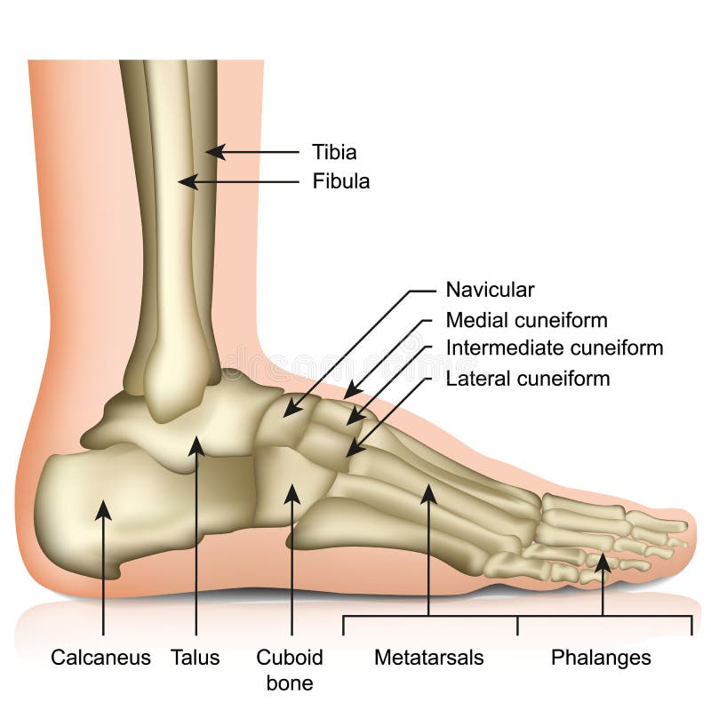
Free with trial Vector illustration of a human leg with denominations of the bones of the foot. Anatomy of lateral or side view of the foot. For advertising or medical publications. EPS 10. Metatarsal vectors Anatomy_bones of the human foot lateral view. Vector illustration of a human leg with denominations of the bones of the foot. Anatomy of lateral or side view of. Vector illustration of a human leg with denominations of the bones of the foot. Anatomy of lateral or side view of the foot. For advertising or medical publications. EPS 10.
Free with trial Most common types of human forefoot shapes with different variations of relative length of the toes. Vector illustration isolated on white background. Metatarsal vectors Types of the forefoot and toes. Most common types of human forefoot shapes with different variations of relative length of the toes. Vector illustration isolated. Most common types of human forefoot shapes with different variations of relative length of the toes. Vector illustration isolated on white background.
Free with trial Vector illustration of Peroneal Tendon Injuries. Longitudinal tear of the peroneus brevis tendon. Lateral ankle injury. Metatarsal vectors Peroneal Tendon Injuries_Longitudinal tear of the peroneus brevis tendon. Vector illustration of Peroneal Tendon Injuries. Longitudinal tear of the peroneus. Vector illustration of Peroneal Tendon Injuries. Longitudinal tear of the peroneus brevis tendon. Lateral ankle injury.
Free with trial Vector illustration of a human leg with denominations of the bones of the foot. Anatomy of medial or side view of the foot. For advertising or medical publications. EPS 10. Metatarsal vectors Anatomy_bones of the human foot medial view. Vector illustration of a human leg with denominations of the bones of the foot. Anatomy of medial or side view of. Vector illustration of a human leg with denominations of the bones of the foot. Anatomy of medial or side view of the foot. For advertising or medical publications. EPS 10.
Free with trial Vector illustration of Peroneal Tendon Injuries. Peroneal tendon dislocation or subluxation when the superior peroneal retinaculum or retinacula is torn. Lateral ankle injury. Metatarsal vectors Peroneal Tendon Injuries_Peroneal tendon dislocation. Vector illustration of Peroneal Tendon Injuries. Peroneal tendon dislocation or subluxation when the. Vector illustration of Peroneal Tendon Injuries. Peroneal tendon dislocation or subluxation when the superior peroneal retinaculum or retinacula is torn. Lateral ankle injury.
Free with trial Morton neuroma, Morton's metatarsalgia, Intermetatarsal neuroma, and Intermetatarsal space neuroma. Neurology. Metatarsal vectors Morton neuroma, Morton's metatarsalgia, Intermetatarsal neuroma, and Intermetatarsal space neuroma. Neurology
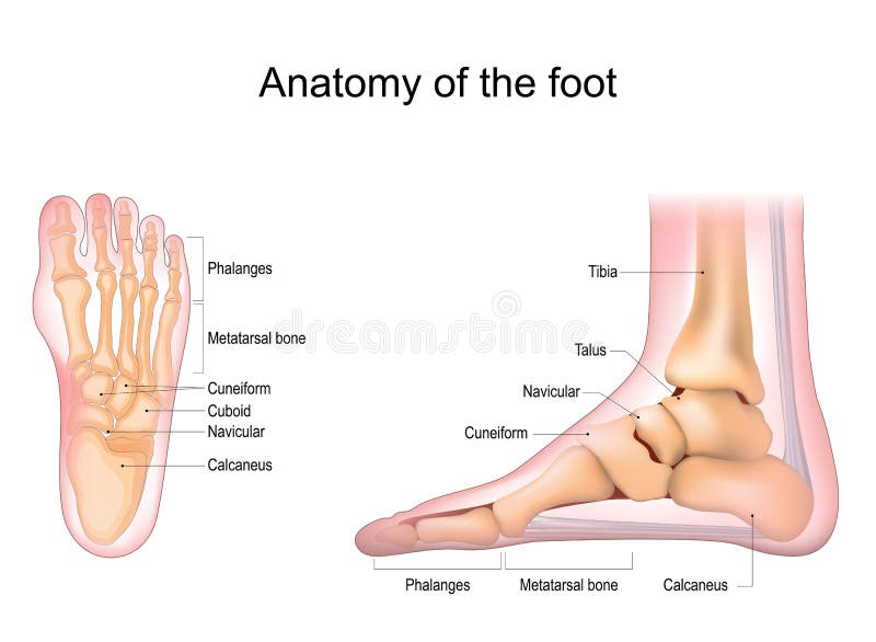
Free with trial Foot bones vector illustration with labels. Medical diagram with tibia, fibula, malleous, talus and navicular. Educational scheme with labeled cuneiforms, cuboid, lateral, calcanius and phalanges. Metatarsal vectors Vector illustration with foot bones. Medical diagram and educational scheme with tibia, fibula, malleous, talus and navicular. Foot bones vector illustration. Foot bones vector illustration with labels. Medical diagram with tibia, fibula, malleous, talus and navicular. Educational scheme with labeled cuneiforms, cuboid, lateral, calcanius and phalanges.
Free with trial Foot anatomy. Human foot with the name and description of all bones and sites. top view and side view. Arches of the feet. skeleton anatomy. Vector illustration. Metatarsal vectors Foot anatomy. Human foot with the name and description of all bones and sites. Top view and side view. Arches of the feet. skeleton anatomy. Vector
Free with trial X-ray elements, vector infographic icons. Metatarsal vectors X-ray elements. Vector infographic icons
Free with trial Vector illustration of a human leg with denominations of the bones of the foot. Anatomy of dorsal and plantar views of the foot. For advertising or medical publications. EPS 10. Metatarsal vectors Anatomy_bones of the human foot dorsal and plantar view. Vector illustration of a human leg with denominations of the bones of the foot. Anatomy of dorsal and. Vector illustration of a human leg with denominations of the bones of the foot. Anatomy of dorsal and plantar views of the foot. For advertising or medical publications. EPS 10.
Free with trial Vector illustration of Peroneal Tendon Injuries. Rupture of the peroneus longus tendon. Lateral ankle injury. Metatarsal vectors Peroneal Tendon Injuries_Rupture of the peroneus longus tendon. Vector illustration of Peroneal Tendon Injuries. Rupture of the peroneus longus tendon. Lateral. Vector illustration of Peroneal Tendon Injuries. Rupture of the peroneus longus tendon. Lateral ankle injury.
Free with trial Foot stress fractures as bone cracking trauma or injury outline diagram. Labeled educational scheme with leg skeletal anatomy and orthopedic problem caused by repetitive overuse vector illustration. Metatarsal vectors Foot stress fractures as bone cracking trauma or injury outline diagram. Labeled educational scheme with leg skeletal anatomy and orthopedic problem caused by
Free with trial Anatomy Bones of the Feet. Superior view. Orthotics for foot Superior view. Metatarsal vectors Anatomy Bones of the Feet. Superior view. Anatomy Bones of the Feet. Orthotics for foot Superior view
Free with trial Vector illustration of Peroneal Tendon Injuries. Degeneration of the peroneus longus and brevis tendons. Tendinosis or tendinopathy. Lateral ankle injury. Metatarsal vectors Peroneal Tendon Injuries_Degeneration of the peroneus longus and. Vector illustration of Peroneal Tendon Injuries. Degeneration of the peroneus longus and brevis. Vector illustration of Peroneal Tendon Injuries. Degeneration of the peroneus longus and brevis tendons. Tendinosis or tendinopathy. Lateral ankle injury.
Free with trial Arthrosis. bone destruction. Vector illustration of arthrosis. destruction of bone tissue. Metatarsal vectors Arthrosis. bone destruction. Vector illustration of arthrosis. destruction of bone tissue
Free with trial Differences arthrosis from arthritis. Vector illustration of the difference between arthrosis and arthritis. Metatarsal vectors Differences arthrosis from arthritis. Vector illustration of the difference between arthrosis and arthritis
Free with trial Jones fracture and foot pinky finger bone broken damage outline diagram. Labeled educational scheme with bone stress or avulsion sections vetor illustration. Peroneus brevis and tertius muscle anatomy. Metatarsal vectors Jones fracture and foot pinky finger bone broken damage outline diagram. Labeled educational scheme with bone stress or avulsion sections vetor illustration
Free with trial Bones of the Foot. Tarsals or tarsus, Metatarsals, Phalanges. Metatarsal vectors Bones of the Foot .Tarsals or tarsus, Metatarsals, Phalanges. Bones of the Foot.Tarsals or tarsus, Metatarsals, Phalanges Anatomy
Free with trial Bunion. Valgus deformity toe. Foot disease. Metatarsal vectors Bunion. Valgus deformity toe. Foot disease
Free with trial Ankle joint fracture Type c medical vector illustration on white background. Eps 10. Metatarsal vectors Ankle joint fracture Type c medical vector illustration on white background. Eps 10
 Sign in