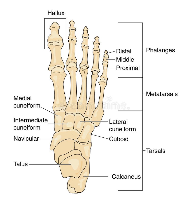
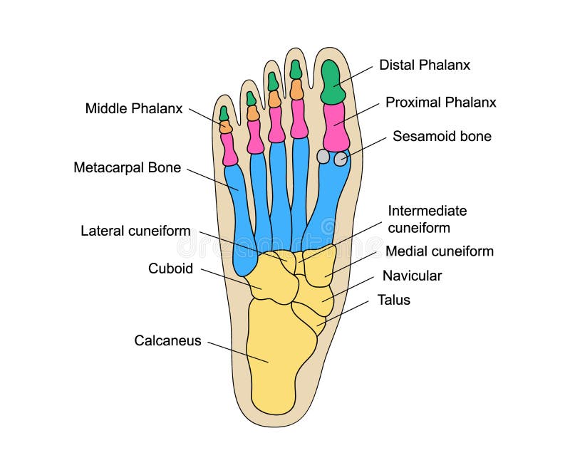
Free with trial Bones of the foot, labeled. Bones of the foot as seen from above (superior aspect), with the bones labeled. Metatarsals vectors Bones of the foot, labeled. Bones of the foot as seen from above (superior aspect), with the bones labeled
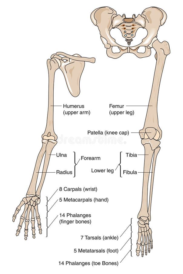
Free with trial Limb bones. Bones of upper and lower human limbs. Metatarsals vectors Limb bones. Bones of upper and lower human limbs
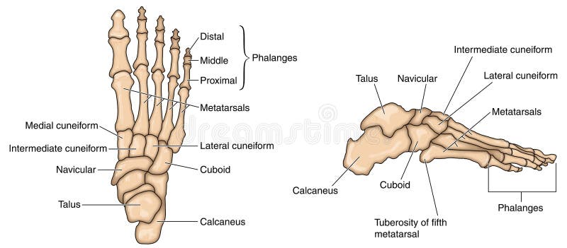
Free with trial Foot bones. Bones of the foot, lateral aspect and dorsal aspect. Created in Adobe Illustrator. Contains transparencies. EPS 10. Metatarsals vectors Foot bones. Bones of the foot, lateral aspect and dorsal aspect. Created in Adobe Illustrator. Contains transparencies. EPS 10
Free with trial Deviation of the first and the fifth metatarsals, transverse flatfoot. Used: gradient, transparency, blend mode. Metatarsals vectors Deviation of the first and the fifth metatarsals transverse flat. Deviation of the first and the fifth metatarsals, transverse flatfoot. Used: gradient. Deviation of the first and the fifth metatarsals, transverse flatfoot. Used: gradient, transparency, blend mode.
Free with trial Bones of the Foot. The bones of the foot as seen from above, unlabeled. Metatarsals vectors Bones of the Foot. The bones of the foot as seen from above, unlabeled
Free with trial Plantar fasciitis vector illustration. Labeled human feet sport disorder diagram. Educational medical scheme with orthopedic leg disease. Painful plantar fascia inflammation and irritation infographic. Metatarsals vectors Plantar fasciitis vector illustration. Labeled human feet disorder diagram. Plantar fasciitis vector illustration. Labeled human feet sport disorder diagram. Plantar fasciitis vector illustration. Labeled human feet sport disorder diagram. Educational medical scheme with orthopedic leg disease. Painful plantar fascia inflammation and irritation infographic
Free with trial Deviation of the first and the fifth metatarsals transverse flat. Foot with orthopedic insole. Used: gradient, transparency, blend mode. Metatarsals vectors Deviation of the first and the fifth metatarsals transverse flat. Foot with orthopedic insole. Used: gradient, transparency, blend mode
Free with trial Cuboid syndrome as orthopedic trauma after foot torn injury outline diagram. Labeled educational scheme with feet bone dislocation and free movement vector illustration. Cuboid subluxation problem. Metatarsals vectors Cuboid syndrome as orthopedic trauma after foot torn injury outline diagram. Labeled educational scheme with feet bone dislocation and free movement vector
Free with trial Anatomy of dog paws with forelimb and hindlimb bones vector illustration. Educational labeled skeleton comparison with zoological inside structure scheme. Animal legs inner closeup examination model. Metatarsals vectors Anatomy of dog paws with forelimb and hindlimb bones vector illustration. Educational labeled skeleton comparison with zoological inside structure scheme
Free with trial Bones of the Foot. Tarsals or tarsus, Metatarsals, Phalanges. Metatarsals vectors Bones of the Foot .Tarsals or tarsus, Metatarsals, Phalanges. Bones of the Foot.Tarsals or tarsus, Metatarsals, Phalanges Anatomy
Free with trial Skeleton Human foot body bones - front Anterior ventral view. Feet, ankle, tarsals metatarsals phalanges flat concept Vector illustration of anatomy isolated on white background. Metatarsals vectors Skeleton Human foot body bones - front Anterior ventral view. Feet, ankle, tarsals metatarsals phalanges flat concept Vector illustration of anatomy isolated
Free with trial Metatarsalgia refers to pain and inflammation in the ball of the foot. This is the area between the arches and toes on the bottom of the foot. Metatarsalgia centers under the five bones at the bases of the toes, the metatarsals. Metatarsals vectors Metatarsalgia Pain in the Ball of the Foot medical educational vector illustration graphic. Metatarsalgia refers to pain and inflammation in the ball of the foot. Metatarsalgia refers to pain and inflammation in the ball of the foot. This is the area between the arches and toes on the bottom of the foot. Metatarsalgia centers under the five bones at the bases of the toes, the metatarsals
Free with trial Skeleton Human foot silhouette body bones - front Anterior ventral view. Feet, ankle, tarsals metatarsals phalanges flat black color concept Vector illustration of anatomy isolated on white background. Metatarsals vectors Skeleton Human foot silhouette body bones - front Anterior ventral view. Feet, ankle, tarsals metatarsals phalanges. Flat black color concept Vector. Skeleton Human foot silhouette body bones - front Anterior ventral view. Feet, ankle, tarsals metatarsals phalanges flat black color concept Vector illustration of anatomy isolated on white background
Free with trial This illustration is abstract parrot bird group sky green color background fly wildflower element graphic. Metatarsals vectors Parrot bird group. This illustration is abstract parrot bird group sky green color background fly wildflower element graphic. This illustration is abstract parrot bird group sky green color background fly wildflower element graphic.
Free with trial Illustration of a skeletal and external view of an ankle. Metatarsals vectors Skeletal and external view of an ankle. Illustration of a skeletal and external view of an ankle. Illustration of a skeletal and external view of an ankle.
Free with trial Ankle joint vector illustration. Labeled educational leg structure scheme. Physiological orthopedics explanation with isolated toe closeup. Cross section with phalanges, tibia, tarsals, ligament graph. Metatarsals vectors Ankle joint vector illustration. Labeled educational leg structure scheme. Physiological orthopedics explanation with isolated toe closeup. Cross section with
Free with trial Foot bones isolated on white vector. Illustration. Metatarsals vectors Foot bones isolated on white vector. Illustration
Free with trial Anatomy Bones of the Feet. Superior view. Orthotics for foot Superior view. Metatarsals vectors Anatomy Bones of the Feet. Superior view. Anatomy Bones of the Feet. Orthotics for foot Superior view
Free with trial Plantigrade, Digitigrade and Unguligrade comparison vector illustration. Educational labeled structure scheme with human, dog and pig legs collection. Bone skeleton parts with location explanation. Metatarsals vectors Plantigrade, Digitigrade and Unguligrade comparison vector illustration. Educational labeled structure scheme with human, dog and pig legs collection. Bone
Free with trial In the dorsal (posterior) view of the bones of the feet, the tarsal bones are visible, including the talus, calcaneus, navicular, cuboid, and the three cuneiform bones. The metatarsal bones, numbering from one to five, can also be observed, extending towards the toes. Additionally, the phalanges, consisting of proximal, middle, and distal phalanges, complete the skeletal structure of the feet in the dorsal view. Metatarsals vectors Bones of feet, dorsal (posterior) view. In the dorsal (posterior) view of the bones of the feet, the tarsal bones are visible, including the talus, calcaneus. In the dorsal (posterior) view of the bones of the feet, the tarsal bones are visible, including the talus, calcaneus, navicular, cuboid, and the three cuneiform bones. The metatarsal bones, numbering from one to five, can also be observed, extending towards the toes. Additionally, the phalanges, consisting of proximal, middle, and distal phalanges, complete the skeletal structure of the feet in the dorsal view.
Free with trial Vector drawing of human foot human foot anatomy drawing labeled. Drawing with layers reedit able. Metatarsals vectors Vector drawing of human foot human foot anatomy drawing labeled. Drawing with layers reedit able
Free with trial Doctor diagnosis for foot and ankle pain in women,heath care page. Hospital digital, Orthopedics technologies concept. Radiology orthopedic,hospital, joint. Blue violet background. Vector illustration. Metatarsals vectors Doctor diagnosis for foot and ankle pain in women,heath care page. Hospital digital, Orthopedics technologies concept. Radiology orthopedic,hospital, joint
Free with trial Bone of the hand and foot anatomy, internal organs body part orthopedic health care, vector illustration cartoon flat character design clip art. Metatarsals vectors Bone of the hand and foot anatomy, internal organs body part orthopedic health care. Vector illustration cartoon flat character design clip art
Free with trial Leg bone. Skeleton anatomy human. Skeletal system cross section. vector illustration. Metatarsals vectors Leg bone. Skeleton anatomy human. Skeletal system cross section. Vector illustration
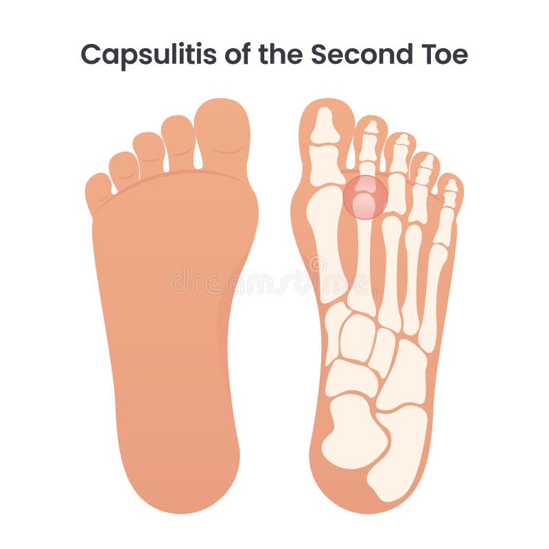
Free with trial Capsulitis of the second toe occurs in the ligaments surrounding the joint at the base of the second toe. These ligaments form a capsule, which helps the joint to function properly. Capsulitis is a condition in which these ligaments have become inflamed. Metatarsals vectors Capsulitis of the Second Bone educational medical vector illustration. Capsulitis of the second toe occurs in the ligaments surrounding the joint at the base of. Capsulitis of the second toe occurs in the ligaments surrounding the joint at the base of the second toe. These ligaments form a capsule, which helps the joint to function properly. Capsulitis is a condition in which these ligaments have become inflamed
Free with trial Illustration human foot bone. on white background. vector about science and medical. Metatarsals vectors Illustration human foot bone. on white background. Illustration of human foot bone. on white background. vector about science and medical
Free with trial Explore the intricate details of the human foot's bone structure, showcasing phalanges and metatarsals for medical and educational applications. Metatarsals vectors Detailed bone structure of a human foot showing phalanges and metatarsals. Explore the intricate details of the human foot's bone structure, showcasing phalanges. Explore the intricate details of the human foot's bone structure, showcasing phalanges and metatarsals for medical and educational applications
Free with trial This image illustrates the intricate structure of the human foot, highlighting the bones and muscles. The foot is composed of numerous bones that work together to provide support, balance, and mobility. The image shows the bones in the toes, the metatarsals, the tarsal bones, and the ankle joint. The muscles are depicted in a way that shows their attachment points and how they contribute to the. Metatarsals vectors Detailed anatomy of the human foot. This image illustrates the intricate structure of the human foot, highlighting the bones and muscles. The foot is composed of. This image illustrates the intricate structure of the human foot, highlighting the bones and muscles. The foot is composed of numerous bones that work together to provide support, balance, and mobility. The image shows the bones in the toes, the metatarsals, the tarsal bones, and the ankle joint. The muscles are depicted in a way that shows their attachment points and how they contribute to the
Free with trial This image depicts a detailed anatomical model of a human foot with surgical implants. The model shows the bones of the foot, including the tarsals, metatarsals, and phalanges. Several metal screws and plates are visible, indicating surgical intervention, likely for fracture repair or stabilization. Metatarsals vectors Anatomical foot with surgical implants. This image depicts a detailed anatomical model of a human foot with surgical implants. The model shows the bones of the. This image depicts a detailed anatomical model of a human foot with surgical implants. The model shows the bones of the foot, including the tarsals, metatarsals, and phalanges. Several metal screws and plates are visible, indicating surgical intervention, likely for fracture repair or stabilization
Free with trial The image depicts an X-ray of a human foot being held by a hand. The X-ray clearly shows the bones of the foot, including the metatarsals and phalanges. The image appears to be used for medical diagnostic purposes, likely to examine the structure and health of the foot bones. Metatarsals vectors Foot x-ray analysis. The image depicts an X-ray of a human foot being held by a hand. The X-ray clearly shows the bones of the foot, including the metatarsals. The image depicts an X-ray of a human foot being held by a hand. The X-ray clearly shows the bones of the foot, including the metatarsals and phalanges. The image appears to be used for medical diagnostic purposes, likely to examine the structure and health of the foot bones
Free with trial Illustration of a human foot skeleton in a side view. The image highlights key bones including the phalanges, metatarsals, tarsals, and heel. The bones appear in a black silhouette against a white background, showcasing the arrangement and articulation of the foots skeletal structure. Metatarsals vectors Illustration of a human foot skeleton in a side view. The image highlights key bones including the phalanges, metatarsals, tarsals, and heel. The bones appear
Free with trial A clear black silhouette icon depicting the intricate human foot skeleton, isolated on a clean white background. This anatomical illustration showcases the structure of the tarsal bones, metatarsals, and phalanges. Ideal for medical, educational, and healthcare-related projects, it can be used in presentations, textbooks, websites, or apps to represent anatomy, orthopedics, podiatry, bone health, or skeletal system concepts. The minimalist design ensures versatility and readability for various applications. Metatarsals vectors Human Foot Skeleton Anatomy Icon. A clear black silhouette icon depicting the intricate human foot skeleton, isolated on a clean white background. This. A clear black silhouette icon depicting the intricate human foot skeleton, isolated on a clean white background. This anatomical illustration showcases the structure of the tarsal bones, metatarsals, and phalanges. Ideal for medical, educational, and healthcare-related projects, it can be used in presentations, textbooks, websites, or apps to represent anatomy, orthopedics, podiatry, bone health, or skeletal system concepts. The minimalist design ensures versatility and readability for various applications.
Free with trial This image is an anatomical diagram of the human foot, showcasing the intricate structure of bones and muscles. The diagram highlights the skeletal framework and the detailed arrangement of muscles that support the foot. It provides a clear view of the bones in the toes, the metatarsals, and the tarsal bones, as well as the muscles that control movement and provide stability. Metatarsals vectors Anatomical diagram of the human foot. This image is an anatomical diagram of the human foot, showcasing the intricate structure of bones and muscles. The diagram. This image is an anatomical diagram of the human foot, showcasing the intricate structure of bones and muscles. The diagram highlights the skeletal framework and the detailed arrangement of muscles that support the foot. It provides a clear view of the bones in the toes, the metatarsals, and the tarsal bones, as well as the muscles that control movement and provide stability
Free with trial A detailed anatomical vector illustration of the human foot bones, featuring the tarsals, metatarsals, and phalanges. Metatarsals vectors Human Foot Bones Anatomy Isolated Vector Illustration. A detailed anatomical vector illustration of the human foot bones, featuring the tarsals, metatarsals, and. A detailed anatomical vector illustration of the human foot bones, featuring the tarsals, metatarsals, and phalanges.
Free with trial A clear, line-art illustration depicting the skeletal structure of the human foot and ankle, showing the tibia, fibula, tarsals, metatarsals, and phalanges. Generated using AI. Metatarsals vectors Anatomical Illustration of the Human Foot and Ankle Bones. A clear, line-art illustration depicting the skeletal structure of the human foot and ankle, showing. A clear, line-art illustration depicting the skeletal structure of the human foot and ankle, showing the tibia, fibula, tarsals, metatarsals, and phalanges. Generated using AI.
Free with trial A professional medical diagram illustrating the complex anatomy of the human foot and lower leg. The visual includes the skeletal system bones like the calcaneus and metatarsals, along with detailed musculature and ligament structures, rendered in a clean style on a white background. Metatarsals vectors Detailed anatomical of the human lower leg and foot showing skeletal structure. A professional medical diagram illustrating the complex anatomy of the human foot. A professional medical diagram illustrating the complex anatomy of the human foot and lower leg. The visual includes the skeletal system bones like the calcaneus and metatarsals, along with detailed musculature and ligament structures, rendered in a clean style on a white background.
Free with trial Minimalist X ray of a human foot and ankle in side profile, showing simplified ankle joint, heel bone, metatarsals and toe bones in a natural anatomical position. Flat black and white monochrome style with crisp contours and clear negative space, suitable for medical education, anatomy materials, healthcare presentations, radiology websites and printed publications. Metatarsals vectors X Ray foot and ankle anatomy. Heel and toe bones. Medical radiology concept. Monochrome. Minimalist X ray of a human foot and ankle in side profile, showing. Minimalist X ray of a human foot and ankle in side profile, showing simplified ankle joint, heel bone, metatarsals and toe bones in a natural anatomical position. Flat black and white monochrome style with crisp contours and clear negative space, suitable for medical education, anatomy materials, healthcare presentations, radiology websites and printed publications.
Free with trial Human Foot Bone Anatomy Icons Black White Skeleton. Human foot bones shown as black and white icons illustrating skeletal anatomy for medical or educational use. Metatarsals vectors Human Foot Bone Anatomy Icons Black White Skeleton. Human foot bones shown as black and white icons illustrating skeletal anatomy for medical or educational use
Free with trial Human foot skeleton icons black and white simple outline for medical or anatomical use. Metatarsals vectors Human Foot Skeleton Icons Black and White Simple Outline. For medical or anatomical use. Human foot skeleton icons black and white simple outline for medical or anatomical use
Free with trial Educational medical line art displaying the skeletal anatomy of the human foot and ankle. The labeled lateral osteology diagram details key skeletal structures including the tibia, talus, calcaneus (heel bone), navicular, cuboid, metatarsals, and phalanges (toe bones). Clean black and white line diagram ideal for podiatry textbooks, orthopedic clinic wall charts, sports injury guides, human osteology worksheets, and medical education presentations. Metatarsals vectors Foot Bone Anatomy Skeletal Structure Line Art Vector Illustration. Educational medical line art displaying the skeletal anatomy of the human foot and ankle. The. Educational medical line art displaying the skeletal anatomy of the human foot and ankle. The labeled lateral osteology diagram details key skeletal structures including the tibia, talus, calcaneus (heel bone), navicular, cuboid, metatarsals, and phalanges (toe bones). Clean black and white line diagram ideal for podiatry textbooks, orthopedic clinic wall charts, sports injury guides, human osteology worksheets, and medical education presentations.
Free with trial Educational medical line art displaying the skeletal anatomy of the human foot and ankle. The labeled lateral osteology diagram details key skeletal structures including the tibia, talus, calcaneus (heel bone), navicular, cuboid, metatarsals, and phalanges (toe bones). Clean black and white line diagram ideal for podiatry textbooks, orthopedic clinic wall charts, sports injury guides, human osteology worksheets, and medical education presentations. Metatarsals vectors Foot Bone Anatomy Skeletal Structure Line Art Vector Illustration. Educational medical line art displaying the skeletal anatomy of the human foot and ankle. The. Educational medical line art displaying the skeletal anatomy of the human foot and ankle. The labeled lateral osteology diagram details key skeletal structures including the tibia, talus, calcaneus (heel bone), navicular, cuboid, metatarsals, and phalanges (toe bones). Clean black and white line diagram ideal for podiatry textbooks, orthopedic clinic wall charts, sports injury guides, human osteology worksheets, and medical education presentations.
Free with trial Detailed Clinical Foot Anterior View Fracture X Ray Displaying Bone Structure, Metatarsals, And Joint Alignment With Clear Fracture Visualization. High Resolution Medical Illustration Supports Orthopedic Evaluation, Radiology Education, And Clinical Assessment. Suitable For Scientific And Healthcare Publications. Metatarsals vectors Detailed Clinical Foot Anterior View Fracture X Ray Isolated On A Transparent Background. Detailed Clinical Foot Anterior View Fracture X Ray Displaying Bone. Detailed Clinical Foot Anterior View Fracture X Ray Displaying Bone Structure, Metatarsals, And Joint Alignment With Clear Fracture Visualization. High Resolution Medical Illustration Supports Orthopedic Evaluation, Radiology Education, And Clinical Assessment. Suitable For Scientific And Healthcare Publications.
Free with trial Anterior Foot X Ray Radiograph Showing Visible Fracture Detail Across Metatarsals And Phalanges With Clear Bone Alignment And Joint Structure. This High Quality Medical Illustration Supports Orthopedic Evaluation, Radiology Training, And Clinical Documentation. Isolated On A Transparent Background For Flexible Use In Medical Design Projects. Metatarsals vectors Anterior Foot X Ray Radiograph With Fracture Detail Isolated On A Transparent Background. Anterior Foot X Ray Radiograph Showing Visible Fracture Detail Across. Anterior Foot X Ray Radiograph Showing Visible Fracture Detail Across Metatarsals And Phalanges With Clear Bone Alignment And Joint Structure. This High Quality Medical Illustration Supports Orthopedic Evaluation, Radiology Training, And Clinical Documentation. Isolated On A Transparent Background For Flexible Use In Medical Design Projects.
Free with trial High quality vector EPS illustration with clean lines and scalable design. Suitable for graphic tees, streetwear apparel, posters, branding, merchandise, logos, stickers, social media, and print projects. Fully editable vector artwork, easy to customize colors and shapes. Ideal for commercial and personal creative use. Metatarsals vectors Dead Inside text with skeletal hands, a gothic and emo design. High quality vector EPS illustration with clean lines and scalable design. Suitable for graphic. High quality vector EPS illustration with clean lines and scalable design. Suitable for graphic tees, streetwear apparel, posters, branding, merchandise, logos, stickers, social media, and print projects. Fully editable vector artwork, easy to customize colors and shapes. Ideal for commercial and personal creative use
Free with trial Medical professional, doctor or physician, examining human foot x-ray for diagnosis of injury, fracture, medical condition. Metatarsals vectors Medical professional, doctor or physician, examining human foot x-ray for diagnosis of injury. Fracture, medical condition
Free with trial Anatomical illustration of the human foot bones, showcasing the complex structure and arrangement of the skeletal system on a dark background. Metatarsals vectors Detailed Foot Bones Anatomy Illustration on Dark Background. Anatomical illustration of the human foot bones, showcasing the complex structure and arrangement of. Anatomical illustration of the human foot bones, showcasing the complex structure and arrangement of the skeletal system on a dark background
Free with trial Detailed vector illustration of a human foot bone structure, perfect for medical websites, educational materials, or presentations. The image features a clear outline of the bones within the foot. Metatarsals vectors Foot Bone Structure, Medical Illustration. Detailed vector illustration of a human foot bone structure, perfect for medical websites, educational materials, or. Detailed vector illustration of a human foot bone structure, perfect for medical websites, educational materials, or presentations. The image features a clear outline of the bones within the foot.
Free with trial Black and white line drawing showing skeletal structure of feet with labeled bones in lateral and dorsal perspectives. Metatarsals vectors Detailed anatomical illustration of human foot bones in side and top views sketch style. Black and white line drawing showing skeletal structure of feet with. Black and white line drawing showing skeletal structure of feet with labeled bones in lateral and dorsal perspectives.
Free with trial Detailed diagram of a human foot showcasing its skeletal structure, perfect for educational materials and anatomical illustrations. It's a great resource for medical or science projects. Metatarsals vectors Human foot bones diagram. Detailed diagram of a human foot showcasing its skeletal structure, perfect for educational materials and anatomical illustrations. It'. Detailed diagram of a human foot showcasing its skeletal structure, perfect for educational materials and anatomical illustrations. It's a great resource for medical or science projects
Free with trial A detailed, monochrome illustration showcasing the anatomical structure of a human foot skeleton, perfect for educational or medical contexts. Metatarsals vectors Detailed Anatomical Illustration of a Human Foot Skeleton. A detailed, monochrome illustration showcasing the anatomical structure of a human foot skeleton. A detailed, monochrome illustration showcasing the anatomical structure of a human foot skeleton, perfect for educational or medical contexts
Free with trial Anterior Foot Fracture X Ray With Detailed Bone Structure Showing Metatarsals, Phalanges, And Joint Alignment With Clear Radiographic Precision. This Medical Illustration Supports Orthopedic Evaluation, Radiology Training, And Clinical Documentation. High Resolution Detail Ensures Accurate Anatomical Representation For Scientific And Healthcare Use. Metatarsals vectors Anterior Foot Fracture X Ray With Detailed Bone Structure Isolated On A Transparent Background. Anterior Foot Fracture X Ray With Detailed Bone Structure Showing. Anterior Foot Fracture X Ray With Detailed Bone Structure Showing Metatarsals, Phalanges, And Joint Alignment With Clear Radiographic Precision. This Medical Illustration Supports Orthopedic Evaluation, Radiology Training, And Clinical Documentation. High Resolution Detail Ensures Accurate Anatomical Representation For Scientific And Healthcare Use.
Free with trial Simple hand-drawn illustration depicting the skeletal structure of a human foot. Perfect for medical, anatomical, or educational purposes. Metatarsals vectors Stylized Foot Bones Anatomy Sketch, Hand Drawn Illustration. Simple hand-drawn illustration depicting the skeletal structure of a human foot. Perfect for medical. Simple hand-drawn illustration depicting the skeletal structure of a human foot. Perfect for medical, anatomical, or educational purposes.
Free with trial Anatomical Illustration of Human Foot Bones Inside a Teal Circle. AI generated. Metatarsals vectors Anatomical Illustration of Human Foot Bones Inside a Teal Circle. AI generated
Free with trial A realistic, light-skinned human foot and ankle, truncated at the lower leg, are centered against a transparent background. Metatarsals vectors Anatomical Model of a Human Foot and Ankle. A realistic, light-skinned human foot and ankle, truncated at the lower leg, are centered against a transparent. A realistic, light-skinned human foot and ankle, truncated at the lower leg, are centered against a transparent background
Free with trial This vector shows a detailed line art of human lower leg and foot bones in a minimalist style, emphasizing anatomical structure, precise and clear. Calm. Generative AI. Metatarsals vectors Human lower leg and foot bone anatomy line art vector illustration medical design. This vector shows a detailed line art of human lower leg and foot bones in a. This vector shows a detailed line art of human lower leg and foot bones in a minimalist style, emphasizing anatomical structure, precise and clear. Calm. Generative AI
Free with trial Illustration of a gray human foot, viewed from the side, outlined in white and black, against transparent background. Metatarsals vectors Gray Foot Illustration on transparent background. Illustration of a gray human foot, viewed from the side, outlined in white and black, against transparent. Illustration of a gray human foot, viewed from the side, outlined in white and black, against transparent background
Free with trial Cuboid syndrome as feet bone disorder condition. orthopedic trauma after foot torn injury. Cuboid subluxation problem. Feet bone dislocation. Medical education poster flat vector illustration. Metatarsals vectors Cuboid syndrome as feet bone disorder condition. orthopedic trauma after foot torn injury. Cuboid subluxation problem. Feet bone. Dislocation. Medical education. Cuboid syndrome as feet bone disorder condition. orthopedic trauma after foot torn injury. Cuboid subluxation problem. Feet bone dislocation. Medical education poster flat vector illustration
Free with trial A human foot skeleton is displayed, focusing on the bones and joints. The foot features bright colors that highlight its structure against a plain background. AI Generative. Metatarsals vectors Detailed View of a Human Foot Skeleton Showing Bones and Joints in Bright Colors on a White Background. A human foot skeleton is displayed, focusing on the bones. A human foot skeleton is displayed, focusing on the bones and joints. The foot features bright colors that highlight its structure against a plain background. AI Generative
Free with trial A human foot skeleton is displayed, focusing on the bones and joints. The foot features bright colors that highlight its structure against a plain background. AI Generative. Metatarsals vectors Detailed View of a Human Foot Skeleton Showing Bones and Joints in Bright Colors on a White Background. A human foot skeleton is displayed, focusing on the bones. A human foot skeleton is displayed, focusing on the bones and joints. The foot features bright colors that highlight its structure against a plain background. AI Generative
Free with trial An detailed silhouette icon detailing the complete bone structure of the human foot and ankle. Metatarsals vectors Human foot bones skeleton x-ray anatomy silhouette icon. An ai generated detailed silhouette icon detailing the complete bone structure of the human foot and. An detailed silhouette icon detailing the complete bone structure of the human foot and ankle
Free with trial Detailed silhouette icon of a human foot skeleton ankle bones anatomy on transparent background. Metatarsals vectors Human foot skeleton ankle tarsal bones anatomy silhouette. AI generated detailed silhouette icon of a human foot skeleton ankle bones anatomy on transparent. detailed silhouette icon of a human foot skeleton ankle bones anatomy on transparent background
Free with trial Illustration of a pale human foot and ankle in profile, with toes facing right, outlined in black against transparent background. Metatarsals vectors Illustrated Right Human Foot and Ankle on transparent background. Illustration of a pale human foot and ankle in profile, with toes facing right, outlined in. Illustration of a pale human foot and ankle in profile, with toes facing right, outlined in black against transparent background
Free with trial Illustration shows human lower leg and foot bones with clear details and shading in vector format. Anatomical and educational, informative mood. Generative AI. Metatarsals vectors Human lower leg and foot bone anatomy detailed vector illustration natural flow. Illustration shows human lower leg and foot bones with clear details and shading. Illustration shows human lower leg and foot bones with clear details and shading in vector format. Anatomical and educational, informative mood. Generative AI
Free with trial This vector shows a human foot skeleton with detailed bone structure in soft beige color, presenting educational and anatomical clarity. Calm. Generative AI. Metatarsals vectors Human Foot Skeleton Bone Anatomy Medical Illustration in Soft Beige Color Vector. This vector shows a human foot skeleton with detailed bone structure in soft. This vector shows a human foot skeleton with detailed bone structure in soft beige color, presenting educational and anatomical clarity. Calm. Generative AI
Free with trial Illustration shows detailed human lower leg and foot bones in line art style for anatomy learning and medical reference. Educational and clear. Generative AI. Metatarsals vectors Human Lower Leg and Foot Bone Anatomy Vector Line Art Illustration for Medical Study. Illustration shows detailed human lower leg and foot bones in line art. Illustration shows detailed human lower leg and foot bones in line art style for anatomy learning and medical reference. Educational and clear. Generative AI
Free with trial Illustration shows the skeletal structure of the human foot with bones clearly defined in black and white. Educational and clinical use. Informative mood. Generative AI. Metatarsals vectors Human foot bone anatomy illustration showing skeletal structure in black and white vector. Illustration shows the skeletal structure of the human foot with bones. Illustration shows the skeletal structure of the human foot with bones clearly defined in black and white. Educational and clinical use. Informative mood. Generative AI
Free with trial Detailed anatomical illustration of a human foot showing bones and joints, emphasizing inflammation in the metatarsophalangeal joint of the big toe. Metatarsals vectors Anatomical illustration of a human foot highlighting the bones and joints with a focus on the metatarsophalangeal joint and. Detailed anatomical illustration of. Detailed anatomical illustration of a human foot showing bones and joints, emphasizing inflammation in the metatarsophalangeal joint of the big toe
Free with trial Detailed anatomical illustration of a human foot highlighting bones, tendons, and ligaments, focusing on the ankle joint and heel area for educational purposes. Metatarsals vectors Anatomical illustration of a human foot showing bones, tendons, and ligaments with a focus on the ankle joint and heel area. Detailed anatomical illustration of. Detailed anatomical illustration of a human foot highlighting bones, tendons, and ligaments, focusing on the ankle joint and heel area for educational purposes
Free with trial Simple black and white illustration showing the bones in a human foot, viewed from above. Useful for educational or medical purposes. Metatarsals vectors Human Foot Bones Anatomy Illustration, Top View. Simple black and white illustration showing the bones in a human foot, viewed from above. Useful for educational. Simple black and white illustration showing the bones in a human foot, viewed from above. Useful for educational or medical purposes.
Free with trial Human foot anatomy infographic diagram. bones phalanges metatarsals tarsal. names joints. front lateral view. cartoon vector drawing. illustration for medical science education. structure parts chart. Metatarsals vectors Human foot anatomy infographic diagram. Bones phalanges metatarsals tarsal. names joints. front lateral view. cartoon vector drawing. illustration for medical
Free with trial Human foot anatomy infographic diagram. bones phalanges metatarsals tarsal. names joints. front lateral view. cartoon vector drawing. illustration for medical science education. structure parts chart. Metatarsals vectors Human foot anatomy infographic diagram. Bones phalanges metatarsals tarsal. names joints. front lateral view. cartoon vector drawing. illustration for medical
Free with trial Tarsalia bearing the corresponding digits with metatarsals and phalanges, vintage line drawing or engraving illustration. Metatarsals vectors Vertebrate Hind Limb, vintage illustration. Tarsalia bearing the corresponding digits with metatarsals and phalanges, vintage line drawing or engraving. Tarsalia bearing the corresponding digits with metatarsals and phalanges, vintage line drawing or engraving illustration
Free with trial Front Left Tarsus of a Penguin showing incomplete fusion of three metatarsals vintage line drawing or engraving illustration. Metatarsals vectors Front Left Tarsus of a Penguin vintage illustration. Front Left Tarsus of a Penguin showing incomplete fusion of three metatarsals vintage line drawing or. Front Left Tarsus of a Penguin showing incomplete fusion of three metatarsals vintage line drawing or engraving illustration
Free with trial Human foot bones anatomy with descriptions. Foot parts structure. Human internal organ illustration. Talus, calcaneus, tarsals, cuboid, metatarsals leg base parts. Metatarsals vectors Human foot bones anatomy with descriptions. Foot parts structure. Human internal organ illustration. Talus, calcaneus, tarsals, cuboid, metatarsals leg base
Free with trial Human foot bones anatomy drawing with a pen on notebook. Foot parts structure diagram with bones description. Human internal organ illustration. Talus, calcaneus, cuboid, metatarsals leg base parts. Metatarsals vectors Human foot bones anatomy drawing with a pen on notebook. Foot parts structure diagram with bones description. Human. Internal organ illustration. Talus. Human foot bones anatomy drawing with a pen on notebook. Foot parts structure diagram with bones description. Human internal organ illustration. Talus, calcaneus, cuboid, metatarsals leg base parts.
Free with trial The forefoot contains the phalanges and metatarsals. The midfoot consists of the five tarsal bones, three cuneiforms, the navicular, and the cuboid. The hindfoot is composed of two tarsal bones, the calcaneus and the talus. Metatarsals vectors Diagram or infographic of the bones that make up a foot from an aerial view with names. The forefoot contains the phalanges and metatarsals. The midfoot consists. The forefoot contains the phalanges and metatarsals. The midfoot consists of the five tarsal bones, three cuneiforms, the navicular, and the cuboid. The hindfoot is composed of two tarsal bones, the calcaneus and the talus.
Free with trial The forefoot contains the phalanges and metatarsals. The midfoot consists of the five tarsal bones, three cuneiforms, the navicular, and the cuboid. The hindfoot is composed of two tarsal bones, the calcaneus and the talus. Metatarsals vectors Diagram or infographic of the bones that make up a foot from an aerial view. The forefoot contains the phalanges and metatarsals. The midfoot consists of the. The forefoot contains the phalanges and metatarsals. The midfoot consists of the five tarsal bones, three cuneiforms, the navicular, and the cuboid. The hindfoot is composed of two tarsal bones, the calcaneus and the talus.
Free with trial Human foot bones anatomy with descriptions. Colored leg base parts structure. Educational diagram with human internal organ illustration. Talus, calcaneus, tarsals, cuboid, metatarsals foot parts. Metatarsals vectors Human foot bones anatomy with descriptions. Educational diagram of internal organ illustration. Calcaneus, tarsals. Human foot bones anatomy with descriptions. Human foot bones anatomy with descriptions. Colored leg base parts structure. Educational diagram with human internal organ illustration. Talus, calcaneus, tarsals, cuboid, metatarsals foot parts.
Free with trial Leg tibia, fibula, Foot, ankle Skeleton Human side view. Set of metatarsals, phalanges 3D Anatomically correct realistic flat natural color concept Vector illustration isolated on white background. Metatarsals vectors Leg tibia, fibula, Foot, ankle Skeleton Human side view. Set of metatarsals, phalanges 3D Anatomically correct realistic. Flat natural color concept Vector. Leg tibia, fibula, Foot, ankle Skeleton Human side view. Set of metatarsals, phalanges 3D Anatomically correct realistic flat natural color concept Vector illustration isolated on white background
Free with trial Medial view of foot bones Diagram. Anatomy. Metatarsals vectors Medial view of foot bones Diagram. Anatomy
 Sign in