Free with trial Vector illustration of a healthy human foot and a foot with lisfranc injury with weight bearing and mechanism of injury, dislocation of the second metatarsal bone. Medial, top view of the foot. EPS 10. Midfoot vectors Lisfranc Injury. Vector illustration of a healthy human foot and a foot with lisfranc injury with weight bearing and mechanism of injury, dislocation of the. Vector illustration of a healthy human foot and a foot with lisfranc injury with weight bearing and mechanism of injury, dislocation of the second metatarsal bone. Medial, top view of the foot. EPS 10
Free with trial Vector illustration of Flat or planovalgus foot without and with orthopedic insole. Side medial and back views. For advertising and medical publications. EPS 10. Midfoot vectors Flat foot with orthopedic insole_Exretnal wiew. Vector illustration of Flat or planovalgus foot without and with orthopedic insole. Side medial and back views. Vector illustration of Flat or planovalgus foot without and with orthopedic insole. Side medial and back views. For advertising and medical publications. EPS 10.
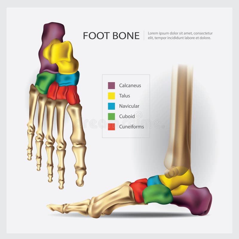
Free with trial Human Anatomy Foot Bone. Vector Illustration. Midfoot vectors Human Anatomy Foot Bone. Vector Illustration
Free with trial Ankle, foot bones. Vector illustration of the ankle bones of the foot. Midfoot vectors Ankle, foot bones. Vector illustration of the ankle bones of the foot
Free with trial Vector illustration of a human leg with denominations of the bones of the foot. Anatomy of lateral or side view of the foot. For advertising or medical publications. EPS 10. Midfoot vectors Anatomy_bones of the human foot lateral view. Vector illustration of a human leg with denominations of the bones of the foot. Anatomy of lateral or side view of. Vector illustration of a human leg with denominations of the bones of the foot. Anatomy of lateral or side view of the foot. For advertising or medical publications. EPS 10.
Free with trial Vector illustration of a human leg with denominations of the bones of the foot. Anatomy of medial or side view of the foot. For advertising or medical publications. EPS 10. Midfoot vectors Anatomy_bones of the human foot medial view. Vector illustration of a human leg with denominations of the bones of the foot. Anatomy of medial or side view of. Vector illustration of a human leg with denominations of the bones of the foot. Anatomy of medial or side view of the foot. For advertising or medical publications. EPS 10.
Free with trial Vector illustration of a human leg with denominations of the bones of the foot. Anatomy of dorsal and plantar views of the foot. For advertising or medical publications. EPS 10. Midfoot vectors Anatomy_bones of the human foot dorsal and plantar view. Vector illustration of a human leg with denominations of the bones of the foot. Anatomy of dorsal and. Vector illustration of a human leg with denominations of the bones of the foot. Anatomy of dorsal and plantar views of the foot. For advertising or medical publications. EPS 10.
Free with trial Foot skeleton. ankle replacement. Vector illustration of foot skeleton. Midfoot vectors Foot skeleton. ankle replacement. Vector illustration of foot skeleton. ankle replacement
Free with trial Human Anatomy Foot Bone. Vector Illustration. Midfoot vectors Human Anatomy Foot Bone. Vector Illustration
Free with trial Foot Bone Anatomy. Vector Illustration. Midfoot vectors Foot Bone Anatomy. Vector Illustration
Free with trial Human Anatomy Foot Bone. Vector Illustration. Midfoot vectors Human Anatomy Foot Bone. Vector Illustration
Free with trial In the dorsal (posterior) view of the bones of the feet, the tarsal bones are visible, including the talus, calcaneus, navicular, cuboid, and the three cuneiform bones. The metatarsal bones, numbering from one to five, can also be observed, extending towards the toes. Additionally, the phalanges, consisting of proximal, middle, and distal phalanges, complete the skeletal structure of the feet in the dorsal view. Midfoot vectors Bones of feet, dorsal (posterior) view. In the dorsal (posterior) view of the bones of the feet, the tarsal bones are visible, including the talus, calcaneus. In the dorsal (posterior) view of the bones of the feet, the tarsal bones are visible, including the talus, calcaneus, navicular, cuboid, and the three cuneiform bones. The metatarsal bones, numbering from one to five, can also be observed, extending towards the toes. Additionally, the phalanges, consisting of proximal, middle, and distal phalanges, complete the skeletal structure of the feet in the dorsal view.
Free with trial Foot bone anatomy concept. Descriptions of the human feet bones and joints. Talus, ligaments, calcaneus, navicular anatomy. Skeletal medical anatomical poster. Ankle X ray isolated vector illustration. Midfoot vectors Foot bones anatomy. Foot bone anatomy concept. Descriptions of the human feet bones and joints. Talus, ligaments, calcaneus, navicular anatomy. Skeletal medical. Foot bone anatomy concept. Descriptions of the human feet bones and joints. Talus, ligaments, calcaneus, navicular anatomy. Skeletal medical anatomical poster. Ankle X ray isolated vector illustration
Free with trial Stylized footprint logo features teal toes, an orange heel, and a green midfoot inside a shield-like outline. Below, "FOOTCARE" is written in blue and green, with "TAGLINE HERE" in orange. The design suggests a focus on foot health or services. Midfoot vectors Stylized footprint logo features teal toes, an orange heel, and a green midfoot inside a shield-like outline. Below, 'FOOTCARE' is written in blue and green. Stylized footprint logo features teal toes, an orange heel, and a green midfoot inside a shield-like outline. Below, "FOOTCARE" is written in blue and green, with "TAGLINE HERE" in orange. The design suggests a focus on foot health or services.
Free with trial Detailed Black Female Footprint Showcasing Realistic Impression Of Individual Toes, Natural Midfoot Arch, And Defined Heel Texture. Subtle Studio Lighting Enhances Surface Depth, Contour Accuracy, And Fine Sole Details For A Premium Stock Photography Appearance. Clean Isolated Composition Ensures Visual Clarity And Professional Presentation. Ideal For Design Projects, Educational Materials, Branding Mockups, And Print Applications Requiring High Resolution Anatomical Foot Impression. Isolated On A Transparent Background For Seamless Integration Into Digital And Print Layouts. Midfoot vectors Detailed Black Female Footprint With Realistic Toe Midfoot And Heel Impression Isolated On A Transparent Background. Detailed Black Female Footprint Showcasing. Detailed Black Female Footprint Showcasing Realistic Impression Of Individual Toes, Natural Midfoot Arch, And Defined Heel Texture. Subtle Studio Lighting Enhances Surface Depth, Contour Accuracy, And Fine Sole Details For A Premium Stock Photography Appearance. Clean Isolated Composition Ensures Visual Clarity And Professional Presentation. Ideal For Design Projects, Educational Materials, Branding Mockups, And Print Applications Requiring High Resolution Anatomical Foot Impression. Isolated On A Transparent Background For Seamless Integration Into Digital And Print Layouts.
Free with trial Vector illustration, diagram. Normal human foot and a foot with talocalcaneal coalition, area of pain. Medial view of the foot, section of the ankle and subtalar joint bones. For medical publications. Midfoot vectors Normal foot and a foot with talocalcaneal coalition. Vector illustration, diagram. Normal human foot and a foot with talocalcaneal coalition, area of pain. Vector illustration, diagram. Normal human foot and a foot with talocalcaneal coalition, area of pain. Medial view of the foot, section of the ankle and subtalar joint bones. For medical publications
Free with trial Illustrated graphic showcasing key elements of efficient running form. The image depicts a runner with annotations for arm drive, high knee lift, midfoot landing, and stride length. This visual aids coaches, athletes, and fitness enthusiasts in understanding proper running mechanics for improved performance and injury prevention. Midfoot vectors Running Technique Elements. Illustrated graphic showcasing key elements of efficient running form. The image depicts a runner with annotations for arm drive. Illustrated graphic showcasing key elements of efficient running form. The image depicts a runner with annotations for arm drive, high knee lift, midfoot landing, and stride length. This visual aids coaches, athletes, and fitness enthusiasts in understanding proper running mechanics for improved performance and injury prevention.
Free with trial A pair of gray sports socks is displayed against a solid white background. They are arranged neatly, with ribbed cuffs and a textured pattern on the midfoot for extra support. The socks feature contrasting horizontal stripes near the toes, enhancing the sporty design. Made of soft, stretchy material that contours to the foot, they are suitable for athletic activities or casual wear. The socks appear to be positioned one slightly in front of the other, suggesting a three-dimensional effect. Midfoot vectors A floating pair of sports socks folded neatly, on a solid white background. A pair of gray sports socks is displayed against a solid white background. They are. A pair of gray sports socks is displayed against a solid white background. They are arranged neatly, with ribbed cuffs and a textured pattern on the midfoot for extra support. The socks feature contrasting horizontal stripes near the toes, enhancing the sporty design. Made of soft, stretchy material that contours to the foot, they are suitable for athletic activities or casual wear. The socks appear to be positioned one slightly in front of the other, suggesting a three-dimensional effect.
Free with trial The forefoot contains the phalanges and metatarsals. The midfoot consists of the five tarsal bones, three cuneiforms, the navicular, and the cuboid. The hindfoot is composed of two tarsal bones, the calcaneus and the talus. Midfoot vectors Diagram or infographic of the bones that make up a foot from an aerial view with names. The forefoot contains the phalanges and metatarsals. The midfoot consists. The forefoot contains the phalanges and metatarsals. The midfoot consists of the five tarsal bones, three cuneiforms, the navicular, and the cuboid. The hindfoot is composed of two tarsal bones, the calcaneus and the talus.
Free with trial The forefoot contains the phalanges and metatarsals. The midfoot consists of the five tarsal bones, three cuneiforms, the navicular, and the cuboid. The hindfoot is composed of two tarsal bones, the calcaneus and the talus. Midfoot vectors Diagram or infographic of the bones that make up a foot from an aerial view. The forefoot contains the phalanges and metatarsals. The midfoot consists of the. The forefoot contains the phalanges and metatarsals. The midfoot consists of the five tarsal bones, three cuneiforms, the navicular, and the cuboid. The hindfoot is composed of two tarsal bones, the calcaneus and the talus.
Free with trial Anatomy of the foot bones. Vector illustration of anatomy of the foot bones. Midfoot vectors Anatomy of the foot bones. Vector illustration of anatomy of the foot bones
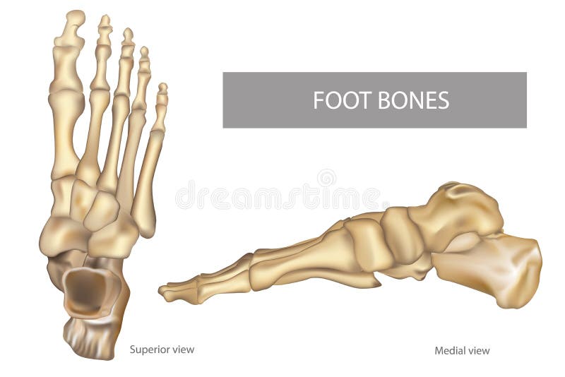
Free with trial Anatomy Bones of the Feet. OrthDiagram. Orthotics for foot Superior view and Medial view. Diagram. Midfoot vectors Anatomy Bones of the Feet. OrthDiagram. Anatomy Bones of the Feet. Orthotics for foot Superior view and Medial view. Diagram
Free with trial Bone structure of the foot. Human anatomy. Vector illustration. Midfoot vectors Bone structure of the foot. Human anatomy. Vector illustration
Free with trial Big toe joint replacement. Vector illustration of big toe joint arthroplasty. Midfoot vectors Big toe joint replacement. Vector illustration of big toe joint arthroplasty
Free with trial Destruction of the joint of the big toe in arthrosis. Vector illustration of the destruction of the joint of the big toe in arthrosis. Midfoot vectors Destruction of the joint of the big toe in arthrosis. Vector illustration of the destruction of the joint of the big toe in arthrosis
Free with trial Arthroplasty of the big toe joint. Vector illustration of partial and complete arthroplasty of the big toe joint. Midfoot vectors Arthroplasty of the big toe joint. Vector illustration of partial and complete arthroplasty of the big toe joint
Free with trial Foot Bone Anatomy. Vector Illustration. Midfoot vectors Foot Bone Anatomy. Vector Illustration
Free with trial Foot Bone Anatomy. Vector Illustration. Midfoot vectors Foot Bone Anatomy. Vector Illustration
Free with trial Bones of the human foot. Illustration of the bones of the human. Midfoot vectors Bones of the human foot. Illustration of the bones of the human
Free with trial The human foot's skeletal structure is composed of 26 bones, including the tarsal, metatarsal, and phalangeal bones. These bones form the arches of the foot, providing support, shock absorption, and flexibility for walking, running, and maintaining balance. Midfoot vectors Bones of the right foot, dorsal view. The human foot's skeletal structure is composed of 26 bones, including the tarsal, metatarsal, and phalangeal bones. These. The human foot's skeletal structure is composed of 26 bones, including the tarsal, metatarsal, and phalangeal bones. These bones form the arches of the foot, providing support, shock absorption, and flexibility for walking, running, and maintaining balance.
Free with trial Knee, ankle and foot bone anatomy. Human leg bones and joints. Doctors appointment in clinic. Skeletal medical anatomical poster for hospital. X ray isolated flat vector illustration. Midfoot vectors Human bones set. Knee, ankle and foot bone anatomy. Human leg bones and joints. Doctors appointment in clinic. Skeletal medical anatomical poster for hospital. X. Knee, ankle and foot bone anatomy. Human leg bones and joints. Doctors appointment in clinic. Skeletal medical anatomical poster for hospital. X ray isolated flat vector illustration.
Free with trial Foot bone anatomy concept. Descriptions of the human feet bones and joints. Talus, ligaments, calcaneus, navicular anatomy. Skeletal medical anatomical poster. Ankle X ray isolated vector illustration. Midfoot vectors Foot bones anatomy. Foot bone anatomy concept. Descriptions of the human feet bones and joints. Talus, ligaments, calcaneus, navicular anatomy. Skeletal medical. Foot bone anatomy concept. Descriptions of the human feet bones and joints. Talus, ligaments, calcaneus, navicular anatomy. Skeletal medical anatomical poster. Ankle X ray isolated vector illustration
Free with trial Foot bone anatomy concept. Descriptions of the human feet bones and joints. Talus, ligaments, calcaneus, navicular anatomy. Skeletal medical anatomical poster. Ankle X ray isolated vector illustration. Midfoot vectors Foot bones anatomy. Foot bone anatomy concept. Descriptions of the human feet bones and joints. Talus, ligaments, calcaneus, navicular anatomy. Skeletal medical. Foot bone anatomy concept. Descriptions of the human feet bones and joints. Talus, ligaments, calcaneus, navicular anatomy. Skeletal medical anatomical poster. Ankle X ray isolated vector illustration
Free with trial Foot bone anatomy concept. Descriptions of the human feet bones and joints. Talus, ligaments, calcaneus, navicular anatomy. Skeletal medical anatomical poster. Ankle X ray isolated vector illustration. Midfoot vectors Foot bones anatomy. Foot bone anatomy concept. Descriptions of the human feet bones and joints. Talus, ligaments, calcaneus, navicular anatomy. Skeletal medical. Foot bone anatomy concept. Descriptions of the human feet bones and joints. Talus, ligaments, calcaneus, navicular anatomy. Skeletal medical anatomical poster. Ankle X ray isolated vector illustration
 Sign in