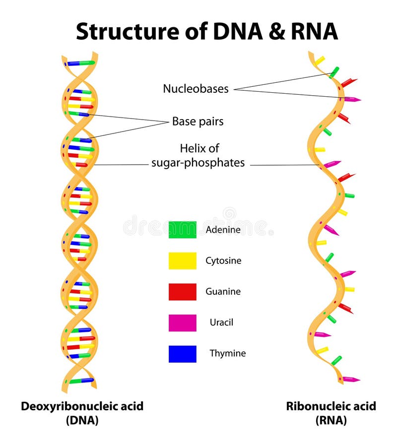
Free with trial Differences between DNA and RNA. There are two differences between DNA and RNA. Each RNA nucleotide contains a ribose sugar (instead of a deoxyribose in DNA), a phosphate, and one of the four nitrogenous bases. The difference between the nitrogenous bases of DNA and that of RNA is that thymine (T) in DNA is translated into Uracil (U) in RNA. Nitrogenous vectors Structure DNA and RNA molecule. Vector. Differences between DNA and RNA. There are two differences between DNA and RNA. Each RNA nucleotide contains a ribose. Differences between DNA and RNA. There are two differences between DNA and RNA. Each RNA nucleotide contains a ribose sugar (instead of a deoxyribose in DNA), a phosphate, and one of the four nitrogenous bases. The difference between the nitrogenous bases of DNA and that of RNA is that thymine (T) in DNA is translated into Uracil (U) in RNA.
Free with trial Structural formulas of purine and pyrimidine nitrogenous bases of DNA and RNA, 2d illustration, vector, isolated on white. Nitrogenous vectors Structural formulas of purine and pyrimidine nitrogenous bases of DNA an RNA. Structural formulas of purine and pyrimidine nitrogenous bases of DNA and RNA, 2d. Structural formulas of purine and pyrimidine nitrogenous bases of DNA and RNA, 2d illustration, vector, isolated on white
Free with trial Ammonia, NH3, molecule model and chemical formula. Chemical compound of nitrogen and hydrogen. A colorless gas. Ball-and-stick model, geometric structure and structural formula. Illustration. Vector. Nitrogenous vectors Ammonia, NH3, molecule model and chemical formula. Chemical compound of nitrogen and hydrogen. A colorless gas. Ball-and-stick model, geometric structure and
Free with trial DNA vs RNA strand infographic diagram with compound comparison chart chain structure parts cytosine guanine adenine thymine and uracil nitrogenous bases vector illustration for biology genetic science education. Nitrogenous vectors DNA vs RNA strand infographic diagram. With compound comparison chart chain structure parts cytosine guanine adenine thymine and uracil nitrogenous bases vector. DNA vs RNA strand infographic diagram with compound comparison chart chain structure parts cytosine guanine adenine thymine and uracil nitrogenous bases vector illustration for biology genetic science education
Free with trial Chemistry - adenine formula and structure. Nitrogenous vectors Chemistry. Adenine formula and structure
Free with trial Antihistamines are drugs which treat allergic rhinitis and other allergies. Affects the perception of the nervous system and brain. Histamine Illustration. Nitrogenous vectors Histaminergic system in the human brain. Histamine Illustration. Antihistamines are drugs which treat allergic rhinitis and other allergies. Affects the. Antihistamines are drugs which treat allergic rhinitis and other allergies. Affects the perception of the nervous system and brain. Histamine Illustration.
Free with trial The kidneys are bean-shaped organs that serve several essential regulatory roles in vertebrates. Their main function is to regulate the balance of electrolytes in the blood, along with maintaining pH homeostasis. Nitrogenous vectors Kidney Structures. Health care education infographic. Vector design. The kidneys are bean-shaped organs that serve several essential regulatory roles in. The kidneys are bean-shaped organs that serve several essential regulatory roles in vertebrates. Their main function is to regulate the balance of electrolytes in the blood, along with maintaining pH homeostasis.
Free with trial Adenosine triphosphate ATP molecule, is intracellular energy transfer and required in the synthesis of RNA. Structural chemical formula and molecule model. Vector illustration. Nitrogenous vectors Adenosine triphosphate ATP molecule, is intracellular energy t. Ransfer and required in the synthesis of RNA. Structural chemical formula and molecule model
Free with trial Chemical formulas of purine and pyrimidine nitrogenous bases. Illustration. Nitrogenous vectors Chemical formulas of purine and pyrimidine nitrogenous bases. Structural formulas of purine and pyrimidine nitrogenous bases. Illustration
Free with trial Nitrogenous base. A nitrogenous base or nitrogen containing base is an organic molecule with a nitrogen atom that has the chemical properties of a base. Nitrogenous vectors Nitrogenous base. A nitrogenous base or nitrogen containing base is an organic molecule with a nitrogen atom that has the chemical properties of a base
Free with trial Trimethylamine is a nitrogenous base and can be protonated to trimethylammonium cation. This colorless, hygroscopic, and flammable tertiary amine has a strong fishy or an ammonia-like odor. Nitrogenous vectors Trimethylamine is a nitrogenous base. And can be protonated to trimethylammonium cation. This colorless, hygroscopic, and flammable tertiary amine has a strong. Trimethylamine is a nitrogenous base and can be protonated to trimethylammonium cation. This colorless, hygroscopic, and flammable tertiary amine has a strong fishy or an ammonia-like odor
Free with trial Histamine molecule. It is amine, nitrogenous compound, stimulant of gastric secretion, vasodilator, and centrally acting neurotransmitter. Structural chemical formula and molecule model. Vector illustration. Nitrogenous vectors Histamine molecule. It is amine, nitrogenous compound, stimulant of gastric secretion, vasodilator, and centrally acting. Neurotransmitter. Structural chemical. Histamine molecule. It is amine, nitrogenous compound, stimulant of gastric secretion, vasodilator, and centrally acting neurotransmitter. Structural chemical formula and molecule model. Vector illustration
Free with trial Histamine molecule. It is amine, nitrogenous compound, stimulant of gastric secretion, vasodilator, and centrally acting neurotransmitter. Structural chemical formula on the dark blue background. Vector illustration. Nitrogenous vectors Histamine molecule. It is amine, nitrogenous compound, stimulant of gastric secretion, vasodilator, and centrally acting. Neurotransmitter. Structural chemical. Histamine molecule. It is amine, nitrogenous compound, stimulant of gastric secretion, vasodilator, and centrally acting neurotransmitter. Structural chemical formula on the dark blue background. Vector illustration
Free with trial Nucleoside vs Nucleotide compound differences comparison outline diagram. Educational labeled pyrimidine base link to sugar example with nitrogenous, phosphate group and pentose vector illustration. Nitrogenous vectors Nucleoside vs Nucleotide compound differences comparison outline diagram. Educational labeled pyrimidine base link to sugar example with nitrogenous, phosphate
Free with trial DNA nucleotides, also known as deoxyribonucleotides, are the building blocks of DNA (deoxyribonucleic acid) molecules. They consist of three main components: a deoxyribose sugar, a phosphate group, and a nitrogenous base. DNA nucleotides can contain one of four nitrogenous bases: adenine (A), cytosine (C), guanine (G), or thymine (T). Adenine and guanine are purine bases, while cytosine and thymine are pyrimidine bases. The nitrogenous base is attached to the 1' carbon atom of the deoxyribose sugar. Nitrogenous vectors DNA nucleotides (deoxyribonucleotides). DNA nucleotides, also known as deoxyribonucleotides, are the building blocks of DNA (deoxyribonucleic acid) molecules. DNA nucleotides, also known as deoxyribonucleotides, are the building blocks of DNA (deoxyribonucleic acid) molecules. They consist of three main components: a deoxyribose sugar, a phosphate group, and a nitrogenous base. DNA nucleotides can contain one of four nitrogenous bases: adenine (A), cytosine (C), guanine (G), or thymine (T). Adenine and guanine are purine bases, while cytosine and thymine are pyrimidine bases. The nitrogenous base is attached to the 1' carbon atom of the deoxyribose sugar.
Free with trial DNA (deoxyribonucleic acid) is a double-stranded molecule that carries the genetic information in all living organisms. It consists of two long strands of nucleotides twisted around each other to form a double helix structure. DNA is composed of individual units called nucleotides. Each nucleotide consists of three components: a deoxyribose sugar molecule, a phosphate group, and one of four nitrogenous bases. The four nitrogenous bases found in DNA are adenine (A), cytosine (C), guanine (G), and thymine (T). Nitrogenous vectors DNA structure. Ladder configuration. DNA (deoxyribonucleic acid) is a double-stranded molecule that carries the genetic information in all living organisms. It. DNA (deoxyribonucleic acid) is a double-stranded molecule that carries the genetic information in all living organisms. It consists of two long strands of nucleotides twisted around each other to form a double helix structure. DNA is composed of individual units called nucleotides. Each nucleotide consists of three components: a deoxyribose sugar molecule, a phosphate group, and one of four nitrogenous bases. The four nitrogenous bases found in DNA are adenine (A), cytosine (C), guanine (G), and thymine (T).
Free with trial ATP (adenosine triphosphate) consists of three main components: a nitrogenous base called adenine, a sugar molecule known as ribose, and a chain of three phosphate groups. ATP serves as the primary energy currency in cells, providing energy for various biological processes through the hydrolysis of its high-energy phosphate bonds. Nitrogenous vectors Adenosine triphosphate (ATP). Chemical formula. ATP (adenosine triphosphate) consists of three main components: a nitrogenous base called adenine, a sugar. ATP (adenosine triphosphate) consists of three main components: a nitrogenous base called adenine, a sugar molecule known as ribose, and a chain of three phosphate groups. ATP serves as the primary energy currency in cells, providing energy for various biological processes through the hydrolysis of its high-energy phosphate bonds.
Free with trial Structure of Cell. From Nucleobase like adenine to Gene, DNA and Chromosome. genome sequence. Molecular biology. Vector poster. Nitrogenous vectors Structure of Cell. From Nucleobase like adenine to Gene, DNA and Chromosome. Genome sequence. Molecular biology. Vector poster
Free with trial Primary nucleobases, chemical formulas and skeletal structures. Adenine, guanine, cytosine, thymine and uracil, represented by letters A, G, C, T and U. Fundamental units of the genetic code. Vector. Nitrogenous vectors Primary nucleobases, chemical formulas and skeletal structures. Adenine, guanine, cytosine, thymine and uracil, represented by letters A, G, C, T and U
Free with trial DNA structure. Base pairing and nucleotide. Cytosine, Thymine, Guanine, Adenine. Vector illustration. Nitrogenous vectors DNA structure. Base pairing and nucleotide. Cytosine, Thymine, Guanine, Adenine. Vector illustration
Free with trial The liver is the only human internal organ capable of natural regeneration of lost tissue. This is however, not true regeneration but rather compensatory growth in mammals. Nitrogenous vectors The liver is the only human internal organ capable of natural re. Generation of lost tissue.This is however, not true regeneration but rather compensatory growth
Free with trial Adenine. Chemical structural formula and model of molecule. C5H5N5. Vector diagram for educational, medical, biological, and scientific use. Nitrogenous vectors Adenine. Chemical structural formula and model of molecule. C5H5N5. Vector diagram for educational, medical, biological, and scientific use
Free with trial The kidneys are bean-shaped organs that serve several essential regulatory roles in vertebrates. Their main function is to regulate the balance of electrolytes in the blood, along with maintaining pH homeostasis. Illustration. Nitrogenous vectors The kidneys are bean-shaped organs. Structures. Illustration. The kidneys are bean-shaped organs that serve several essential regulatory roles in vertebrates. The kidneys are bean-shaped organs that serve several essential regulatory roles in vertebrates. Their main function is to regulate the balance of electrolytes in the blood, along with maintaining pH homeostasis. Illustration.
Free with trial VR wireframe headset vector banner. Polygonal man wearing virtual reality glasses, with holographic of atp acid. Science, diagnostics, virtual analytics, analysis. VR games. Thank you for watching. Nitrogenous vectors VR wireframe headset vector banner. Polygonal man wearing virtual reality glasses, with holographic of atp acid. Science. Diagnostics, virtual analytics. VR wireframe headset vector banner. Polygonal man wearing virtual reality glasses, with holographic of atp acid. Science, diagnostics, virtual analytics, analysis. VR games. Thank you for watching
Free with trial Types and Functions of Neurotransmitter. A Neurotransmitter are chemical messengers in the body,Their working is to transmit signals from nerve cells to target cells. Nitrogenous vectors Types and Functions of Neurotransmitter. A Neurotransmitter are chemical messengers in the body,Their working is to transmit signals from nerve cells to target
Free with trial Acute Headache. Diagram learning The Acute Headache as a result a process of the brain and consequences expanded the blood vessel. Diagram. Nitrogenous vectors Acute Headache. Diagram learning The Acute Headache as a result a process of the brain and consequences expanded the blood vessel. Acute Headache. Diagram
Free with trial A genome sciences educational diagram that make up the differences between RNA and DNA illustrating the different nucleic acids and phosphate sugars. Nitrogenous vectors DNA versus RNA genomic educational diagram. A genome sciences educational diagram that make up the differences between RNA and DNA illustrating the different. A genome sciences educational diagram that make up the differences between RNA and DNA illustrating the different nucleic acids and phosphate sugars
Free with trial Chemical organic formula of histamine diagram hand drawn schematic vector illustration. Medical science educational illustration. Nitrogenous vectors Chemical formula histamine diagram medical science. Chemical organic formula of histamine diagram hand drawn schematic vector illustration. Medical science. Chemical organic formula of histamine diagram hand drawn schematic vector illustration. Medical science educational illustration
Free with trial Guanosine phosphates guanosine triphosphate, guanosine diphosphate, guanosine monophosphate, cyclic guanosine monophosphate. Structural chemical formula of phosphate nucleotides that are the building blocks of nucleic acids. Vector illustration. Nitrogenous vectors Guanosine phosphates guanosine triphosphate, guanosine diphosph. Ate, guanosine monophosphate, cyclic guanosine monophosphate. Structural chemical formula of. Guanosine phosphates guanosine triphosphate, guanosine diphosphate, guanosine monophosphate, cyclic guanosine monophosphate. Structural chemical formula of phosphate nucleotides that are the building blocks of nucleic acids. Vector illustration
Free with trial Chemical structural formulas Adenosine phosphates nucleotides : adenosine monophosphate AMP, adenosine diphosphate ADP , adenosine triphosphate ATP , cyclic adenosine monophosphate cAMP. Building blocks of nucleic acids. Vector illustration. Nitrogenous vectors Chemical structural formulas Adenosine phosphates nucleotides :. Adenosine monophosphate AMP, adenosine diphosphate ADP , adenosine triphosphate ATP , cyclic. Chemical structural formulas Adenosine phosphates nucleotides : adenosine monophosphate AMP, adenosine diphosphate ADP , adenosine triphosphate ATP , cyclic adenosine monophosphate cAMP. Building blocks of nucleic acids. Vector illustration
Free with trial The four bases of DNA (adenine, thymine, guanine, cytosine). Nitrogenous vectors The four bases of DNA. (adenine, thymine, guanine, cytosine
Free with trial Chemical structural formulas Adenosine phosphates nucleotides : adenosine monophosphate AMP, adenosine diphosphate ADP , adenosine triphosphate ATP , cyclic adenosine monophosphate cAMP. Sheet of paper in a cage. Vector illustration. Nitrogenous vectors Chemical structural formulas Adenosine phosphates nucleotides :. Adenosine monophosphate AMP, adenosine diphosphate ADP , adenosine triphosphate ATP , cyclic. Chemical structural formulas Adenosine phosphates nucleotides : adenosine monophosphate AMP, adenosine diphosphate ADP , adenosine triphosphate ATP , cyclic adenosine monophosphate cAMP. Sheet of paper in a cage. Vector illustration
Free with trial Adenosine - purine nucleoside molecule, is important part of ATP, ADP, cAMP , RNA, DNA. Structural chemical formula and molecule model. Vector illustration Adenosine - purine nucleoside molecule, is important part of ATP, ADP, cAMP , RNA, DNA. Vector illustration. Nitrogenous vectors Adenosine - purine nucleoside molecule, is important part of ATP. ADP, cAMP , RNA, DNA. Structural chemical formula and molecule model. Vector illustration. Adenosine - purine nucleoside molecule, is important part of ATP, ADP, cAMP , RNA, DNA. Structural chemical formula and molecule model. Vector illustration Adenosine - purine nucleoside molecule, is important part of ATP, ADP, cAMP , RNA, DNA. Structural chemical formula and molecule model. Vector illustration
Free with trial Adenosine monophosphate AMP molecule, it is an ester of phosphoric acid and the nucleoside adenosine monomer in the production RNA. Structural chemical formula and molecule model. Vector illustration. Nitrogenous vectors Adenosine monophosphate AMP molecule, it is an ester of phosph. Oric acid and the nucleoside adenosine monomer in the production RNA . Structural chemical. Adenosine monophosphate AMP molecule, it is an ester of phosphoric acid and the nucleoside adenosine monomer in the production RNA . Structural chemical formula and molecule model. Vector illustration
Free with trial Adenosine monophosphate AMP molecule, it is an ester of phosphoric acid and the nucleoside adenosine monomer in the production RNA. Sheet of paper in a cage. Structural chemical formula and molecule model. Vector illustration. Nitrogenous vectors Adenosine monophosphate AMP molecule, it is an ester of phosph. Oric acid and the nucleoside adenosine monomer in the production RNA . Sheet of paper in a cage. Adenosine monophosphate AMP molecule, it is an ester of phosphoric acid and the nucleoside adenosine monomer in the production RNA . Sheet of paper in a cage. Structural chemical formula and molecule model. Vector illustration
Free with trial Deoxyribonucleic acid and Ribonucleic acid spirals with nucleobases molecular formulas. DNA and RNA molecule with nucleobases structure description - cytosine, guanine, adenine, uracil, thymine. Nitrogenous vectors Deoxyribonucleic acid and Ribonucleic acid with nucleobases formulas. DNA and RNA nucleobases structure - cytosine. Deoxyribonucleic acid and Ribonucleic acid. Deoxyribonucleic acid and Ribonucleic acid spirals with nucleobases molecular formulas. DNA and RNA molecule with nucleobases structure description - cytosine, guanine, adenine, uracil, thymine.
Free with trial Pyrimidine nucleic acid, nitrogenous base, nitrogen and hydrogen formula. Nitrogen and hydrogen biology molecular structure, DNA nitrogenous base microbiology molecule or nucleic acid vector formula. Nitrogenous vectors Pyrimidine nucleic acid, nitrogenous base formula. Pyrimidine nucleic acid, nitrogenous base, nitrogen and hydrogen formula. Nitrogen and hydrogen biology. Pyrimidine nucleic acid, nitrogenous base, nitrogen and hydrogen formula. Nitrogen and hydrogen biology molecular structure, DNA nitrogenous base microbiology molecule or nucleic acid vector formula
Free with trial Nitrogenous bases are nitrogen-containing molecules that are crucial components of DNA and RNA that form nucleotides, ultimately coding for all of life. Nitrogenous vectors Nitrogenous bases of DNA and RNA. Nitrogenous bases are nitrogen-containing molecules that are crucial components of DNA and RNA that form nucleotides. Nitrogenous bases are nitrogen-containing molecules that are crucial components of DNA and RNA that form nucleotides, ultimately coding for all of life
Free with trial Nitrogenous bases are nitrogen-containing molecules that are crucial components of DNA and RNA that form nucleotides, ultimately coding for all of life. Nitrogenous vectors Nitrogenous bases of DNA and RNA. Nitrogenous bases are nitrogen-containing molecules that are crucial components of DNA and RNA that form nucleotides. Nitrogenous bases are nitrogen-containing molecules that are crucial components of DNA and RNA that form nucleotides, ultimately coding for all of life
Free with trial Adenine nucleic acid, nitrogenous base, nitrogen and hydrogen formula. Nitrogen and hydrogen science formula, nucleic acid molecular structure or DNA nitrogenous base research vector molecule. Nitrogenous vectors Adenine nucleic acid, nitrogenous base formula. Adenine nucleic acid, nitrogenous base, nitrogen and hydrogen formula. Nitrogen and hydrogen science formula. Adenine nucleic acid, nitrogenous base, nitrogen and hydrogen formula. Nitrogen and hydrogen science formula, nucleic acid molecular structure or DNA nitrogenous base research vector molecule
Free with trial Cytosine nucleic acid, nitrogenous base, nitrogen and hydrogen formula. DNA nitrogenous base molecule, nitrogen and hydrogen biology science formula or nucleic acid molecular vector structure. Nitrogenous vectors Cytosine nucleic acid, nitrogenous base formula. Cytosine nucleic acid, nitrogenous base, nitrogen and hydrogen formula. DNA nitrogenous base molecule, nitrogen. Cytosine nucleic acid, nitrogenous base, nitrogen and hydrogen formula. DNA nitrogenous base molecule, nitrogen and hydrogen biology science formula or nucleic acid molecular vector structure
Free with trial Nucleic acids or nitrogenous bases, nitrogen and hydrogen formulas, vector molecule structure. Purine, adenine, guanine and thymine with cytosine or pyrimidine chemical formula and molecular structure. Nitrogenous vectors Nucleic acids, nitrogenous bases molecule formulas. Nucleic acids or nitrogenous bases, nitrogen and hydrogen formulas, vector molecule structure. Purine. Nucleic acids or nitrogenous bases, nitrogen and hydrogen formulas, vector molecule structure. Purine, adenine, guanine and thymine with cytosine or pyrimidine chemical formula and molecular structure
Free with trial Different types of nitrogenous base molecule types structure with diagram form chain. Vector illustration. Labeled pyrimidines, purines, adenine, guanine, thymine, cytosine, uracil. Nitrogen molecule. Nitrogenous vectors Different types of nitrogenous base molecule types structure with diagram form chain. Vector illustration. Labeled pyrimidines,. Purines, adenine, guanine. Different types of nitrogenous base molecule types structure with diagram form chain. Vector illustration. Labeled pyrimidines, purines, adenine, guanine, thymine, cytosine, uracil. Nitrogen molecule
Free with trial Complementary base pairing hydrogen bonds diagram form chain. Labeled nitrogenous bases adenine, uracil pairs, thymine, cytosine, and guanine. Chemical educational poster vector illustration. Nitrogenous vectors Complementary base pairing hydrogen bonds diagram form chain. Labeled nitrogenous bases adenine, uracil pairs, thymine, cytosine,. And guanine. Chemical. Complementary base pairing hydrogen bonds diagram form chain. Labeled nitrogenous bases adenine, uracil pairs, thymine, cytosine, and guanine. Chemical educational poster vector illustration
Free with trial DNA nucleotides (deoxyribonucleoside monophosphates): dAMP, dGMP, dCMP, and dTMP. Each molecule is shown with its three main componentsâ€'phosphate group, deoxyribose sugar, and nitrogenous base (adenine, guanine, cytosine, and thymine. Nitrogenous vectors DNA nucleotides molecules showing dAMP, dGMP, dCMP, and dTMP Showing Phosphate Group, Deoxyribose Sugar, and Nitrogenous Bases. DNA nucleotides (. DNA nucleotides (deoxyribonucleoside monophosphates): dAMP, dGMP, dCMP, and dTMP. Each molecule is shown with its three main componentsâ€'phosphate group, deoxyribose sugar, and nitrogenous base (adenine, guanine, cytosine, and thymine
Free with trial Vector Illustration of nitrogenous base molecules types. Adeline, Thymine, Guanine, Cytosine, Uracil. Nitrogenous vectors Vector Illustration of nitrogenous base molecules types. Adeline, Thymine, Guanine, Cytosine, Uracil
Free with trial Collection of science and laboratory elements in clean flat style, including scientific equipment, laboratory tools, research symbols, chemistry and biology objects, educational science icons, and medical research elements. Isolated on a white background, suitable for education, laboratory, research, healthcare, science projects, textbooks, presentations, websites, infographics, and stock design. Nitrogenous vectors DNA vs RNA: Structural Comparison and Nitrogenous Bases. Collection of science and laboratory elements in clean flat style, including scientific equipment. Collection of science and laboratory elements in clean flat style, including scientific equipment, laboratory tools, research symbols, chemistry and biology objects, educational science icons, and medical research elements. Isolated on a white background, suitable for education, laboratory, research, healthcare, science projects, textbooks, presentations, websites, infographics, and stock design.
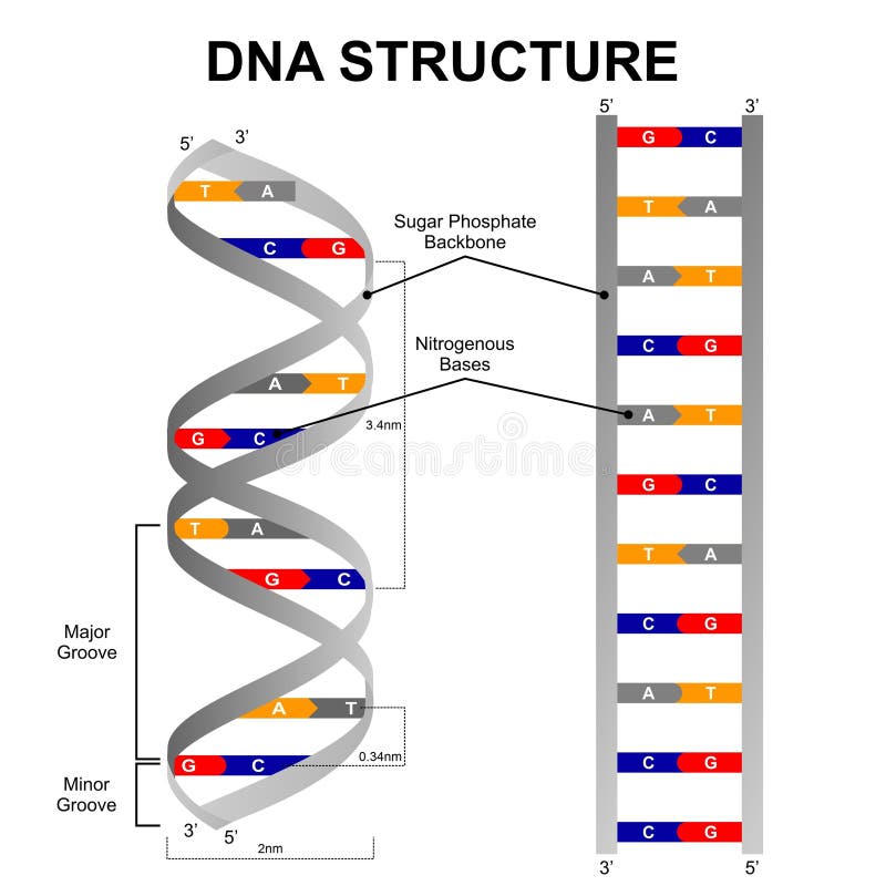
Free with trial Illustration of a DNA double helix with labeled components. The gray sugar-phosphate backbone twists around nitrogenous bases: Adenine (A) pairs with Thymine (T), and Cytosine (C) pairs with Guanine (G) using hydrogen bonds. The structure shows the major and minor grooves, with distances marked as 3. 4 nm per helical turn and 0. 34 nm per base pair. The diagram includes 5' to 3' directional indicators, emphasizing the sequential alignment of bases along the structure. Nitrogenous vectors DNA Structure Illustration. Illustration of a DNA double helix with labeled components. The gray sugar-phosphate backbone twists around nitrogenous bases. Illustration of a DNA double helix with labeled components. The gray sugar-phosphate backbone twists around nitrogenous bases: Adenine (A) pairs with Thymine (T), and Cytosine (C) pairs with Guanine (G) using hydrogen bonds. The structure shows the major and minor grooves, with distances marked as 3.4 nm per helical turn and 0.34 nm per base pair. The diagram includes 5' to 3' directional indicators, emphasizing the sequential alignment of bases along the structure.
Free with trial Illustration of chemistry, Purines and Pyrimidines models, Nucleotides: The difference between pyrimidines and purines, Purine and pyrimidine nitrogenous bases - chemical formulas. Nitrogenous vectors Illustration of chemistry, Purines and Pyrimidines models, Nucleotides. The difference between pyrimidines and purines, Purine and pyrimidine nitrogenous bases
Free with trial A minimal scientific icon illustrating a single-stranded RNA (Ribonucleic Acid) molecule with exposed nitrogenous bases. Perfect for genetics textbook graphics, biochemistry courseware, biotechnology design assets, and educational microbiology infographics. Nitrogenous vectors Single Stranded RNA Molecule Vector Icon for Molecular Biology. A minimal scientific icon illustrating a single-stranded RNA (Ribonucleic Acid) molecule with. A minimal scientific icon illustrating a single-stranded RNA (Ribonucleic Acid) molecule with exposed nitrogenous bases. Perfect for genetics textbook graphics, biochemistry courseware, biotechnology design assets, and educational microbiology infographics.
Free with trial This image depicts a simplified illustration of the double helix structure of a DNA molecule. The DNA is shown with its characteristic twisted ladder shape, featuring two strands wound around each other. Each strand is represented by a sugar-phosphate backbone, connected by pairs of nitrogenous bases in the center. The illustration emphasizes the iconic helical nature of DNA, a fundamental. Nitrogenous vectors Double helix structure of deoxyribonucleic acid (dna) molecule. This image depicts a simplified illustration of the double helix structure of a DNA molecule. The. This image depicts a simplified illustration of the double helix structure of a DNA molecule. The DNA is shown with its characteristic twisted ladder shape, featuring two strands wound around each other. Each strand is represented by a sugar-phosphate backbone, connected by pairs of nitrogenous bases in the center. The illustration emphasizes the iconic helical nature of DNA, a fundamental
Free with trial A comprehensive educational molecular biology diagram comparing Deoxyribonucleic Acid (DNA) and Ribonucleic Acid (RNA). Detailing double-stranded vs single-stranded structures, deoxyribose vs ribose sugars, thymine (T) vs uracil (U) nitrogenous bases, and primary functions in genetic information storage versus gene expression and protein synthesis. Ideal for genetics textbooks, molecular biology courseware, biotechnology guides, and scientific infographics. Nitrogenous vectors DNA and RNA Comparison Molecular Biology Diagram Showing Structural Differences. A comprehensive educational molecular biology diagram comparing Deoxyribonucleic. A comprehensive educational molecular biology diagram comparing Deoxyribonucleic Acid (DNA) and Ribonucleic Acid (RNA). Detailing double-stranded vs single-stranded structures, deoxyribose vs ribose sugars, thymine (T) vs uracil (U) nitrogenous bases, and primary functions in genetic information storage versus gene expression and protein synthesis. Ideal for genetics textbooks, molecular biology courseware, biotechnology guides, and scientific infographics.
Free with trial A genome sciences educational figure of RNA, or ribonucleic acid using ribose sugar and the nitrogenous base uracil instead of deoxyribose and thymine. Nitrogenous vectors Genomic science illustration of the components of an RNA molecule. A genome sciences educational figure of RNA, or ribonucleic acid using ribose sugar and the. A genome sciences educational figure of RNA, or ribonucleic acid using ribose sugar and the nitrogenous base uracil instead of deoxyribose and thymine
Free with trial Thymine and uracil nitrogenous bases have a very similar chemical structure. The only difference between them is the presence of an extra methyl group in the thymine structure which is depicted in this diagram. Nitrogenous vectors Thymine versus Uracil genomic educational graphic illustration. Thymine and uracil nitrogenous bases have a very similar chemical structure. The only difference. Thymine and uracil nitrogenous bases have a very similar chemical structure. The only difference between them is the presence of an extra methyl group in the thymine structure which is depicted in this diagram
Free with trial A genome sciences educational figure of RNA, or ribonucleic acid using ribose sugar and the nitrogenous base uracil instead of deoxyribose and thymine. Nitrogenous vectors Genomic science illustration of the components of an RNA molecule. A genome sciences educational figure of RNA, or ribonucleic acid using ribose sugar and the. A genome sciences educational figure of RNA, or ribonucleic acid using ribose sugar and the nitrogenous base uracil instead of deoxyribose and thymine
Free with trial Illustration of biology, DNA structure consists of two strands of nucleotides that are held together by hydrogen bonds, four nitrogenous bases: adenine , thymine, guanine, cytosine. Nitrogenous vectors Illustration of biology, DNA structure consists of two strands of nucleotides. That are held together by hydrogen bonds, four nitrogenous bases: adenine. illustration of biology, DNA structure consists of two strands of nucleotides that are held together by hydrogen bonds, four nitrogenous bases: adenine , thymine, guanine, cytosine
Free with trial Educational DNA nucleotide vector illustration showing the structure of deoxyguanosine monophosphate (dGMP). Diagram highlights the three major components of a DNA nucleotide including phosphate group, deoxyribose sugar, and guanine nitrogenous base. Ideal for biology education, genetics, molecular biology, biochemistry, scientific presentations, textbooks, healthcare publications, and academic learning materials. Nitrogenous vectors DNA Nucleotide Structure dGMP Deoxyguanosine Monophosphate with Phosphate Deoxyribose and Guanine Educational Vector Illustration. Educational DNA nucleotide. Educational DNA nucleotide vector illustration showing the structure of deoxyguanosine monophosphate (dGMP). Diagram highlights the three major components of a DNA nucleotide including phosphate group, deoxyribose sugar, and guanine nitrogenous base. Ideal for biology education, genetics, molecular biology, biochemistry, scientific presentations, textbooks, healthcare publications, and academic learning materials.
Free with trial Thymine and uracil nitrogenous bases have a very similar chemical structure. The only difference between them is the presence of an extra methyl group in the thymine structure which is depicted in this diagram. Nitrogenous vectors Molecular structure differences between thymine and Uracil. educational graphic illustration. Thymine and uracil nitrogenous bases have a very similar chemical. Thymine and uracil nitrogenous bases have a very similar chemical structure. The only difference between them is the presence of an extra methyl group in the thymine structure which is depicted in this diagram
Free with trial Isolated on a white background the three components that make up DNA, the nitrogenous base, pentose sugar, and a phosphate group. Nitrogenous vectors Chemical structure of DNA nucleotides and phosphate sugar backbone. Isolated on a white background the three components that make up DNA, the nitrogenous base. Isolated on a white background the three components that make up DNA, the nitrogenous base, pentose sugar, and a phosphate group
Free with trial Isolated on a white background the three components that make up DNA, the nitrogenous base, pentose sugar, and a phosphate group. Nitrogenous vectors Chemical structure of DNA nucleotides and phosphate sugar backbone. Isolated on a white background the three components that make up DNA, the nitrogenous base. Isolated on a white background the three components that make up DNA, the nitrogenous base, pentose sugar, and a phosphate group
Free with trial Isolated on a white background the three components that make up DNA, the nitrogenous base, pentose sugar, and a phosphate group. Nitrogenous vectors Chemical structure of DNA nucleotides and phosphate sugar backbone. Isolated on a white background the three components that make up DNA, the nitrogenous base. Isolated on a white background the three components that make up DNA, the nitrogenous base, pentose sugar, and a phosphate group
Free with trial A clear, educational diagram precisely illustrates the molecular structure of a short section of a DNA double helix. The visual focuses on the complementary arrangement of four base pairs, distinctly highlighting the key chemical components: the deoxyribose sugars, the phosphate backbone, and the central nitrogenous bases (Adenine, Thymine, Guanine, Cytosine). This image is an essential resource for teaching molecular biology and genetics, fundamentally explaining the elegant, stable structure that stores hereditary information. Nitrogenous vectors Simple diagram illustrating the structure of four base pairs in a DNA molecule. Section of the double helix with phosphate. A clear, educational diagram. A clear, educational diagram precisely illustrates the molecular structure of a short section of a DNA double helix. The visual focuses on the complementary arrangement of four base pairs, distinctly highlighting the key chemical components: the deoxyribose sugars, the phosphate backbone, and the central nitrogenous bases (Adenine, Thymine, Guanine, Cytosine). This image is an essential resource for teaching molecular biology and genetics, fundamentally explaining the elegant, stable structure that stores hereditary information.
Free with trial A clear, educational diagram precisely illustrates the molecular structure of a short section of a DNA double helix. The visual focuses on the complementary arrangement of four base pairs, distinctly highlighting the key chemical components: the deoxyribose sugars, the phosphate backbone, and the central nitrogenous bases (Adenine, Thymine, Guanine, Cytosine). This image is an essential resource for teaching molecular biology and genetics, fundamentally explaining the elegant, stable structure that stores hereditary information. Nitrogenous vectors Simple diagram illustrating the structure of four base pairs in a DNA molecule. Section of the double helix with phosphate. A clear, educational diagram. A clear, educational diagram precisely illustrates the molecular structure of a short section of a DNA double helix. The visual focuses on the complementary arrangement of four base pairs, distinctly highlighting the key chemical components: the deoxyribose sugars, the phosphate backbone, and the central nitrogenous bases (Adenine, Thymine, Guanine, Cytosine). This image is an essential resource for teaching molecular biology and genetics, fundamentally explaining the elegant, stable structure that stores hereditary information.
Free with trial Nucleotides are organic molecules composed of a nitrogenous base, a pentose sugar and a phosphate. They serve as monomeric units of the nucleic acid polymers. Nitrogenous vectors Nucleotide scientific biochemistry vector illustration infographic. Nucleotides are organic molecules composed of a nitrogenous base, a pentose sugar and a. Nucleotides are organic molecules composed of a nitrogenous base, a pentose sugar and a phosphate. They serve as monomeric units of the nucleic acid polymers
Free with trial Thymine and uracil nitrogenous bases have a very similar chemical structure. The only difference between them is the presence of an extra methyl group in the thymine structure which is depicted in this diagram. Nitrogenous vectors Thymine versus Uracil genomic educational graphic illustration. Thymine and uracil nitrogenous bases have a very similar chemical structure. The only difference. Thymine and uracil nitrogenous bases have a very similar chemical structure. The only difference between them is the presence of an extra methyl group in the thymine structure which is depicted in this diagram
Free with trial Illustration of biology, DNA structure consists of two strands of nucleotides that are held together by hydrogen bonds, four nitrogenous bases: adenine , thymine, guanine, cytosine. Nitrogenous vectors DNA structure consists of two strands of nucleotides that are held together by hydrogen bonds. Illustration of biology, DNA structure consists of two strands of. illustration of biology, DNA structure consists of two strands of nucleotides that are held together by hydrogen bonds, four nitrogenous bases: adenine , thymine, guanine, cytosine
Free with trial Cell Structure. Gene, DNA and Chromosome. Vector illustration. Nitrogenous vectors Cell Structure. Gene, DNA and Chromosome. Vector illustration
Free with trial Ammonia molecule, NH3, space-filling model. Inorganic chemical compound of nitrogen and hydrogen. Stable binary hydride and simplest pnictogen hydride, used in fertilizers, refrigerants, etc. Vector. Nitrogenous vectors Ammonia molecule, NH3, space-filling model and chemical formula. Ammonia molecule, NH3, space-filling model. Inorganic chemical compound of nitrogen and hydrogen. Ammonia molecule, NH3, space-filling model. Inorganic chemical compound of nitrogen and hydrogen. Stable binary hydride and simplest pnictogen hydride, used in fertilizers, refrigerants, etc. Vector
Free with trial Stylized black and white 3d rendering of adenine, the purine nucleobase component of dna and rna, isolated on white to emphasize molecular structure and scientific concept. Nitrogenous vectors Adenine dna molecule chemical structure icon. Stylized black and white 3d rendering of adenine, the purine nucleobase component of dna and rna, isolated on white. Stylized black and white 3d rendering of adenine, the purine nucleobase component of dna and rna, isolated on white to emphasize molecular structure and scientific concept
Free with trial Thymine is one of the bases of the DNA. Molecule. Nitrogenous vectors Thymine is one of the bases of the DNA. Molecule
Free with trial Guanine is one of the bases of the DNA. Molecule. Nitrogenous vectors Guanine is one of the bases of the DNA. Molecule
Free with trial Cytosine is one of the bases of the DNA. Molecule. Nitrogenous vectors Cytosine is one of the bases of the DNA. Molecule
Free with trial Science and laboratory elements collection in flat style, featuring scientific equipment, research tools, chemistry, biology, and educational science icons isolated on a white background. Nitrogenous vectors DNA Nucleotide Molecular Structure Diagram Illustration. Science and laboratory elements collection in flat style, featuring scientific equipment, research tools. Science and laboratory elements collection in flat style, featuring scientific equipment, research tools, chemistry, biology, and educational science icons isolated on a white background.
Free with trial Educational vector illustration showing the structure of DNA, including the double helix, nucleotide components, complementary base pairing, sugar-phosphate backbone, and hydrogen bonds. The diagram explains adenine, thymine, cytosine, and guanine interactions for genetics, molecular biology, biotechnology, and science education. Nitrogenous vectors DNA Structure and Nucleotide Base Pairing Educational Medical Vector Illustration. Educational vector illustration showing the structure of DNA, including the. Educational vector illustration showing the structure of DNA, including the double helix, nucleotide components, complementary base pairing, sugar-phosphate backbone, and hydrogen bonds. The diagram explains adenine, thymine, cytosine, and guanine interactions for genetics, molecular biology, biotechnology, and science education.
Free with trial The image depicts a detailed molecular model of a double-stranded DNA helix. The structure highlights the sugar-phosphate backbone and paired nitrogenous bases (adenine, thymine, cytosine, and guanine) that form the genetic code. The visualization emphasizes the helical twist and the complementary base pairing between strands, showcasing the fundamental architecture of DNA molecules. The atoms are. Nitrogenous vectors Molecular structure of a double-stranded dna helix with base pairs. The image depicts a detailed molecular model of a double-stranded DNA helix. The structure. The image depicts a detailed molecular model of a double-stranded DNA helix. The structure highlights the sugar-phosphate backbone and paired nitrogenous bases (adenine, thymine, cytosine, and guanine) that form the genetic code. The visualization emphasizes the helical twist and the complementary base pairing between strands, showcasing the fundamental architecture of DNA molecules. The atoms are
Free with trial This image depicts a stylized and simplified representation of the DNA double helix structure. The illustration shows the classic twisted ladder formation, composed of two long strands that wind around each other. Each strand is made of a series of sugar-phosphate backbones, connected by pairs of nitrogenous bases. The iconic spiral pattern symbolizes the genetic information carrier found in all. Nitrogenous vectors The iconic double helix structure of deoxyribonucleic acid (dna) molecule. This image depicts a stylized and simplified representation of the DNA double helix. This image depicts a stylized and simplified representation of the DNA double helix structure. The illustration shows the classic twisted ladder formation, composed of two long strands that wind around each other. Each strand is made of a series of sugar-phosphate backbones, connected by pairs of nitrogenous bases. The iconic spiral pattern symbolizes the genetic information carrier found in all
Free with trial This image depicts a detailed illustration of a DNA double helix structure in a gradient of colors. The DNA molecule is shown in a twisted ladder-like structure, with the sugar and phosphate molecules forming the backbone and the nitrogenous bases projecting inward from the backbone and pairing with each other in a complementary manner. The gradient of colors gives the image a sense of depth and dimensionality, highlighting the intricate structure of the DNA molecule. This image could be used in a variety of scientific and educational contexts to illustrate the structure and function of DNA. Nitrogenous vectors A detailed illustration of a DNA double helix structure in a gradient of colors. This image depicts a detailed illustration of a DNA double helix structure in a. This image depicts a detailed illustration of a DNA double helix structure in a gradient of colors. The DNA molecule is shown in a twisted ladder-like structure, with the sugar and phosphate molecules forming the backbone and the nitrogenous bases projecting inward from the backbone and pairing with each other in a complementary manner. The gradient of colors gives the image a sense of depth and dimensionality, highlighting the intricate structure of the DNA molecule. This image could be used in a variety of scientific and educational contexts to illustrate the structure and function of DNA.
Free with trial This image depicts a detailed illustration of a DNA double helix structure with light effects. The DNA molecule is shown in a spiral shape, with two strands twisted together. The image highlights the intricate details of the molecular structure, showcasing the sugar and phosphate molecules that make up the backbone of the DNA, as well as the nitrogenous bases that project inward from the backbone and pair with each other in a complementary manner. The image is often used in scientific and educational contexts to represent the fundamental concept of genetics and molecular biology. Nitrogenous vectors A detailed illustration of a DNA double helix structure with light effects. This image depicts a detailed illustration of a DNA double helix structure with light. This image depicts a detailed illustration of a DNA double helix structure with light effects. The DNA molecule is shown in a spiral shape, with two strands twisted together. The image highlights the intricate details of the molecular structure, showcasing the sugar and phosphate molecules that make up the backbone of the DNA, as well as the nitrogenous bases that project inward from the backbone and pair with each other in a complementary manner. The image is often used in scientific and educational contexts to represent the fundamental concept of genetics and molecular biology.
Free with trial This image presents a detailed close-up of a DNA double helix structure, showcasing its twisted spiral shape in a green color scheme. The DNA molecule is a fundamental component of life, carrying genetic information that determines the characteristics of living organisms. The double helix structure is composed of two complementary strands of nucleotides, with sugar and phosphate molecules forming the backbone and nitrogenous bases projecting inward from the backbone and pairing with each other in a complementary manner. This iconic structure was first described by James Watson and Francis Crick in 1953, revolutionizing our understanding of genetics and paving the way for major advances in fields such as genetic engineering, gene therapy, and personalized medicine. Nitrogenous vectors A detailed close-up of a DNA double helix structure with a green color scheme. This image presents a detailed close-up of a DNA double helix structure. This image presents a detailed close-up of a DNA double helix structure, showcasing its twisted spiral shape in a green color scheme. The DNA molecule is a fundamental component of life, carrying genetic information that determines the characteristics of living organisms. The double helix structure is composed of two complementary strands of nucleotides, with sugar and phosphate molecules forming the backbone and nitrogenous bases projecting inward from the backbone and pairing with each other in a complementary manner. This iconic structure was first described by James Watson and Francis Crick in 1953, revolutionizing our understanding of genetics and paving the way for major advances in fields such as genetic engineering, gene therapy, and personalized medicine.
Free with trial This image depicts a detailed illustration of a DNA double helix structure in a warm golden color. The DNA molecule is shown in a twisted ladder-like structure, with the sugar and phosphate molecules forming the backbone and the nitrogenous bases projecting inward from the backbone and pairing with each other in a complementary manner. The image is a digital illustration and is suitable for use in educational materials, scientific publications, and medical websites. Nitrogenous vectors A detailed illustration of a DNA double helix structure in a warm golden color. This image depicts a detailed illustration of a DNA double helix structure in a. This image depicts a detailed illustration of a DNA double helix structure in a warm golden color. The DNA molecule is shown in a twisted ladder-like structure, with the sugar and phosphate molecules forming the backbone and the nitrogenous bases projecting inward from the backbone and pairing with each other in a complementary manner. The image is a digital illustration and is suitable for use in educational materials, scientific publications, and medical websites.
 Sign in