Free with trial Osteoarthritis of the Knee. Medical illustration. Patella vectors Osteoarthritis of the Knee. Medical illustration
Free with trial Osteoarthritis of knee joint. Knee joint anatomy normal and with arthritis, eps10. Patella vectors Osteoarthritis of knee joint. Knee joint anatomy normal and with arthritis, eps10
Free with trial Common types of arthritis. Eps8, gradient and mesh printing compatible. Patella vectors Common types of arthritis. Eps8, gradient and mesh printing compatible
Free with trial Osteoarthritis. Arthritis or pain within a joint. degenerative joint disease. Cartilage becomes worn. This results in inflammation, swelling, and pain in the joint. Patella vectors Osteoarthritis or arthritis. Osteoarthritis. Arthritis or pain within a joint. degenerative joint disease. Cartilage becomes worn. This results in inflammation. Osteoarthritis. Arthritis or pain within a joint. degenerative joint disease. Cartilage becomes worn. This results in inflammation, swelling, and pain in the joint.
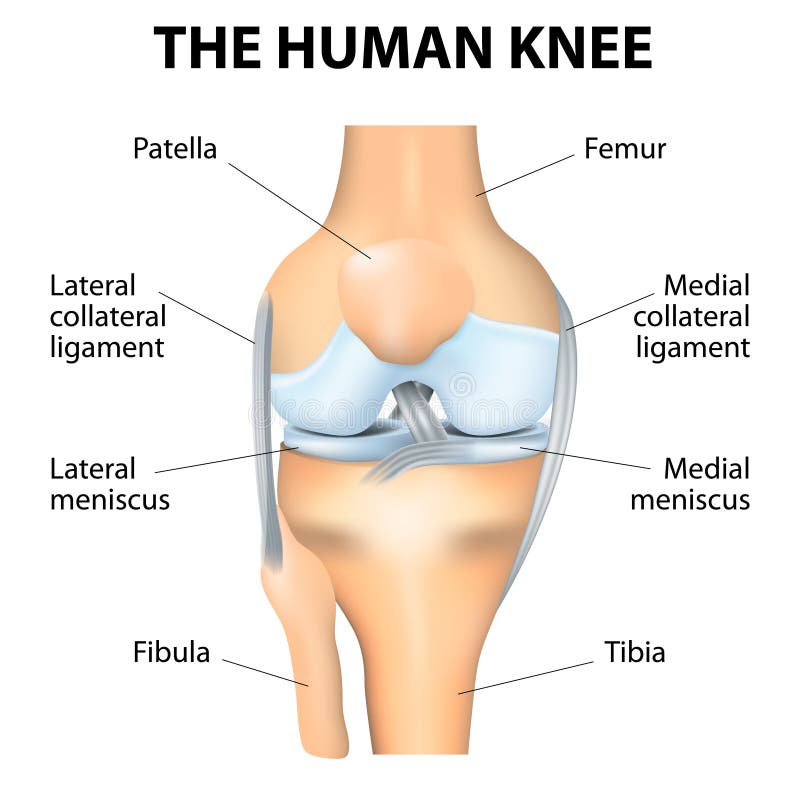
Free with trial Human Knee Anatomy. Human Knee joint anatomy. Vector. Patella vectors Human Knee Anatomy. Human Knee joint anatomy. Vector
Free with trial Typical bone fractures. Vector scheme. Patella vectors Typical bone fractures. Vector scheme
Free with trial Jumper's knee anatomy. Diagram showing mechanism of knee injury, eps8, gradient and mesh printing compatible. Patella vectors Jumper's knee anatomy. Diagram showing mechanism of knee injury, eps8, gradient and mesh printing compatible
Free with trial Anatomy of the Right Knee. Medical illustration. Patella vectors Anatomy of the Right Knee. Medical illustration
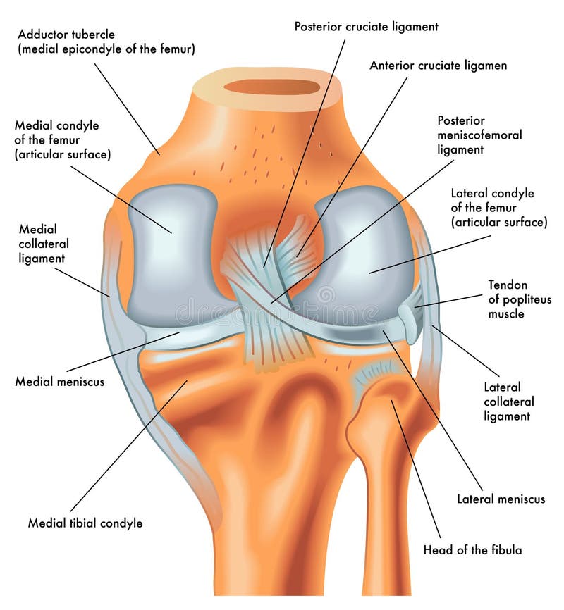
Free with trial Posterior view of the right knee in extension. Anatomy of the posterior of the right knee in extension. Patella vectors Posterior view of the right knee in extension. Anatomy of the posterior of the right knee in extension
Free with trial Chondromalacia. Patellae or soft cartilage under the kneecap condition. Patella vectors Chondromalacia. Patellae or soft cartilage under the kneecap condition
Free with trial Human knee joint. Anterior view of Human knee joint, eps8. Patella vectors Human knee joint. Anterior view of Human knee joint, eps8
Free with trial Menisci of the knee. Superior view of human knee with menisci, eps8. Patella vectors Menisci of the knee. Superior view of human knee with menisci, eps8
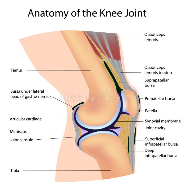
Free with trial Anatomy of the Knee Joint. Anatomy of human Knee Joint, eps8. Patella vectors Anatomy of the Knee Joint. Anatomy of human Knee Joint, eps8
Free with trial Synovial bursa of the human knee. Synovial bursa is a sac filled with lubricating fluid, located between tissues such as bone, muscle, tendons, and skin, that decreases rubbing. Patella vectors Synovial bursa of the human knee. Synovial bursa is a sac filled with lubricating fluid, located between tissues such as bone, muscle, tendons, and skin, that
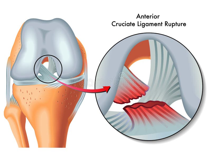
Free with trial Anterior cruciate ligament rupture. Medical Illustration of anterior cruciate ligament rupture. Patella vectors Anterior cruciate ligament rupture. Medical Illustration of anterior cruciate ligament rupture
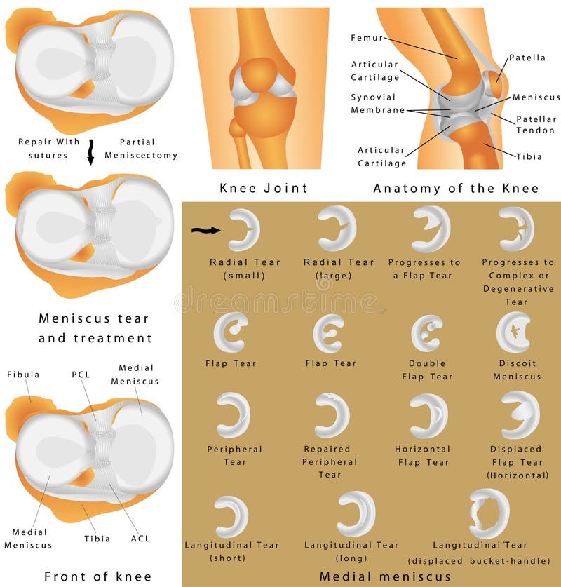
Free with trial Meniscus tear and surgery treatment. Types of Meniscus tear and surgery treatment, eps8. Patella vectors Meniscus tear and surgery treatment. Types of Meniscus tear and surgery treatment, eps8
Free with trial Posterior cruciate ligament rupture. Medical Illustration of posterior cruciate ligament ruptur. Patella vectors Posterior cruciate ligament rupture. Medical Illustration of posterior cruciate ligament ruptur
Free with trial Front view of the right knee in flexion. Anatomy of the front of the right knee in flexion. Patella vectors Front view of the right knee in flexion. Anatomy of the front of the right knee in flexion
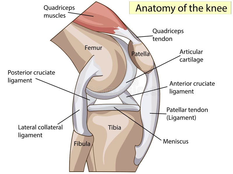
Free with trial Anatomy of the knee, illustration (for basic medical education, for clinics & Schools). Patella vectors Anatomy of the knee. Illustration (for basic medical education, for clinics & Schools
Free with trial Anatomy of the Knee. Human Knee Joint. Menisci of the knee. Medial meniscus. Lateral meniscus. Meniscus tear and surgery. Patella vectors Anatomy of the Knee. Human Knee Joint. Anatomy of the Knee. Menisci of the knee. Medial meniscus. Lateral meniscus. Meniscus tear and surgery
Free with trial Anatomy. Knee Joint Cross Section Showing the major parts which made the knee joint For Basic Medical Education Also for clinics. Patella vectors Anatomy. Structure knee joint vector. Anatomy. Knee Joint Cross Section Showing the major parts which made the knee joint For Basic Medical Education Also for. Anatomy. Knee Joint Cross Section Showing the major parts which made the knee joint For Basic Medical Education Also for clinics
Free with trial Luxating patella. Healthy and unhealthy dog`s knee joint, the medial luxating patella or knee cap dislocation illustration. Patella vectors Luxating patella. Healthy and unhealthy dog`s knee joint, the medial luxating patella or knee cap dislocation illustration
Free with trial Knee. Art illustration of leg with a highlight for the kneecap (patella. Patella vectors Knee. Art illustration of leg with a highlight for the kneecap (patella
Free with trial Shin splints vector illustration. Leg muscle sport trauma and bone pain labeled diagram. Isolated femur, patella, fibula, tibia and foot bones with shown injury location. Patella vectors Shin splints vector illustration. Leg muscle sport trauma and bone pain. Shin splints vector illustration. Leg muscle sport trauma and bone pain labeled diagram. Shin splints vector illustration. Leg muscle sport trauma and bone pain labeled diagram. Isolated femur, patella, fibula, tibia and foot bones with shown injury location.
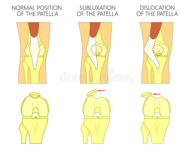
Free with trial Patellar dislocation. Normal position of kneecap and Patella displaced. Anatomy of the Knee. Patella vectors Patellar dislocation. Normal position of kneecap and Patella displaced. Anatomy of the Knee
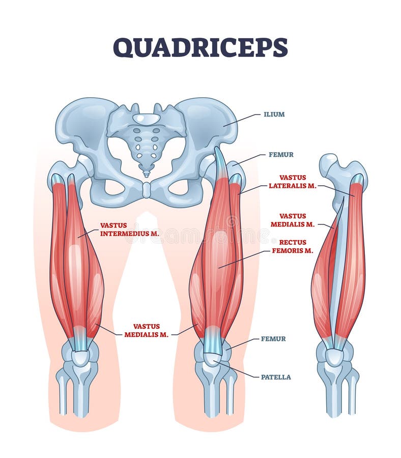
Free with trial Quadriceps muscle and quads leg muscular or bone anatomy outline diagram. Labeled educational medical scheme with vastus intermedius, medialis, lateralis or rectus femoris location vector illustration. Patella vectors Quadriceps muscle or quads leg muscular anatomical structure outline diagram. Quadriceps muscle and quads leg muscular or bone anatomy outline diagram. Labeled. Quadriceps muscle and quads leg muscular or bone anatomy outline diagram. Labeled educational medical scheme with vastus intermedius, medialis, lateralis or rectus femoris location vector illustration
Free with trial Bursitis is the inflammation of bursae. Bursitis. inflammation of bursae synovial fluid. Prepatellar bursitis housemaid`s knee and Infrapatellar bursitis. Patella vectors Bursitis is the inflammation of bursae. Bursitis. inflammation of bursae synovial fluid. Prepatellar bursitis housemaid`s knee and Infrapatellar bursitis
Free with trial Burning knee, painful knee and normal knee joint, abstract design. Human legs on a blue checkered background. Patella vectors Burning knee. Painful knee and normal knee joint, abstract design. Human legs on a blue checkered background
Free with trial Trigeminal neuralgia. neuroscience. Illustration of a male head. Patella vectors Trigeminal neuralgia. neuroscience. Illustration of a male head. trigeminal neuralgia. neuroscience
Free with trial Synovial bursa. Synovial Joint. Human anatomy. vector. Patella vectors Synovial bursa. Synovial Joint. Human anatomy. vector
Free with trial Knee joint. Human anatomy. knee joint on a white background. Patella vectors Knee joint. Human anatomy. knee joint on a white background
Free with trial Knee Joint Labeled Diagram. A knee joint with detailed labels. Patella vectors Knee Joint Labeled Diagram. A knee joint with detailed labels
Free with trial Bakers cyst. A complication of rheumatoid arthritis. Patella vectors Bakers cyst. A complication of rheumatoid arthritis
Free with trial Components of a Knee Replacement. Total Knee Replacement. Knee implant. Vector. Patella vectors Components of a Knee Replacement. Total Knee Replacement. Knee implant. Vector
Free with trial Medial collateral ligament rupture. Medical Illustration of medial collateral ligament rupture. Patella vectors Medial collateral ligament rupture. Medical Illustration of medial collateral ligament rupture
Free with trial The Iliotibial Band is a longitudinal fibrous reinforcement of the fascia lata in a knee muscle. Part of anatomy human body. Illustration. Patella vectors Iliotibial Band Anatomy. Illustration. The Iliotibial Band is a longitudinal fibrous reinforcement of the fascia lata in a knee muscle. Part of anatomy human. The Iliotibial Band is a longitudinal fibrous reinforcement of the fascia lata in a knee muscle. Part of anatomy human body. Illustration
Free with trial Human joints, knee joint, elbow joint, ankle joint, wrist. Medical orthopedic vector of set. Anatomy orthopedic human joint and illustration icon leg and hand joint. Patella vectors Human joints, knee and elbow, ankle wrist. Medical orthopedic vector set. Human joints, knee joint, elbow joint, ankle joint, wrist. Medical orthopedic vector of. Human joints, knee joint, elbow joint, ankle joint, wrist. Medical orthopedic vector of set. Anatomy orthopedic human joint and illustration icon leg and hand joint
Free with trial Ligaments of the knee. Anterior and Posterior cruciate ligaments, Patellar and Quadriceps, tendons, Medial and Lateral collateral ligaments. joint anatomy. Vector illustration for biological, medical, science and educational use. Patella vectors Ligaments of the knee. Anterior and Posterior cruciate ligaments, Patellar and Quadriceps, tendons, Medial and Lateral collateral ligaments. joint anatomy
Free with trial Bone fracture healing process. Fracture and repair. Vector scheme. Patella vectors Bone fracture healing process. Fracture and repair. Vector scheme
Free with trial Human knee joint icons - illustration for the web. Patella vectors Human knee joint icons. Illustration for the web
Free with trial Vector illustration of a healthy human knee joint and unhealthy knee with Baker`s cyst. Anatomy of human knee, sagittal section of the knee. for advertising and medical publications. EPS 10. Patella vectors Anatomy of the knee_Baker`s cyst. Vector illustration of a healthy human knee joint and unhealthy knee with Baker`s cyst. Anatomy of human knee, sagittal section. Vector illustration of a healthy human knee joint and unhealthy knee with Baker`s cyst. Anatomy of human knee, sagittal section of the knee. for advertising and medical publications. EPS 10.
Free with trial Anterior cruciate ligament injury. joint anatomy. Vector illustration for biological, medical, science and educational use. Patella vectors Anterior cruciate ligament injury. Joint anatomy. Vector illustration for biological, medical, science and educational use
Free with trial Vector illustration. Anatomy, front x-ray of an arthritic knee joint and a knee after unicompartmental or partial knee replacement. For advertising and medical publications. EPS 10. Patella vectors Meniscus _Arthritic knee and Partial knee replacement. Vector illustration. Anatomy, front x-ray of an arthritic knee joint and a knee after unicompartmental or. Vector illustration. Anatomy, front x-ray of an arthritic knee joint and a knee after unicompartmental or partial knee replacement. For advertising and medical publications. EPS 10
Free with trial Knee Tendons medical vector illustration scheme, anatomical diagram. Educational information. Patella vectors Knee Tendons medical vector illustration scheme, anatomical diagram. Educational information
Free with trial Quad leg muscles anatomy labeled diagram, vector illustration fitness poster. Sports physiotherapy educational information. Healthy muscular structure and bones. Vastus femoris, lateralis and medialis. Patella vectors Quad leg muscles anatomy labeled diagram, vector illustration fitness poster. Sports physiotherapy educational information. Healthy muscular structure and
Free with trial Knee Dislocations - medical vector illustration diagrams. Anatomical knee injury types scheme. Physiotherapy educational information. Patella vectors Knee Dislocations - medical vector illustration diagrams. Anatomical knee injury types scheme. Physiotherapy educational information
Free with trial Structure of the human knee. Vector drawing anatomy of the knee. Patella vectors Structure of the human knee. Vector drawing anatomy of the knee
Free with trial Cartilage vector illustration. Medical scheme of chondrocytes, collagen fibers, calcified cartilage, tide mark, subchondral and cancellous bone. Diagram of superficial, transition and deep zone. Patella vectors Cartilage vector illustration. Scheme of chondrocytes, collagen fibers, calcified, subchondral cancellous bone and superficial. Cartilage vector illustration. Cartilage vector illustration. Medical scheme of chondrocytes, collagen fibers, calcified cartilage, tide mark, subchondral and cancellous bone. Diagram of superficial, transition and deep zone.
Free with trial Anatomy of the knee joint side view, template for training a medical surgical poster, traumatology pages, vector illustration. Patella vectors Anatomy of the knee joint side view, template for training a medical surgical poster, traumatology page. Vector illustration. Anatomy of the knee joint side. Anatomy of the knee joint side view, template for training a medical surgical poster, traumatology pages, vector illustration.
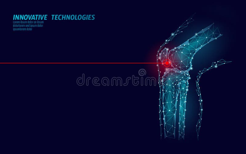
Free with trial Human knee joint 3d model vector illustration. Low poly design future technology cure pain treatment. Blue background and red injury man body leg medicine template art. Patella vectors Human knee joint 3d model vector illustration. Low poly design future technology cure pain treatment. Blue background. And red injury man body leg medicine
Free with trial Anatomy. Knee Joint Cross Section Showing the major parts which made the knee joint For Basic Medical Education Also for clinics. Patella vectors Anatomy. Structure knee joint vector. Anatomy. Knee Joint Cross Section Showing the major parts which made the knee joint For Basic Medical Education Also for. Anatomy. Knee Joint Cross Section Showing the major parts which made the knee joint For Basic Medical Education Also for clinics
Free with trial Anatomy. Knee Joint Cross Section Showing the major parts which made the knee joint For Basic Medical Education Also for clinics. Patella vectors Anatomy. Structure knee joint vector. Anatomy. Knee Joint Cross Section Showing the major parts which made the knee joint For Basic Medical Education Also for. Anatomy. Knee Joint Cross Section Showing the major parts which made the knee joint For Basic Medical Education Also for clinics
Free with trial Illustration diagram of normal leg with a healthy knee and a leg with over pronation of the foot arch and chondromalacia patella. Used: gradient, blend mode, transparency. Patella vectors Knee Pain and chondromalacia patella. Illustration diagram of normal leg with a healthy knee and a leg with over pronation of the foot arch and chondromalacia. Illustration diagram of normal leg with a healthy knee and a leg with over pronation of the foot arch and chondromalacia patella. Used: gradient, blend mode, transparency.
Free with trial Illustration diagram of normal knee joint and a knee with chondromalacia patella. Flexed bent knee. Front view. Patella vectors Chondromalacia patella. Flattened femoral groove. Illustration diagram of normal knee joint and a knee with chondromalacia patella. Flexed bent knee. Front view. Illustration diagram of normal knee joint and a knee with chondromalacia patella. Flexed bent knee. Front view.
Free with trial Vector illustration of a healthy human knee joint and unhealthy knees with problem. Subluxation and dislocation of the patella or kneecap. Anatomy of human knee joint, front view of straight and bent knee. Patella vectors Knee joint problem_Dislocation of the patella. Vector illustration of a healthy human knee joint and unhealthy knees with problem. Subluxation and dislocation of. Vector illustration of a healthy human knee joint and unhealthy knees with problem. Subluxation and dislocation of the patella or kneecap. Anatomy of human knee joint, front view of straight and bent knee.
Free with trial Rupture of the quadriceps tendon. Knee joint with patella and Tendon Tears. Patella vectors Rupture of the quadriceps tendon. Knee joint with patella and Tendon Tears
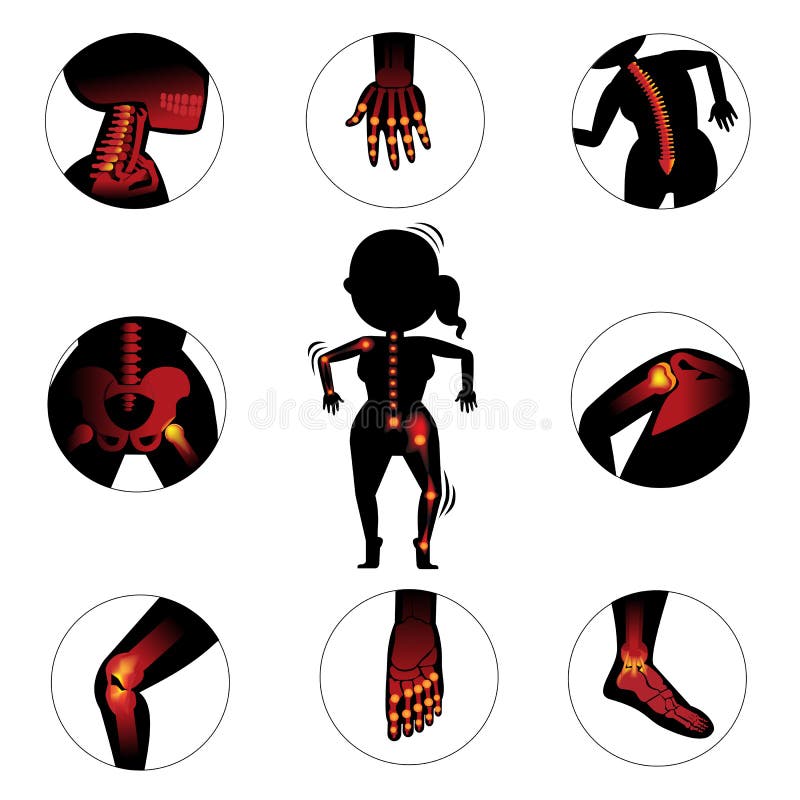
Free with trial Bones and joints. In the circle. Vector Illustration. Patella vectors Bones and joints. In the circle .Vector Illustration
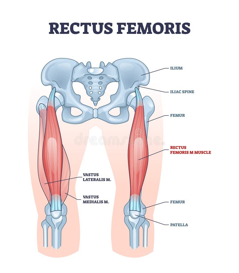
Free with trial Rectus femoris muscle as one of quadriceps muscular group outline diagram. Labeled educational scheme with skeletal upper leg anatomy vector illustration. Body vastus lateralis and medialis location. Patella vectors Rectus femoris muscle as one of quadriceps muscular group outline diagram. Labeled educational scheme with skeletal upper leg anatomy vector illustration. Body
Free with trial Knee pain. A man pain his knee from Osteoarthritis of the knee. Patella vectors Knee pain. A man pain his knee from Osteoarthritis of the knee
Free with trial Knee joint anatomy labeled. Patella vectors Knee. Joint anatomy labeled. Knee joint anatomy labeled .
Free with trial Broken Knee Fracture. A knee joint with a break and high levels of detail. Patella vectors Broken Knee Fracture. A knee joint with a break and high levels of detail
Free with trial Meniscus tear and surgery. Vector illustration of a Meniscus tear and surgery. Patella vectors Meniscus tear and surgery. Vector illustration of a Meniscus tear and surgery
Free with trial Knee joints healthy and unhealthy. Normal leg knee joint at the blue background and unhealthy joint at the red background. Patella vectors Knee joints healthy and unhealthy. Normal leg knee joint at the blue background and unhealthy joint at the red background
Free with trial Healthy knee and knee with osteoarthritis. On a white background. Patella vectors Healthy knee and knee with osteoarthritis. On a white background
Free with trial Anatomy of the canine knee. Anatomy of the canine dog`s knee joint colorful design, healthy joint info poster illustration. Patella vectors Anatomy of the canine knee. Anatomy of the canine dog`s knee joint colorful design, healthy joint info poster illustration
Free with trial Vector illustration anatomy of human legs and diagram of human bones isolated on white background. For advertising and medical publications. EPS 10. Patella vectors Bone fracture_Human leg anatomy and skeleton. Vector illustration anatomy of human legs and diagram of human bones isolated on white background. For advertising. Vector illustration anatomy of human legs and diagram of human bones isolated on white background. For advertising and medical publications. EPS 10.
Free with trial ILLUSTRATED TWO MAIN FACTORS NECESSARY FOR THE OCCURRENCE OF A SEIZURE. THE CENTER OF EXCITEMENT AND GENERAL CONVULSIVE READINESS OF THE BRAIN. Patella vectors EPILEPSY, factors of emergence. ILLUSTRATED TWO MAIN FACTORS NECESSARY FOR THE OCCURRENCE OF A SEIZURE. THE CENTER OF EXCITEMENT AND GENERAL CONVULSIVE READINESS. ILLUSTRATED TWO MAIN FACTORS NECESSARY FOR THE OCCURRENCE OF A SEIZURE. THE CENTER OF EXCITEMENT AND GENERAL CONVULSIVE READINESS OF THE BRAIN.
Free with trial Knee dislocation. Lateral trauma to the knee, torn collateral ligaments, cruciate ligament injury and meniscus injury. An obvious deformity of the knee. Human anatomy. Patella vectors Knee dislocation. Lateral trauma to the knee, torn collateral ligaments, cruciate ligament injury and meniscus injury. An obvious deformity of the knee. Human
Free with trial X-ray. Nurse and man in X-ray machine. Patella vectors X-ray. Nurse and man in X-ray machine
Free with trial Human leg, knee anatomy, abstract grey mesh background. Patella vectors Human leg, knee anatomy. Abstract grey mesh background
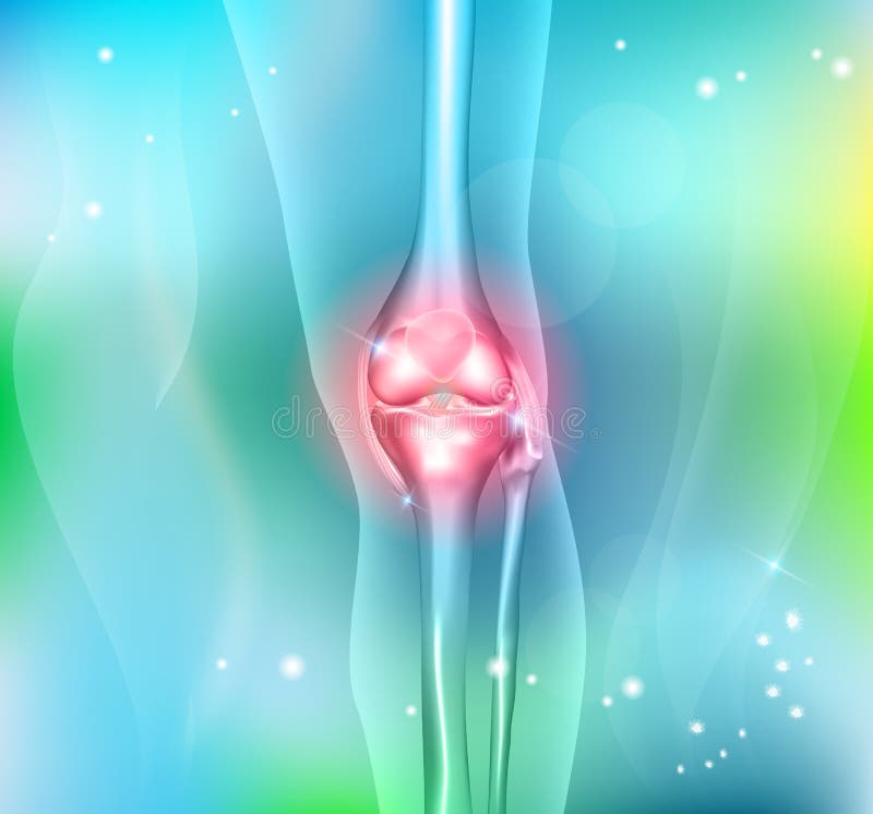
Free with trial Human leg and knee joint. Human leg knee joint anatomy on a beautiful light blue background. Patella vectors Human leg and knee joint. Human leg knee joint anatomy on a beautiful light blue background
Free with trial Knee joint endoprosthesis. Vector illustration of a total knee replacement. Patella vectors Knee joint endoprosthesis. Vector illustration of a total knee replacement
Free with trial Vector illustration. Anatomy, front x-ray of an arthritic knee joint and a knee after total knee replacement. For advertising and medical publications. EPS 10. Patella vectors Meniscus _Arthritic knee and Total knee replacement. Vector illustration. Anatomy, front x-ray of an arthritic knee joint and a knee after total knee replacement. Vector illustration. Anatomy, front x-ray of an arthritic knee joint and a knee after total knee replacement. For advertising and medical publications. EPS 10
Free with trial A medical illustration of a knee with an inflamed prepatellar bursa in comparison with a healthy knee. Patella vectors Prepatellar bursitis. A medical illustration of a knee with an inflamed prepatellar bursa in comparison with a healthy knee. A medical illustration of a knee with an inflamed prepatellar bursa in comparison with a healthy knee.
Free with trial Tear of a meniscus is a rupturing of one or more of the fibrocartilage strips in the knee. Human Joint, and Traumatic force in sports or physical exertion. Torn meniscus. Patella vectors Tear of a meniscus. Torn meniscus. Tear of a meniscus is a rupturing of one or more of the fibrocartilage strips in the knee. Human Joint, and Traumatic force in. Tear of a meniscus is a rupturing of one or more of the fibrocartilage strips in the knee. Human Joint, and Traumatic force in sports or physical exertion. Torn meniscus
Free with trial Knee joint anatomy isolated on white vector. Knee joint anatomy isolated on white photo-realistic vector illustration. Patella vectors Knee joint anatomy isolated on white vector. Knee joint anatomy isolated on white photo-realistic vector illustration
Free with trial Knee joint anatomy isolated on white vector. Knee joint anatomy isolated on white photo-realistic vector illustration. Patella vectors Knee joint anatomy isolated on white vector. Knee joint anatomy isolated on white photo-realistic vector illustration
Free with trial Knee joint pain abstract triangle background. Patella vectors Knee joint pain abstract triangle background. Knee joint pain abstract blue triangle background
 Sign in