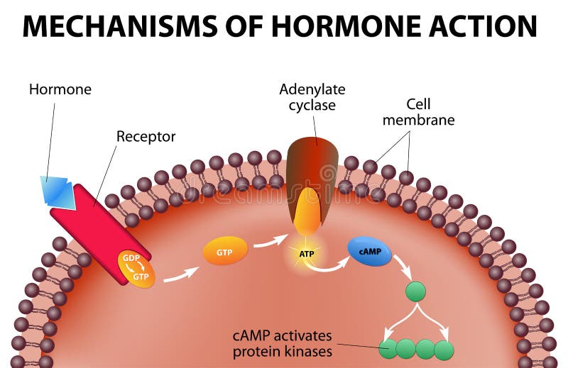
Free with trial Hormones bind to receptors on the plasma membrane. The hormone itself is the first messenger. Binding to the receptors activates a second messenger inside the cell. The second messenger causes intracellular effects. Secretory vectors Mechanisms of hormone action. Hormones bind to receptors on the plasma membrane. The hormone itself is the first messenger. Binding to the receptors activates a. Hormones bind to receptors on the plasma membrane. The hormone itself is the first messenger. Binding to the receptors activates a second messenger inside the cell. The second messenger causes intracellular effects
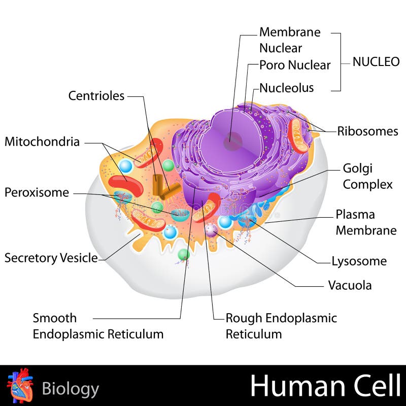
Free with trial Human Cell. Easy to edit vector illustration of human cell structure. Secretory vectors Human Cell. Easy to edit vector illustration of human cell structure
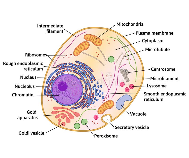
Free with trial Vector human cell structure. Cell structure, cross section of the cell detailed anatomy with description. Secretory vectors Vector human cell structure. Cell structure, cross section of the cell detailed anatomy with description
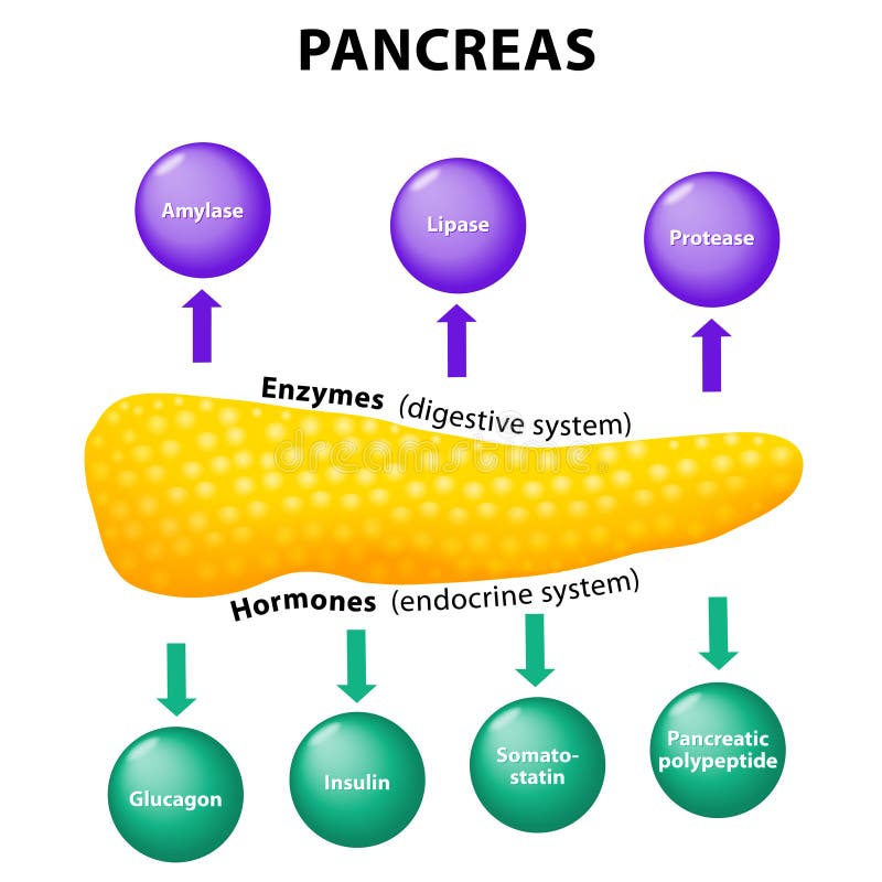
Free with trial Pancreas Physiology. secretory function. Enzymes: amylase, lipase, protease (digestive system) and Hormones: pancreatic polypeptide, somatostatin, insulin, glucagon (endocrine system). Human anatomy. Secretory vectors Pancreas. secretory function. Pancreas Physiology. secretory function. Enzymes: amylase, lipase, protease (digestive system) and Hormones: pancreatic polypeptide. Pancreas Physiology. secretory function. Enzymes: amylase, lipase, protease (digestive system) and Hormones: pancreatic polypeptide, somatostatin, insulin, glucagon (endocrine system). Human anatomy
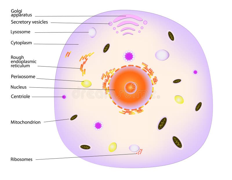
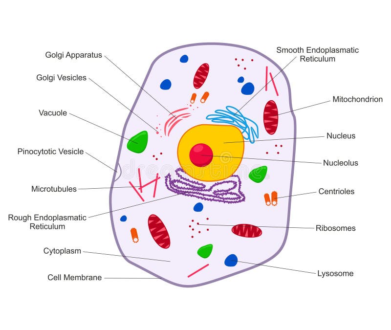
Free with trial A typical cell, labeled. Major components of an animal cell, eps10. Secretory vectors A typical cell, labeled. Major components of an animal cell, eps10
Free with trial Menstrual cycle. endometrium and hormone. Menstruation, Follicle phase, Ovulation and Corpus luteum phase. Secretory vectors Menstrual cycle. endometrium and hormone. Menstrual cycle. Menstruation, Follicle phase, Ovulation and Corpus luteum phase. endometrium and hormone
Free with trial Structure of a typical animal cell. Vector. Secretory vectors Animal cell. Structure of a typical animal cell. Vector. Structure of a typical animal cell. Vector.
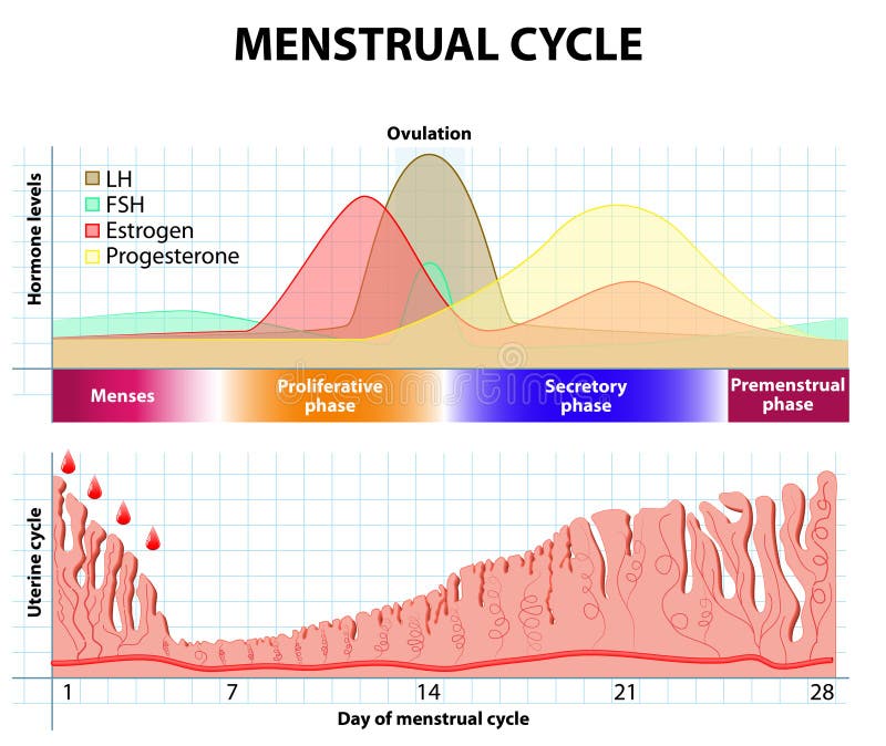
Free with trial Scheme of the menstrual cycle. Ovarian cycle phase, level of hormones female period, changes in the endometrium, uterine cycle. Secretory vectors Scheme of the menstrual cycle. Ovarian cycle phase, level of hormones female period, changes in the endometrium, uterine cycle
Free with trial Gastric pits and glands, plus secretory cells of the stomach lining. Created in Adobe Illustrator. EPS 10. Secretory vectors Detail of stomach mucous membrane. Gastric pits and glands, plus secretory cells of the stomach lining. Created in Adobe Illustrator. EPS 10. Gastric pits and glands, plus secretory cells of the stomach lining. Created in Adobe Illustrator. EPS 10.
Free with trial Set the cell organelles. Nucleus, endoplasmic reticulum, Golgi apparatus, mitochondria, centrosome, lysosome, the ribosome. Infographics. Vector illustration on isolated background. Secretory vectors Set the cell organelles. Nucleus, endoplasmic reticulum, Golgi a. Pparatus, mitochondria, centrosome, lysosome, the ribosome. Infographics. Vector illustration. Set the cell organelles. Nucleus, endoplasmic reticulum, Golgi apparatus, mitochondria, centrosome, lysosome, the ribosome. Infographics. Vector illustration on isolated background.
Free with trial Golgi apparatus vector illustration. Labeled microscopic scheme with cisternae, lumen, cis or trans face, cell, secretory and newly forming vesicle. Closeup diagram with receiving and shipping side. Secretory vectors Golgi apparatus vector illustration. Labeled microscopic scheme and diagram with cisternae, lumen, secretory forming vesicle. Golgi apparatus vector. Golgi apparatus vector illustration. Labeled microscopic scheme with cisternae, lumen, cis or trans face, cell, secretory and newly forming vesicle. Closeup diagram with receiving and shipping side.
Free with trial Insulin secretion vector illustration. Biological pancreas function labeled scheme. Full cycle diagram with glucose transporter, metabolism, ATP and potassium channel. Secretory vectors Insulin secretion vector illustration. Biological pancreas function. Insulin secretion vector illustration. Biological pancreas function labeled scheme. Full. Insulin secretion vector illustration. Biological pancreas function labeled scheme. Full cycle diagram with glucose transporter, metabolism, ATP and potassium channel.
Free with trial Types of gland. Anatomy of an Endocrine and exocrine glands. different of glands secretion. cross-section. Vector diagram for educational, medical, biological and science use. Secretory vectors Anatomy of an Endocrine and exocrine glands. Types of gland. Anatomy of an Endocrine and exocrine glands. different of glands secretion. cross-section. Vector. Types of gland. Anatomy of an Endocrine and exocrine glands. different of glands secretion. cross-section. Vector diagram for educational, medical, biological and science use
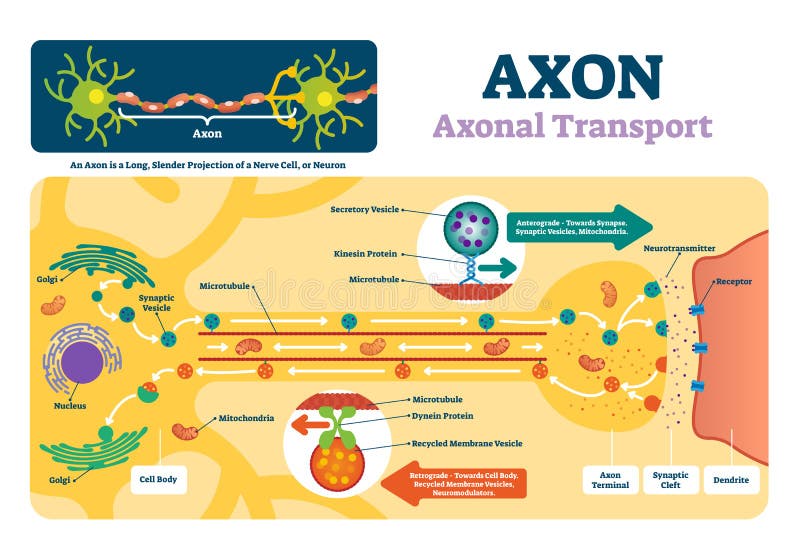
Free with trial Axon vector illustration. Labeled diagram with explanation and structure. Closeup with cell body, terminal, synaptic cleft and dendrite. Nerve cell projection or neuron. Secretory vectors Axon vector illustration. Labeled diagram with explanation and structure. Closeup with cell body, terminal, synaptic cleft and dendrite. Nerve cell projection
Free with trial The pineal gland, conarium, or epiphysis cerebri. Diagram of pituitary and pineal glands in the human brain. Secretory vectors Diagram of pituitary and pineal glands in the human brain. The pineal gland, conarium, or epiphysis cerebri. Diagram of pituitary and pineal glands in the human. The pineal gland, conarium, or epiphysis cerebri. Diagram of pituitary and pineal glands in the human brain
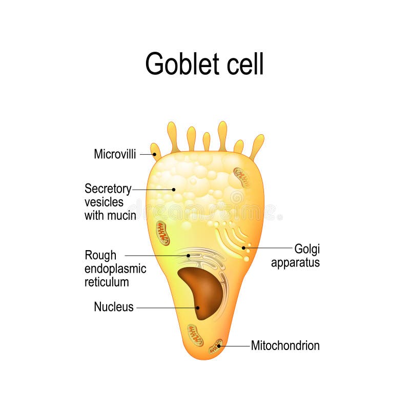
Free with trial Goblet cell. simple columnar epithelial cell for secrete mucus. They are found inside the trachea, bronchi, small and large intestine, and conjunctiva in the eyes. Structure cell nucleus and other organelles. Secretory vectors Goblet cell. Structure cell nucleus and other organelles. Goblet cell. simple columnar epithelial cell for secrete mucus. They are found inside the trachea. Goblet cell. simple columnar epithelial cell for secrete mucus. They are found inside the trachea, bronchi, small and large intestine, and conjunctiva in the eyes. Structure cell nucleus and other organelles.
Free with trial Secretory vesicle vector illustration. Labeled closeup infographic scheme with constitutive and regulated secretion. Diagram with golgi apparatus and secreted proteins. Secretory vectors Secretory vesicle vector illustration. Labeled closeup infographic scheme. Secretory vesicle vector illustration. Labeled closeup infographic scheme with. Secretory vesicle vector illustration. Labeled closeup infographic scheme with constitutive and regulated secretion. Diagram with golgi apparatus and secreted proteins.
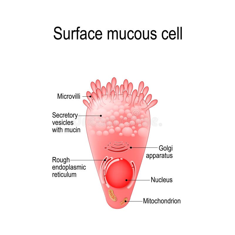
Free with trial Surface mucous cell is a foveolar mucus-producing cell that covering the inside of the stomach. Structure cell: golgi apparatus, secretory vesicle, mucin, nucleus, mitochondrion, microvilli. Secretory vectors Surface mucous cell. Is a foveolar mucus-producing cell that covering the inside of the stomach. Structure cell: golgi apparatus, secretory vesicle, mucin. Surface mucous cell is a foveolar mucus-producing cell that covering the inside of the stomach. Structure cell: golgi apparatus, secretory vesicle, mucin, nucleus, mitochondrion, microvilli
Free with trial Menstrual cycle. Ovarian cycle: follicular phase and luteal phase. Uterine cycle: Secretory phase, proliferative phase and menstruation. Vector Diagram showing the progression of the menstrual cycle. Secretory vectors Menstrual cycle. Ovarian cycle: follicular phase and luteal phase. Uterine cycle: Secretory phase, proliferative phase and menstruation. Vector Diagram showing
Free with trial Hallmarks of ageing. Cellular senescence, Telomere and DNA damaged, mitochondrial dysfunction, Chronic inflammation, Stem cell exhaustion. Anti-aging therapy. Anti aging medicine. vector illustration. Secretory vectors Hallmarks of ageing. Cellular senescence. Anti-aging therapy. Hallmarks of ageing. Cellular senescence, Telomere and DNA damaged, mitochondrial dysfunction. Hallmarks of ageing. Cellular senescence, Telomere and DNA damaged, mitochondrial dysfunction, Chronic inflammation, Stem cell exhaustion. Anti-aging therapy. Anti aging medicine. vector illustration
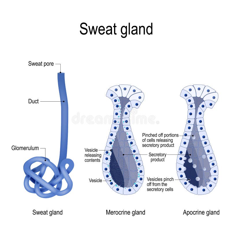
Free with trial Sweat gland. Merocrine and apocrine. different of manner of secretion. cross-section of the human skin, with the sweat gland. Close-up of dark and clear cells, lumen, sweat duct, glomerulum and pore. labeled Vector diagram for educational, medical, biological and science use. Secretory vectors Sweat gland. cross-section. Sweat gland. Merocrine and apocrine. different of manner of secretion. cross-section of the human skin, with the sweat gland. Close. Sweat gland. Merocrine and apocrine. different of manner of secretion. cross-section of the human skin, with the sweat gland. Close-up of dark and clear cells, lumen, sweat duct, glomerulum and pore. labeled Vector diagram for educational, medical, biological and science use
Free with trial Animal cell anatomy. Cartoon vector illustration of structure of animal cell. Illustration showing the animal cell anatomy. Secretory vectors Animal cell anatomy. Cartoon vector illustration of structure of animal cell. Illustration showing the animal cell anatomy
Free with trial A vector illustration of skin and hair, normal and greasy or with excessive sebum secretion. Secretory vectors Normal and greasy hair. A vector illustration of skin and hair, normal and greasy or with excessive sebum secretion. A vector illustration of skin and hair, normal and greasy or with excessive sebum secretion.
Free with trial Fibroblast is a dermis cell vital to the skin`s strength and elasticity. Structure of Fibroblast cell. Secretory vectors Fibroblast is vital to the skin`s strength and elasticity. Struc. Fibroblast is a dermis cell vital to the skin`s strength and elasticity. Structure of. Fibroblast is a dermis cell vital to the skin`s strength and elasticity. Structure of Fibroblast cell.
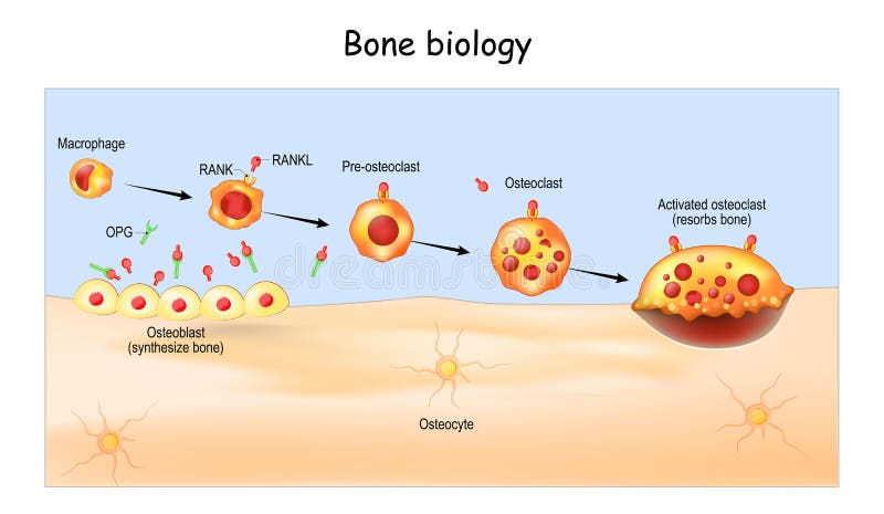
Free with trial Bone Biology. Role of RANK, RANKL, and OPG. bone remodeling. Bone is broken down by osteoclasts, and rebuilt by osteoblasts. Receptor activator of RANKL is the mediator of bone resorption. Osteoprotegerin OPG. Paracrine and endocrine actions of bone. functions of secretory proteins from osteoblasts, osteocytes, and osteoclasts. Secretory vectors Bone Biology. Role of RANK, RANKL, and OPG. bone remodeling. Bone is broken down by osteoclasts, and rebuilt by osteoblasts. Receptor activator of RANKL is the
Free with trial Anatomy of the female breast side view. Structure of the Milk ducts and Lobules of the mammary gland. Mammary Alveoli and Myoepithelial cell. Milk producing organs. Secretory vectors Anatomy of the female breast side view. Structure of the Milk ducts and Lobules of the mammary gland. Mammary Alveoli. And Myoepithelial cell. Milk producing
Free with trial Planaria with excretory system protonephridia isolated on white background. Flatworm flame cell. Protonephridia of planaria and other flatworm. Secretory vectors Planaria with excretory system protonephridia isolated on white background. Flatworm flame cell. Protonephridia of planaria and other flatworm
Free with trial Animal Cell structure. Cross section of the cell detailed colorful anatomy with description. Animal cell in flat style on white background. Secretory vectors Animal Cell structure. Cross section of the cell detailed colorful anatomy with description. Animal cell in flat style. On white background
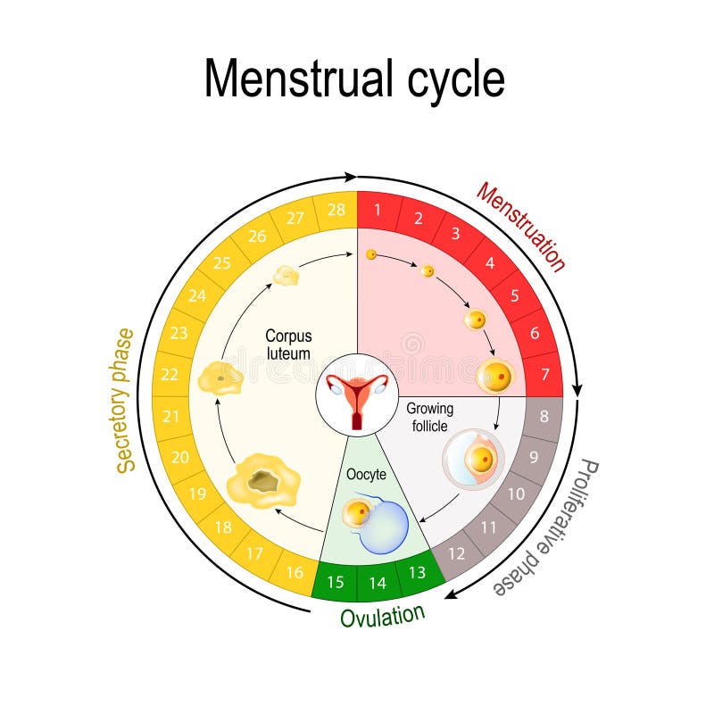
Free with trial Menstrual cycle. Luteal and Follicular phase. Growing follicle, Oocyte and Corpus luteum. From Menses and Proliferative phase, to Ovulation and Secretory phase. Vector poster for education and medical use. Secretory vectors Menstrual cycle. menses and Proliferative phase, Ovulation and Secretory phase. Menstrual cycle. Luteal and Follicular phase. Growing follicle, Oocyte and Corpus. Menstrual cycle. Luteal and Follicular phase. Growing follicle, Oocyte and Corpus luteum. From Menses and Proliferative phase, to Ovulation and Secretory phase. Vector poster for education and medical use
Free with trial Ovarian cycle. Menstrual cycle diagram. Menstrual, proliferative ovulation and secretory phases. Luteal and Follicular phase. Growing follicle, Oocyte and Corpus luteum. Medical illustration vector. Secretory vectors Ovarian cycle. Menstrual cycle diagram. Menstrual, proliferative ovulation and secretory phases. Luteal and Follicular phase. Growing follicle, Oocyte and
Free with trial Endocrine vs exocrine glands brief comparison shows hormone release to capillaries vs enzyme flow through ducts, key objects, duct, capillary, secretory cells. Outline diagram. Secretory vectors Endocrine vs exocrine glands brief comparison shows hormone release to ... Endocrine vs exocrine glands brief comparison shows hormone release to capillaries vs. Endocrine vs exocrine glands brief comparison shows hormone release to capillaries vs enzyme flow through ducts, key objects, duct, capillary, secretory cells. Outline diagram
Free with trial Mucous neck cell is a foveolar mucus-producing cell that covering the inside of the stomach. Structure cell: golgi apparatus, secretory vesicle, nucleus, mitochondrion, and microvilli. Secretory vectors Mucous neck cell. Is a foveolar mucus-producing cell that covering the inside of the stomach. Structure cell: golgi apparatus, secretory vesicle, nucleus. Mucous neck cell is a foveolar mucus-producing cell that covering the inside of the stomach. Structure cell: golgi apparatus, secretory vesicle, nucleus, mitochondrion, and microvilli
Free with trial Types of antibodies. Structure of IgA, IgE, IgD, IgG, IgM. Immunoglobulin anatomy. Disulfide bonds, Heavy and Light chains, Joining chain, and Secretory component. Detailed Vector poster. Secretory vectors Types of antibodies. Structure of IgA, IgE, IgD, IgG, IgM. Immunoglobulin anatomy. Disulfide bonds, Heavy and Light chains, Joining chain, and Secretory
Free with trial Menstrual cycle. Ovarian cycle: follicular phase and luteal phase. Uterine cycle: Secretory phase, proliferative phase and menstruation. Vector Diagram showing the progression of the menstrual cycle. Secretory vectors Menstrual cycle. Ovarian cycle: follicular phase and luteal phase. Uterine cycle: Secretory phase, proliferative phase and menstruation. Vector Diagram showing
Free with trial Cellular senescence. Functions of the SASP. Senescence-Associated Secretory Phenotype. changes senescent cells During ageing. spread the senescence phenotype in a paracrine manner to surrounding cells. Vector illustration. Secretory vectors Cellular senescence. Functions of the SASP. Senescence-Associated Secretory Phenotype. changes senescent cells During ageing. spread the senescence phenotype
Free with trial Microscopic view of the parotid gland's secretory and ductal system, culminating in saliva delivery to the oral cavity. Secretory vectors Culminating in saliva delivery to the oral cavity. Microscopic view of the parotid gland's secretory and ductal system, culminating in saliva delivery to the. Microscopic view of the parotid gland's secretory and ductal system, culminating in saliva delivery to the oral cavity
Free with trial Set of office and school supplies , painted in watercolor - the brush , pen , pencil, pen, secretory marker , scissors, stationery knife, chtikery , tape , ruler. Secretory vectors Set of office and school supplies , painted in watercolor - the. Brush , pen , pencil, pen, secretory marker , scissors, stationery knife, chtikery , tape
Free with trial Menstrual cycle. changes in the endometrium during the menstrual cycle. Uterus lining from Menstruation, Proliferative phase to Ovulation and Secretory phase. Luteal and Follicular phase. Vector poster. Secretory vectors Menstrual cycle. changes in the endometrium during the menstrual cycle. Uterus lining from Menstruation, Proliferative phase to Ovulation and Secretory phase
Free with trial Inflammaging. Ageing process. Chronic inflammation that develops with advanced age. Anti-aging therapy. Anti aging medicine. Vector illustration. Secretory vectors Inflammaging. Chronic inflammation. Ageing process. Inflammaging. Ageing process. Chronic inflammation that develops with advanced age. Anti-aging therapy. Anti. Inflammaging. Ageing process. Chronic inflammation that develops with advanced age. Anti-aging therapy. Anti aging medicine. Vector illustration
Free with trial Immunoglobulin classes overview showing antibody isotypes, IgG, IgA, IgM dominate, with labeled structures and functions for immunity and B-cell roles. Outline diagram. Secretory vectors Immunoglobulin classes overview showing antibody isotypes, IgG, IgA, IgM dominate, ... Immunoglobulin classes overview showing antibody isotypes, IgG, IgA, IgM. Immunoglobulin classes overview showing antibody isotypes, IgG, IgA, IgM dominate, with labeled structures and functions for immunity and B-cell roles. Outline diagram
Free with trial Enteric nervous system overview shows gut-brain axis with myenteric. Plexus, submucosal plexus, and neurons regulating motility and secretion. Outline diagram. Secretory vectors Enteric nervous system overview shows gut-brain axis with myenteric. Plexus, submucosal plexus, and neurons regulating motility and secretion. Outline diagram
Free with trial Sweat gland anatomy and morphology. Cross section of a skin with Apocrine sweat gland and Eccrine sweat gland. Close-up of cells. Exocrine and Merocrine secretion. Thermoregulation. Vector illustration. Medical diagram. Secretory vectors Skin with Apocrine sweat gland and Eccrine sweat gland. comparison. Sweat gland anatomy and morphology. Cross section of a skin with Apocrine sweat gland and. Sweat gland anatomy and morphology. Cross section of a skin with Apocrine sweat gland and Eccrine sweat gland. Close-up of cells. Exocrine and Merocrine secretion. Thermoregulation. Vector illustration. Medical diagram
Free with trial Punctal Occlusion Therapy. Punctal Plugs for Treatment of dry eye syndrome. Illustration of eye. Secretory vectors Punctal Occlusion Therapy. Punctal Plugs for Treatment of dry eye syndrome. Illustration of eye
Free with trial Inflammaging. Hallmarks of ageing. Chronic inflammation. That develops with advanced age. Anti-aging therapy. Anti aging medicine. Vector illustration. Secretory vectors Inflammaging. Hallmarks of ageing. Chronic inflammation. That develops with advanced age. Anti-aging therapy. Anti aging medicine. Vector illustration
Free with trial Anatomy of a G-cell. This cells are located in the gastric glands into stomach. G cell is a type of cell in the stomach and duodenum that secretes hormone gastrin. Secretory vectors Anatomy of a G-cell. gastrin. Anatomy of a G-cell. This cells are located in the gastric glands into stomach. G cell is a type of cell in the stomach and. Anatomy of a G-cell. This cells are located in the gastric glands into stomach. G cell is a type of cell in the stomach and duodenum that secretes hormone gastrin.
Free with trial Exocrine gland. Method of secretion. Apocrine gland cells secrete by budding off a portion of their cytoplasm. Merocrine gland cells excrete their substances by exocytosis. Vector illustration. Medical poster. Schematic diagram. Secretory vectors Exocrine gland. Apocrine gland cells and Merocrine cells. Exocrine gland. Method of secretion. Apocrine gland cells secrete by budding off a portion of their. Exocrine gland. Method of secretion. Apocrine gland cells secrete by budding off a portion of their cytoplasm. Merocrine gland cells excrete their substances by exocytosis. Vector illustration. Medical poster. Schematic diagram
Free with trial Detailed medical diagram of Pituitary Gland functions in the human body, complete with annotations. Secretory vectors Pituitary Gland functions. Detailed medical diagram of Pituitary Gland functions in the human body, complete with annotations. Detailed medical diagram of Pituitary Gland functions in the human body, complete with annotations.
Free with trial Paneth cell it is the epithelium cells of the small intestine. their granules consist of anti-microbial compounds for important in immunity. Structure of the Paneth cell nucleus, mitochondria and other organelles. Secretory vectors Paneth cell. It is the epithelium cells of the small intestine. their granules consist of anti-microbial compounds for important in immunity. Structure of the. Paneth cell it is the epithelium cells of the small intestine. their granules consist of anti-microbial compounds for important in immunity. Structure of the Paneth cell nucleus, mitochondria and other organelles.
Free with trial Hallmarks of aging. Chromosomes with Telomeres before and after division of new and senescent cell. Cell division will cease once telomeres shorten to a critical length. Cellular aging. Vector illustration. Secretory vectors Hallmarks of aging. Chromosomes with Telomeres before and after division of new and senescent cell. Cell division will cease once telomeres shorten to a
Free with trial Scheme of the menstrual cycle. Ovarian cycle phase, level of hormones female period, changes in the endometrium, uterine cycle. Secretory vectors Scheme of the menstrual cycle. Ovarian cycle phase, level of hormones female period, changes in the endometrium, uterine cycle
Free with trial Gastric chief peptic, gastric zymogenic cell. release the zymogen enzyme. this cells are located in the gastric glands into stomach. Structure of a chief cell: golgi apparatus, nucleus, mitochondrion, microvilli. Secretory vectors Gastric chief peptic, gastric zymogenic cell. Release the zymogen enzyme. this cells are located in the gastric glands into stomach. Structure of a chief cell
Free with trial Menstrual cycle chart. increase and decrease of the hormones. The graph also depicts the growth of the follicle. Fluctuation of hormones that occurs during menstruation cycle. Vector illustration for educational, biological, science and medical use. Secretory vectors Menstrual cycle chart. Increase and decrease of the hormones. The graph also depicts the growth of the follicle. Fluctuation of hormones that occurs during
Free with trial Illustration of the Golgi apparatus structure. Vector infographics. Secretory vectors Illustration of the Golgi apparatus structure. Vector infographics.
Free with trial Fibroblast anatomy. structure of cell. Diagram with golgi apparatus, nucleus, mitochondrion and ribosomes. vector illustration. Poster. Secretory vectors Fibroblast anatomy. structure of cell. Diagram with golgi apparatus, nucleus, mitochondrion and ribosomes. vector illustration. Poster
Free with trial Bone Biology. Role of RANK, RANKL, and OPG. bone remodeling. Bone is broken down by osteoclasts, and rebuilt by osteoblasts. Receptor activator of RANKL is the mediator of bone resorption. Osteoprotegerin OPG. Paracrine and endocrine actions of bone. functions of secretory proteins from osteoblasts, osteocytes, and osteoclasts. Secretory vectors Bone Biology. Role of RANK, RANKL, and OPG. bone remodeling. Bone is broken down by osteoclasts, and rebuilt by osteoblasts. Receptor activator of RANKL is the
Free with trial Pituitary gland anatomy. Hormones. location of hypophysis. vector illustration. diagram for education. medical poster about human`s brain and endocrine system. Secretory vectors Pituitary gland anatomy. Hormones. location of hypophysis. Vector illustration. diagram for education. medical poster about human`s brain and endocrine system
Free with trial Animal cell anatomy. Cartoon vector illustration of structure of animal cell. Illustration showing the animal cell anatomy. Secretory vectors Animal cell anatomy. Cartoon vector illustration of structure of animal cell. Illustration showing the animal cell anatomy
Free with trial Purple Golgi apparatus structure. Vector Illustration. Secretory vectors Purple Golgi apparatus structure. Vector Illustration.
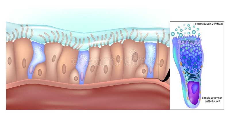
Free with trial Simple columnar epithelial cells that secrete mucin 2 in the lower gastrointestinal tract. Structure of Simple Columnar Epithelial Cell in Stomach. Mucin 2 Production in Gastric Epithelial Cells. Secretory vectors Simple columnar epithelial cells that secrete mucin 2 in the lower gastrointestinal tract. Structure of Simple Columnar. Epithelial Cell in Stomach. Mucin 2. Simple columnar epithelial cells that secrete mucin 2 in the lower gastrointestinal tract. Structure of Simple Columnar Epithelial Cell in Stomach. Mucin 2 Production in Gastric Epithelial Cells
Free with trial Diagram of animal cell eukaryotic cell labeled. Animal cell eukaryotic cell diagram, A typical cell labeled vector illustration on white background. Secretory vectors Diagram of animal cell eukaryotic cell labeled. Animal cell eukaryotic cell diagram, A typical cell labeled vector illustration on white background
Free with trial Lacrimal Apparatus system. Nasolacrimal duct, Lacrimal sac, Sacrimal canals, Lacrimal punctums, Lacrimal gland. Secretory vectors Lacrimal Apparatus system. Nasolacrimal duct, Lacrimal sac, Sacrimal canals, Lacrimal punctums, gland. Lacrimal Apparatus system .Nasolacrimal duct, Lacrimal. Lacrimal Apparatus system .Nasolacrimal duct, Lacrimal sac, Sacrimal canals, Lacrimal punctums, Lacrimal gland.
Free with trial Exocrine glands structure for anatomical secrete substances outline diagram. Labeled educational scheme with duct and secretary portion microbiological parts vector illustration. Tubular and acinar. Secretory vectors Exocrine glands structure for anatomical secrete substances outline diagram. Labeled educational scheme with duct and secretary portion microbiological parts
Free with trial Pancreas function. internal organ of a digestive and endocrine systems. pancreatic juice and hormones of a human pancreas. Enzymes: trypsinogen, chymotrypsinogen, elastase, carboxypeptidase, pancreatic lipase, nucleases and Hormones: pancreatic polypeptide, somatostatin, insulin, glucagon. vector poster. Secretory vectors Pancreas function. pancreatic juice and hormones of a human pancreas. Pancreas function. internal organ of a digestive and endocrine systems. pancreatic juice. Pancreas function. internal organ of a digestive and endocrine systems. pancreatic juice and hormones of a human pancreas. Enzymes: trypsinogen, chymotrypsinogen, elastase, carboxypeptidase, pancreatic lipase, nucleases and Hormones: pancreatic polypeptide, somatostatin, insulin, glucagon. vector poster
Free with trial Anatomy of trichome with biological model structure closeup outline diagram. Labeled educational scheme with microscopic side view of cuticle, striae, stipe, basal cells and bract vector illustration. Secretory vectors Anatomy of trichome with biological model structure closeup outline diagram. Labeled educational scheme with microscopic side view of cuticle, striae, stipe
Free with trial Vector illustration. Fibroblast is a dermis cell. Structure of Fibroblast cell. Secretory vectors Fibroblast is a dermis cell. Structure of Fibroblast cell. Vector illustration. Fibroblast is a dermis cell. Structure of Fibroblast cell. Vector illustration. Fibroblast is a dermis cell. Structure of Fibroblast cell.
Free with trial Vector illustration. Fibroblast is a dermis cell. Structure of Fibroblast cell. Secretory vectors Fibroblast is a dermis cell. Structure of Fibroblast cell. Vector illustration. Fibroblast is a dermis cell. Structure of Fibroblast cell. Vector illustration. Fibroblast is a dermis cell. Structure of Fibroblast cell.
Free with trial Nervous, Reproductive, Digestive and Excretory system of planaria flatworm. Isolated on white background. Secretory vectors Nervous, Reproductive, Digestive and Excretory system of planaria flatworm. Isolated on white background
Free with trial Diarrhea or stomach pain, character design -. Illustration. Secretory vectors Diarrhea or stomach pain, character design -. Illustration
Free with trial Planaria with excretory system protonephridia isolated on white background. Flatworm flame cell. Protonephridia of planaria. And other flatworm. Secretory vectors Planaria with excretory system protonephridia isolated on white background. Flatworm flame cell. Protonephridia of planaria. And other flatworm
Free with trial Protonephridia of planaria and other flatworm. Structure of excretory system. Of flatworm. Secretory vectors Protonephridia of planaria and other flatworm. Structure of excretory system. Of flatworm
Free with trial Protonephridia of planaria and other flatworm. Structure of excretory system. Of flatworm. Secretory vectors Protonephridia of planaria and other flatworm. Structure of excretory system. Of flatworm
Free with trial Vector illustration. Fibroblast is a dermis cell. Structure of Fibroblast cell. Isolated. Secretory vectors Vector illustration. Fibroblast is a dermis cell. Structure of Fibroblast cell. Isolated
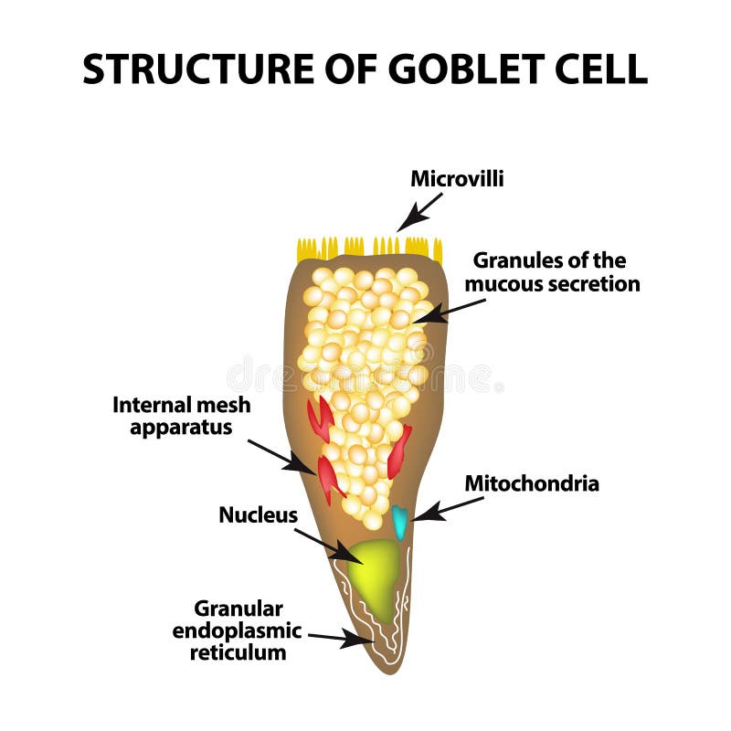
Free with trial Structure Goblet cells of the intestine. Infographics. Vector illustration on isolated background. Secretory vectors Structure Goblet cells of the intestine. Infographics. Vector illustration on isolated background.
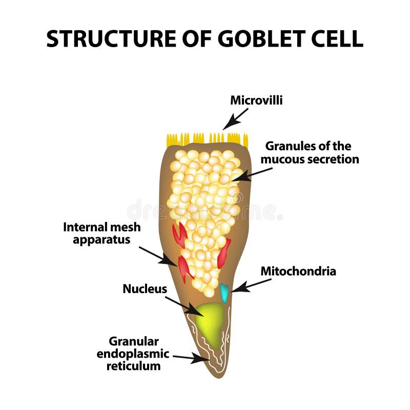
Free with trial Structure Goblet cells of the intestine. Infographics. Vector illustration on isolated background. Secretory vectors Structure Goblet cells of the intestine. Infographics. Vector illustration on isolated background.
Free with trial The structure of Paneth cells. Davidoff`s cell. fographics. Vector illustration on isolated background. Secretory vectors The structure of Paneth cells. Davidoff`s cell. fographics. Vector illustration on isolated background. The structure of Paneth cells. Davidoff`s cell
Free with trial Flatworm flame cell. Structure of element of excretory system with title. Protonephridia of planaria. And other flatworm. Secretory vectors Flatworm flame cell. Structure of element of excretory system with title. Protonephridia of planaria. And other flatworm
Free with trial Pineal gland and epithalamus anatomy in the human brain. Endocrine system. Production of melatonin, serotonin derived hormone. Neuroendocrine secretory circumventricular organ 3D vector illustration. Secretory vectors Pineal gland anatomy. Pineal gland and epithalamus anatomy in the human brain. Endocrine system. Production of melatonin, serotonin derived hormone. Pineal gland and epithalamus anatomy in the human brain. Endocrine system. Production of melatonin, serotonin derived hormone. Neuroendocrine secretory circumventricular organ 3D vector illustration
Free with trial Pineal gland and epithalamus anatomy in the human brain. Production of melatonin, serotonin derived hormone. Endocrine system. Neuroendocrine secretory circumventricular organ flat vector illustration. Secretory vectors Pineal gland anatomy. Pineal gland and epithalamus anatomy in the human brain. Production of melatonin, serotonin derived hormone. Endocrine system. Pineal gland and epithalamus anatomy in the human brain. Production of melatonin, serotonin derived hormone. Endocrine system. Neuroendocrine secretory circumventricular organ flat vector illustration
Free with trial Vector isolated illustration of Pineal gland in man head. Human brain components detailed anatomy. Medical infographics for poster, educational, science and medical use. Sagittal view of the brain. Secretory vectors Vector isolated illustration of Pineal gland. In man head. Human brain components detailed anatomy. Medical infographics for poster, educational, science and. Vector isolated illustration of Pineal gland in man head. Human brain components detailed anatomy. Medical infographics for poster, educational, science and medical use. Sagittal view of the brain
Free with trial Vector isolated illustration of Pineal gland in woman head. Human brain components detailed anatomy. Medical infographics for poster, educational, science and medical use. Sagittal view of the brain. Secretory vectors Vector isolated illustration of Pineal gland. In woman head. Human brain components detailed anatomy. Medical infographics for poster, educational, science and. Vector isolated illustration of Pineal gland in woman head. Human brain components detailed anatomy. Medical infographics for poster, educational, science and medical use. Sagittal view of the brain
 Sign in