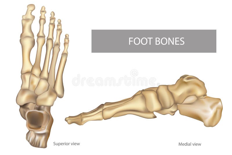
Free with trial Human foot bones front and side view anatomy. Illustration Of Human foot bones front and side view anatomy. Tarsal bones vectors Human foot bones front and side view anatomy. Illustration Of Human foot bones front and side view anatomy
Free with trial In the dorsal (posterior) view of the bones of the feet, the tarsal bones are visible, including the talus, calcaneus, navicular, cuboid, and the three cuneiform bones. The metatarsal bones, numbering from one to five, can also be observed, extending towards the toes. Additionally, the phalanges, consisting of proximal, middle, and distal phalanges, complete the skeletal structure of the feet in the dorsal view. Tarsal bones vectors Bones of feet, dorsal (posterior) view. In the dorsal (posterior) view of the bones of the feet, the tarsal bones are visible, including the talus, calcaneus. In the dorsal (posterior) view of the bones of the feet, the tarsal bones are visible, including the talus, calcaneus, navicular, cuboid, and the three cuneiform bones. The metatarsal bones, numbering from one to five, can also be observed, extending towards the toes. Additionally, the phalanges, consisting of proximal, middle, and distal phalanges, complete the skeletal structure of the feet in the dorsal view.
Free with trial Severs disease. calcaneus apophysitis. inflammation at the back of the heel growth plate. Foot anatomy. side view of a Human foot with the name of bones. Arches of the feet. skeleton anatomy. realistic Vector poster. Tarsal bones vectors Severs disease. calcaneus apophysitis. inflammation at the back of the heel growth plate. Foot anatomy. side view of a Human foot with the name of bones
Free with trial Anatomy Bones of the Feet. Superior view. Orthotics for foot Superior view. Tarsal bones vectors Anatomy Bones of the Feet. Superior view. Anatomy Bones of the Feet. Orthotics for foot Superior view
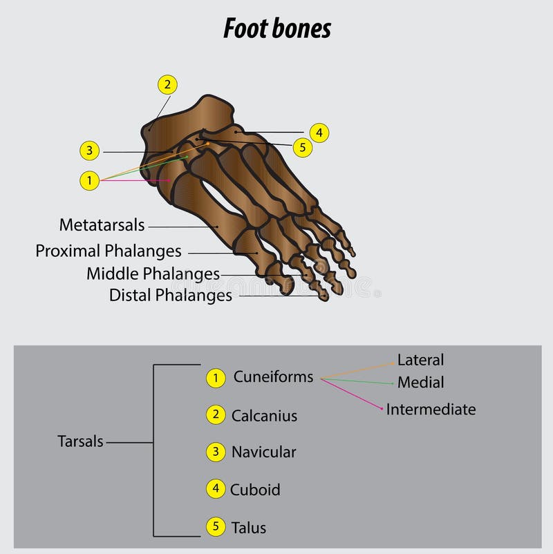
Free with trial Bones of the Foot. Tarsals or tarsus, Metatarsals, Phalanges. Tarsal bones vectors Bones of the Foot .Tarsals or tarsus, Metatarsals, Phalanges. Bones of the Foot.Tarsals or tarsus, Metatarsals, Phalanges Anatomy
Free with trial Fracture of the metatarsal bones in the foot. Anatomical structure of the foot. Skeleton. Broken bones. Vector illustration on isolated background. Tarsal bones vectors Fracture of the metatarsal bones in the foot. Anatomical structure of the foot. Skeleton. Broken bones. Vector. Illustration on isolated background
Free with trial Vector illustration of foot bones with explanation. Skeleton part for medical or health care design. Anatomy guide skeletal human foot. Tarsal bones vectors Foot bones with explanation. Vector illustration of foot bones with explanation. Skeleton part for medical or health care design. Anatomy guide skeletal human. Vector illustration of foot bones with explanation. Skeleton part for medical or health care design. Anatomy guide skeletal human foot.
Free with trial Front view foot bones. Hand drawn illustration of the foot bones on white, artistic anatomy graphic study. Tarsal bones vectors Front view foot bones. Hand drawn illustration of the foot bones on white, artistic anatomy graphic study
Free with trial Human foot bones and joints skeleton vector sketch body anatomy icon. Isolated symbol of feet and toes limbs structure of leg organ for anatomical orthopedic or medical surgery design element. Tarsal bones vectors Vector sketch icon of human foot bones or joints. Human foot bones and joints skeleton vector sketch body anatomy icon. Isolated symbol of feet and toes limbs. Human foot bones and joints skeleton vector sketch body anatomy icon. Isolated symbol of feet and toes limbs structure of leg organ for anatomical orthopedic or medical surgery design element
Free with trial Vector illustration of a human leg with denominations of the bones of the foot. Anatomy of lateral or side view of the foot. For advertising or medical publications. EPS 10. Tarsal bones vectors Anatomy_bones of the human foot lateral view. Vector illustration of a human leg with denominations of the bones of the foot. Anatomy of lateral or side view of. Vector illustration of a human leg with denominations of the bones of the foot. Anatomy of lateral or side view of the foot. For advertising or medical publications. EPS 10.
Free with trial Vector illustration of a human leg with denominations of the bones of the foot. Anatomy of medial or side view of the foot. For advertising or medical publications. EPS 10. Tarsal bones vectors Anatomy_bones of the human foot medial view. Vector illustration of a human leg with denominations of the bones of the foot. Anatomy of medial or side view of. Vector illustration of a human leg with denominations of the bones of the foot. Anatomy of medial or side view of the foot. For advertising or medical publications. EPS 10.
Free with trial Vector illustration of a human leg with denominations of the bones of the foot. Anatomy of dorsal and plantar views of the foot. For advertising or medical publications. EPS 10. Tarsal bones vectors Anatomy_bones of the human foot dorsal and plantar view. Vector illustration of a human leg with denominations of the bones of the foot. Anatomy of dorsal and. Vector illustration of a human leg with denominations of the bones of the foot. Anatomy of dorsal and plantar views of the foot. For advertising or medical publications. EPS 10.
Free with trial Vector drawing of human foot human foot anatomy drawing labeled. Drawing with layers reedit able. Tarsal bones vectors Vector drawing of human foot human foot anatomy drawing labeled. Drawing with layers reedit able
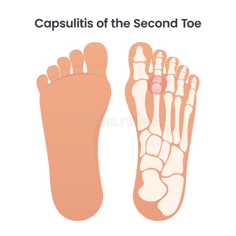
Free with trial Capsulitis of the second toe occurs in the ligaments surrounding the joint at the base of the second toe. These ligaments form a capsule, which helps the joint to function properly. Capsulitis is a condition in which these ligaments have become inflamed. Tarsal bones vectors Capsulitis of the Second Bone educational medical vector illustration. Capsulitis of the second toe occurs in the ligaments surrounding the joint at the base of. Capsulitis of the second toe occurs in the ligaments surrounding the joint at the base of the second toe. These ligaments form a capsule, which helps the joint to function properly. Capsulitis is a condition in which these ligaments have become inflamed
Free with trial Realistic 3D render of human tarsal bones slightly rotated showing talus calcaneus navicular cuboid and cuneiforms isolated on white background with ultra sharp professional focus. Tarsal bones vectors Realistic 3D Render Human Tarsal Bones Detailed Ankle Anatomy. Realistic 3D render of human tarsal bones slightly rotated showing talus calcaneus navicular. Realistic 3D render of human tarsal bones slightly rotated showing talus calcaneus navicular cuboid and cuneiforms isolated on white background with ultra sharp professional focus
Free with trial Illustration of a horses lower leg anatomy detailing key bones and joints. It includes the cannon bone (third metacarpal tarsal bone), the splint bone (second and fourth metacarpal tarsal bones), the fetlock joint (metacarpophalangeal metatarsophalangeal joint), the pastern bones (proximal and middle phalanges), and the coffin bone (distal phalanx). The image serves as an educational tool for understanding the structure and function of a horses limb. Tarsal bones vectors Illustration of a horses lower leg anatomy detailing key bones and joints. It includes the cannon bone (third metacarpal tarsal bone), the splint bone (second
Free with trial Detailed silhouette icon of a human foot skeleton ankle bones anatomy on transparent background. Tarsal bones vectors Human foot skeleton ankle tarsal bones anatomy silhouette. AI generated detailed silhouette icon of a human foot skeleton ankle bones anatomy on transparent. detailed silhouette icon of a human foot skeleton ankle bones anatomy on transparent background
Free with trial A black-and-white anatomical diagram presents eight labeled bones of the foot: the calcaneus, talus, navicular, cuboid, medial, intermediate and lateral cuneiforms, and first metatarsal. Each bone is shown separately as a simplified, outlined with shaded areas suggesting its shape and contours; the first metatarsal appears elongated, while the tarsal bones are more compact and irregular. The clean layout on a white background emphasizes identification and comparison, with bold labels beneath each drawing. Tarsal bones vectors A black-and-white anatomical diagram presents eight labeled bones of the foot: the calcaneus, talus, navicular, cuboid, medial. Intermediate and lateral. A black-and-white anatomical diagram presents eight labeled bones of the foot: the calcaneus, talus, navicular, cuboid, medial, intermediate and lateral cuneiforms, and first metatarsal. Each bone is shown separately as a simplified, outlined with shaded areas suggesting its shape and contours; the first metatarsal appears elongated, while the tarsal bones are more compact and irregular. The clean layout on a white background emphasizes identification and comparison, with bold labels beneath each drawing.
Free with trial Human Ankle Joint Bones Skeleton Icons. Presented in outline and solid black styles for medical or anatomical representation. Tarsal bones vectors Human Ankle Joint Bones Skeleton Icons. Presented in outline and solid black styles for medical or anatomical representation
Free with trial This image illustrates the intricate structure of the human foot, highlighting the bones and muscles. The foot is composed of numerous bones that work together to provide support, balance, and mobility. The image shows the bones in the toes, the metatarsals, the tarsal bones, and the ankle joint. The muscles are depicted in a way that shows their attachment points and how they contribute to the. Tarsal bones vectors Detailed anatomy of the human foot. This image illustrates the intricate structure of the human foot, highlighting the bones and muscles. The foot is composed of. This image illustrates the intricate structure of the human foot, highlighting the bones and muscles. The foot is composed of numerous bones that work together to provide support, balance, and mobility. The image shows the bones in the toes, the metatarsals, the tarsal bones, and the ankle joint. The muscles are depicted in a way that shows their attachment points and how they contribute to the
Free with trial Human foot bones illustration. Detailed anatomical illustration of a human foot showcasing its bones and structure, ideal for medical or educational purposes. Tarsal bones vectors Human foot bones illustration. Detailed anatomical illustration of a human foot showcasing its bones and structure, ideal for medical or educational purposes
Free with trial Detailed anatomical illustration of human feet bones in a vintage style, set against a dark background, showcasing skeletal structure and medical detail. Tarsal bones vectors Anatomical Illustration of Human Feet Bones on Dark Background. Detailed anatomical illustration of human feet bones in a vintage style, set against a dark. Detailed anatomical illustration of human feet bones in a vintage style, set against a dark background, showcasing skeletal structure and medical detail
Free with trial This is a detailed illustration of a human foot skeleton, showcasing the intricate structure of the skeletal system. The image features a gray foot skeleton with a transparent background, allowing for clear visibility of the bones. The skeleton is composed of multiple bones, including the phalanges, metatarsal, tarsal, calcaneus, talus, cuboid, navicular, and cuneiform. This image is ideal for medical, scientific, and educational purposes, providing a comprehensive view of the human foot anatomy. Tarsal bones vectors A detailed illustration of a human foot skeleton on transparent background. This is a detailed illustration of a human foot skeleton, showcasing the intricate. This is a detailed illustration of a human foot skeleton, showcasing the intricate structure of the skeletal system. The image features a gray foot skeleton with a transparent background, allowing for clear visibility of the bones. The skeleton is composed of multiple bones, including the phalanges, metatarsal, tarsal, calcaneus, talus, cuboid, navicular, and cuneiform. This image is ideal for medical, scientific, and educational purposes, providing a comprehensive view of the human foot anatomy.
Free with trial Human foot and ankle joint skeleton vector illustration isolated on black background. X-ray style side view of foot bones, anatomy tarsal tibia fibula for medical poster, sprain injury design. Tarsal bones vectors Human foot and ankle joint skeleton vector illustration isolated on black background. X-ray style side view of foot bones, anatomy. Tarsal tibia fibula for. Human foot and ankle joint skeleton vector illustration isolated on black background. X-ray style side view of foot bones, anatomy tarsal tibia fibula for medical poster, sprain injury design
Free with trial This image is an anatomical diagram of the human foot, showcasing the intricate structure of bones and muscles. The diagram highlights the skeletal framework and the detailed arrangement of muscles that support the foot. It provides a clear view of the bones in the toes, the metatarsals, and the tarsal bones, as well as the muscles that control movement and provide stability. Tarsal bones vectors Anatomical diagram of the human foot. This image is an anatomical diagram of the human foot, showcasing the intricate structure of bones and muscles. The diagram. This image is an anatomical diagram of the human foot, showcasing the intricate structure of bones and muscles. The diagram highlights the skeletal framework and the detailed arrangement of muscles that support the foot. It provides a clear view of the bones in the toes, the metatarsals, and the tarsal bones, as well as the muscles that control movement and provide stability
Free with trial Illustration shows human foot bones with highlighted ankle joints in a clean medical style vector image. Educational and informative mood. Generative AI. Tarsal bones vectors Human foot bones skeletal anatomy illustration with highlighted ankle joints in medical style vector. Illustration shows human foot bones with highlighted ankle. Illustration shows human foot bones with highlighted ankle joints in a clean medical style vector image. Educational and informative mood. Generative AI
Free with trial A clean side-view anatomical shows the bones and soft tissues of a human foot and lower ankle, with the heel at left and toes extending to the right. Cream-colored bones trace the ankle, arch, and five toes, while white tendons and ligaments cross the pink and red muscle structures, all outlined in dark ink. The uncluttered white background and warm colors make the layered anatomy easy to distinguish, giving the image a clear educational reference quality. Tarsal bones vectors Human Foot Bones and Tendons Anatomy Illustration – Side View Skeletal Structure with Ankle, Heel and Ligaments. A clean side-view anatomical shows the bones. A clean side-view anatomical shows the bones and soft tissues of a human foot and lower ankle, with the heel at left and toes extending to the right. Cream-colored bones trace the ankle, arch, and five toes, while white tendons and ligaments cross the pink and red muscle structures, all outlined in dark ink. The uncluttered white background and warm colors make the layered anatomy easy to distinguish, giving the image a clear educational reference quality.
Free with trial This detailed anatomical shows the human foot and ankle skeletal structure, highlighting the complex arrangement of bones and joints in a clear side profile view. Tarsal bones vectors Anatomical of the human foot and ankle skeletal structure showing bones and joints in a side profile view for medical. This detailed anatomical shows the human. This detailed anatomical shows the human foot and ankle skeletal structure, highlighting the complex arrangement of bones and joints in a clear side profile view
Free with trial A high-quality, detailed anatomical of the human foot skeleton, showcasing the intricate structure of bones including the calcaneus, talus, and phalanges. Tarsal bones vectors Detailed anatomical diagram of human foot bones showing skeletal structure with labels for medical education isolated on white. A high-quality, detailed. A high-quality, detailed anatomical of the human foot skeleton, showcasing the intricate structure of bones including the calcaneus, talus, and phalanges
Free with trial Detailed diagram of a human foot showcasing its skeletal structure and individual bones, perfect for educational materials or medical illustrations. This image has a transparent background. Tarsal bones vectors Human foot bones diagram with transparent background. Detailed diagram of a human foot showcasing its skeletal structure and individual bones, perfect for. Detailed diagram of a human foot showcasing its skeletal structure and individual bones, perfect for educational materials or medical illustrations. This image has a transparent background
Free with trial A clear black silhouette icon depicting the intricate human foot skeleton, isolated on a clean white background. This anatomical illustration showcases the structure of the tarsal bones, metatarsals, and phalanges. Ideal for medical, educational, and healthcare-related projects, it can be used in presentations, textbooks, websites, or apps to represent anatomy, orthopedics, podiatry, bone health, or skeletal system concepts. The minimalist design ensures versatility and readability for various applications. Tarsal bones vectors Human Foot Skeleton Anatomy Icon. A clear black silhouette icon depicting the intricate human foot skeleton, isolated on a clean white background. This. A clear black silhouette icon depicting the intricate human foot skeleton, isolated on a clean white background. This anatomical illustration showcases the structure of the tarsal bones, metatarsals, and phalanges. Ideal for medical, educational, and healthcare-related projects, it can be used in presentations, textbooks, websites, or apps to represent anatomy, orthopedics, podiatry, bone health, or skeletal system concepts. The minimalist design ensures versatility and readability for various applications.
Free with trial A clear, line-art illustration depicting the skeletal structure of the human foot and ankle, showing the tibia, fibula, tarsals, metatarsals, and phalanges. Generated using AI. Tarsal bones vectors Anatomical Illustration of the Human Foot and Ankle Bones. A clear, line-art illustration depicting the skeletal structure of the human foot and ankle, showing. A clear, line-art illustration depicting the skeletal structure of the human foot and ankle, showing the tibia, fibula, tarsals, metatarsals, and phalanges. Generated using AI.
Free with trial Vector illustration, diagram. Normal human foot and a foot with talocalcaneal coalition, area of pain. Medial view of the foot, section of the ankle and subtalar joint bones. For medical publications. Tarsal bones vectors Normal foot and a foot with talocalcaneal coalition. Vector illustration, diagram. Normal human foot and a foot with talocalcaneal coalition, area of pain. Vector illustration, diagram. Normal human foot and a foot with talocalcaneal coalition, area of pain. Medial view of the foot, section of the ankle and subtalar joint bones. For medical publications
Free with trial Anatomy of bone ankle toe talus, concept Vector illustration of anatomy isolated on white background. Tarsal bones vectors Anatomy of bone ankle toe talus. Concept Vector illustration of anatomy isolated on white background
Free with trial Detailed anatomical illustration of a human foot skeleton, showcasing the bones and structure, set against a stark black background for emphasis. Tarsal bones vectors Anatomical Illustration of a Human Foot Skeleton on Black Background. Detailed anatomical illustration of a human foot skeleton, showcasing the bones and. Detailed anatomical illustration of a human foot skeleton, showcasing the bones and structure, set against a stark black background for emphasis
Free with trial Stylized cartoon illustration of a skeletal foot and ankle on a pink sparkly background. AI generated. Tarsal bones vectors Stylized cartoon illustration of a skeletal foot and ankle on a pink sparkly background. AI generated
Free with trial Detailed anatomical illustration of human feet showcasing the skeletal structure, perfect for medical or educational resources. This image has a transparent background. Tarsal bones vectors Foot bones diagram with transparent background. Detailed anatomical illustration of human feet showcasing the skeletal structure, perfect for medical or. Detailed anatomical illustration of human feet showcasing the skeletal structure, perfect for medical or educational resources. This image has a transparent background
Free with trial Healthy limb posture section element diagnostic concept on white text space. Black line hand drawn hip medial care logo pictogram in doodle art vintage style. Analysis abstract frontal above top view. Tarsal bones vectors Bones of the foot. Vector drawing. Healthy limb posture section element diagnostic concept on white text space. Black line hand drawn hip medial care logo. Healthy limb posture section element diagnostic concept on white text space. Black line hand drawn hip medial care logo pictogram in doodle art vintage style. Analysis abstract frontal above top view
Free with trial Skeleton of foot, tarsal and metatarsal bones. Foot bones skeleton isolated sketch. Vector detailed human anatomy, metatarsal and tarsal bones. Tarsal bones vectors Skeleton of foot, tarsal and metatarsal bones. Foot bones skeleton isolated sketch. Vector detailed human anatomy, metatarsal and tarsal bones
Free with trial The human foot's skeletal structure is composed of 26 bones, including the tarsal, metatarsal, and phalangeal bones. These bones form the arches of the foot, providing support, shock absorption, and flexibility for walking, running, and maintaining balance. Tarsal bones vectors Bones of the right foot, dorsal view. The human foot's skeletal structure is composed of 26 bones, including the tarsal, metatarsal, and phalangeal bones. These. The human foot's skeletal structure is composed of 26 bones, including the tarsal, metatarsal, and phalangeal bones. These bones form the arches of the foot, providing support, shock absorption, and flexibility for walking, running, and maintaining balance.
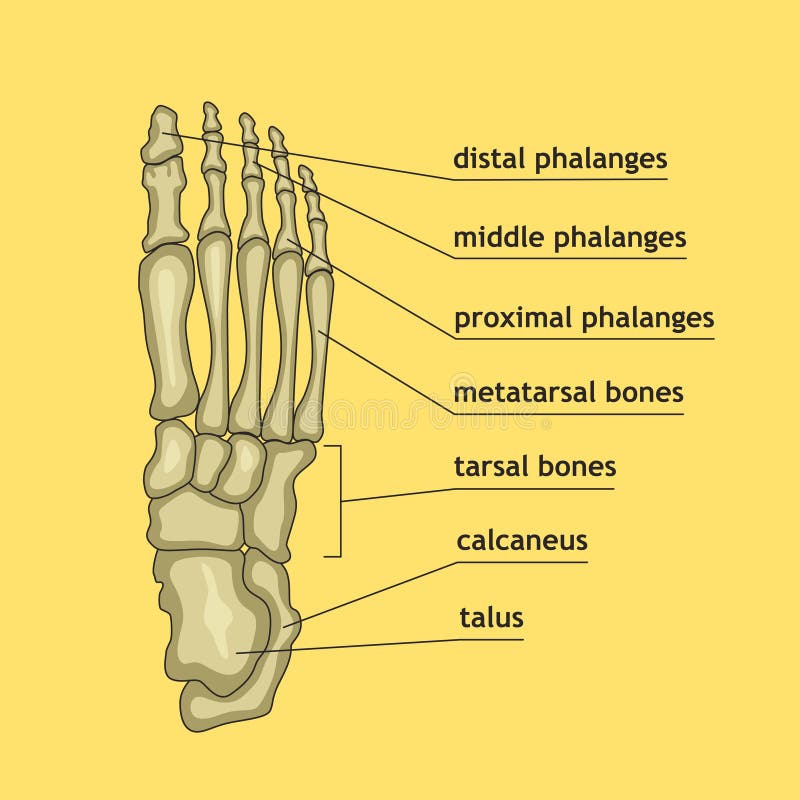
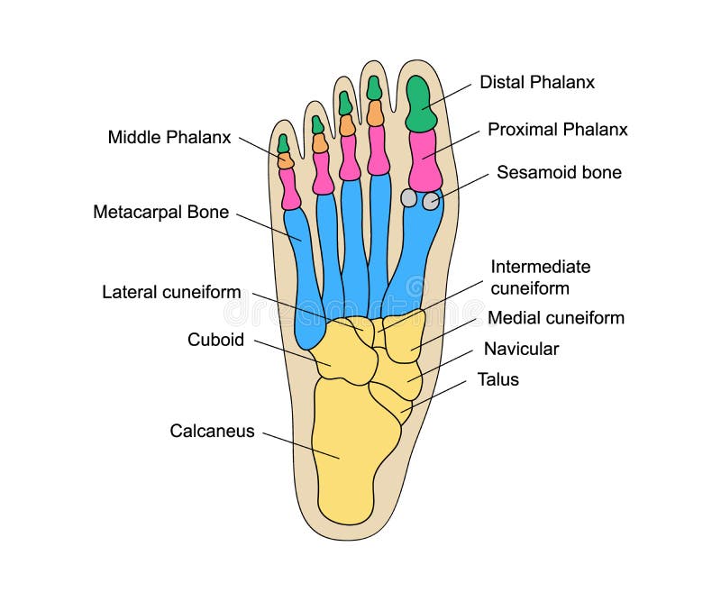
Free with trial The forefoot contains the phalanges and metatarsals. The midfoot consists of the five tarsal bones, three cuneiforms, the navicular, and the cuboid. The hindfoot is composed of two tarsal bones, the calcaneus and the talus. Tarsal bones vectors Diagram or infographic of the bones that make up a foot from an aerial view with names. The forefoot contains the phalanges and metatarsals. The midfoot consists. The forefoot contains the phalanges and metatarsals. The midfoot consists of the five tarsal bones, three cuneiforms, the navicular, and the cuboid. The hindfoot is composed of two tarsal bones, the calcaneus and the talus.
Free with trial The forefoot contains the phalanges and metatarsals. The midfoot consists of the five tarsal bones, three cuneiforms, the navicular, and the cuboid. The hindfoot is composed of two tarsal bones, the calcaneus and the talus. Tarsal bones vectors Diagram or infographic of the bones that make up a foot from an aerial view. The forefoot contains the phalanges and metatarsals. The midfoot consists of the. The forefoot contains the phalanges and metatarsals. The midfoot consists of the five tarsal bones, three cuneiforms, the navicular, and the cuboid. The hindfoot is composed of two tarsal bones, the calcaneus and the talus.
Free with trial Foot bones anatomy with descriptions. Colored leg base parts structure. Educational diagram with internal organ illustration. Distal, proximal and middle phalanges, metacarpal, tarsal bones foot parts. Tarsal bones vectors Foot bones anatomy with descriptions. Educational diagram of internal organ. Distal, proximal and middle phalanges. Foot bones anatomy with descriptions. Colored. Foot bones anatomy with descriptions. Colored leg base parts structure. Educational diagram with internal organ illustration. Distal, proximal and middle phalanges, metacarpal, tarsal bones foot parts
Free with trial Anatomy Bones of the Feet. OrthDiagram. Orthotics for foot Superior view and Medial view. Diagram. Tarsal bones vectors Anatomy Bones of the Feet. OrthDiagram. Anatomy Bones of the Feet. Orthotics for foot Superior view and Medial view. Diagram
Free with trial Foot bones vector sketch of human anatomy, orthopedics medicine design. Side view of skeleton leg with phalange, metatarsal, tarsal and calcaneus, cuneiform, navicular and tibia bones diagram. Tarsal bones vectors Foot bones, sketch of human anatomy. Foot bones vector sketch of human anatomy, orthopedics medicine design. Side view of skeleton leg with phalange, metatarsal. Foot bones vector sketch of human anatomy, orthopedics medicine design. Side view of skeleton leg with phalange, metatarsal, tarsal and calcaneus, cuneiform, navicular and tibia bones diagram
Free with trial Foot with bones, ankle joint inflammation in circle, logo design. Healthcare, medicine, rheumatology and traumatology, vector desi. Gn and illustration. Tarsal bones vectors Foot with bones, ankle joint inflammation in circle, logo design. Healthcare, medicine, rheumatology and traumatology, vector desi. Gn and illustration
Free with trial Bones of foot of human skeleton vector flat illustration isolated on white background. Anatomical fingers element for scientist and halloween horror design. Tarsal bones vectors Bones of human foot vector flat illustration isolated on white background. Bones of foot of human skeleton vector flat illustration isolated on white background. Bones of foot of human skeleton vector flat illustration isolated on white background. Anatomical fingers element for scientist and halloween horror design.
Free with trial Object isolated on white background. Bones of the foot with main parts labeled. From above, lateral and medial views. Vector illustration. Tarsal bones vectors Object isolated on white background. Bones of the foot with main parts labeled. From above, lateral and medial views. Vector illustration
Free with trial Fracture of the metatarsal bones in the foot. Anatomical structure of the foot. Skeleton. Broken bones. Vector illustration on isolated background. Tarsal bones vectors Fracture of the metatarsal bones in the foot. Anatomical structure of the foot. Skeleton. Broken bones. Vector. Illustration on isolated background
Free with trial Fracture of the metatarsal bones in the foot. Anatomical structure of the foot. Skeleton. Broken bones. Vector illustration on isolated background. Tarsal bones vectors Fracture of the metatarsal bones in the foot. Anatomical structure of the foot. Skeleton. Broken bones. Vector. Illustration on isolated background
Free with trial Foot with bones, ankle joint inflammation in circle, logo design. Healthcare medicine, rheumatology and traumatology, vector design and illustration. Tarsal bones vectors Foot with bones, ankle joint inflammation in circle, logo design. Healthcare medicine. Rheumatology and traumatology, vector design and illustration
Free with trial Pop art background. Bones of the foot with main parts labeled. From above, lateral and medial views. Vector. Illustration. Tarsal bones vectors Pop art background. Bones of the foot with main parts labeled. From above, lateral and medial views. Vector. Illustration
Free with trial Human foot bones. Leg and foot step anatomical structure with captions, front view. Healthy skeleton scheme and foot deformities, bunions stages. Flat vector illustration. Tarsal bones vectors Human foot bones. Footstep anatomical structure with captions,. Human foot bones. Leg and foot step anatomical structure with captions, front view. Healthy. Human foot bones. Leg and foot step anatomical structure with captions, front view. Healthy skeleton scheme and foot deformities, bunions stages. Flat vector illustration
Free with trial Human foot bones. Footstep anatomical structure with captions, front and side views. Healthy skeleton scheme, distal and proximal phalanges, tarsal bones, calcaneus. Flat vector illustration. Tarsal bones vectors Human foot bones. Footstep anatomical structure with captions,. Front and side views. Healthy skeleton scheme, distal and proximal phalanges, tarsal bones. Human foot bones. Footstep anatomical structure with captions, front and side views. Healthy skeleton scheme, distal and proximal phalanges, tarsal bones, calcaneus . Flat vector illustration
Free with trial Illustration foot bone. the bones of the foot. vector about science and medical. Tarsal bones vectors Illustration foot bone. the bones of the foot. Vector about science and medical
Free with trial Human foot bones anatomy with descriptions. Colored leg base parts structure. Educational diagram with human internal organ illustration. Talus, calcaneus, tarsals, cuboid, metatarsals foot parts. Tarsal bones vectors Human foot bones anatomy with descriptions. Educational diagram of internal organ illustration. Calcaneus, tarsals. Human foot bones anatomy with descriptions. Human foot bones anatomy with descriptions. Colored leg base parts structure. Educational diagram with human internal organ illustration. Talus, calcaneus, tarsals, cuboid, metatarsals foot parts.
Free with trial Medial view of foot bones Diagram. Anatomy. Tarsal bones vectors Medial view of foot bones Diagram. Anatomy
Free with trial Human foot anatomy infographic diagram. bones phalanges metatarsals tarsal. names joints. front lateral view. cartoon vector drawing. illustration for medical science education. structure parts chart. Tarsal bones vectors Human foot anatomy infographic diagram. Bones phalanges metatarsals tarsal. names joints. front lateral view. cartoon vector drawing. illustration for medical
Free with trial Human foot anatomy infographic diagram. bones phalanges metatarsals tarsal. names joints. front lateral view. cartoon vector drawing. illustration for medical science education. structure parts chart. Tarsal bones vectors Human foot anatomy infographic diagram. Bones phalanges metatarsals tarsal. names joints. front lateral view. cartoon vector drawing. illustration for medical
Free with trial Human foot bones anatomy sketch design of vector orthopedics medicine. Skeleton leg ankle joints and toe phalanges, cuboid, metatarsal, navicular and cuneiform bones, hand drawn dorsal view of foot. Tarsal bones vectors Human foot bones anatomy vector sketch. Human foot bones anatomy sketch design of vector orthopedics medicine. Skeleton leg ankle joints and toe phalanges. Human foot bones anatomy sketch design of vector orthopedics medicine. Skeleton leg ankle joints and toe phalanges, cuboid, metatarsal, navicular and cuneiform bones, hand drawn dorsal view of foot
Free with trial Below view foot bones. Hand drawn illustration of the foot bones on white, artistic anatomy graphic study. Tarsal bones vectors Below view foot bones. Hand drawn illustration of the foot bones on white, artistic anatomy graphic study
Free with trial Internal lateral view foot bones color. Hand drawn illustration of the foot bones isolated on white, artistic anatomy graphic study. Tarsal bones vectors Internal lateral view foot bones color. Hand drawn illustration of the foot bones isolated on white, artistic anatomy graphic study
Free with trial Lateral view foot bones color. Hand drawn illustration of the foot bones isolated on white, artistic anatomy graphic study. Tarsal bones vectors Lateral view foot bones color. Hand drawn illustration of the foot bones isolated on white, artistic anatomy graphic study
Free with trial Human leg bones icons. Chest image in a flat style. Vector illustration in beige colour isolated on a dark grey background. Tarsal bones vectors Human skeleton bones. Human leg bones icons. Chest image in a flat style. Vector illustration in beige colour isolated on a dark grey background. Human leg bones icons. Chest image in a flat style. Vector illustration in beige colour isolated on a dark grey background.
Free with trial Internal lateral view foot bones. Hand drawn illustration of the foot bones on white, artistic anatomy graphic study. Tarsal bones vectors Internal lateral view foot bones. Hand drawn illustration of the foot bones on white, artistic anatomy graphic study
Free with trial Bones of the human foot, vector hand drawn illustration isolated on a white background, orthopedics medicine anatomy sketch. Tarsal bones vectors Bones of the human foot, vector hand drawn illustration isolated on a white background. Orthopedics medicine anatomy sketch
Free with trial Lateral view foot bones. Hand drawn illustration of the foot bones on white, artistic anatomy graphic study. Tarsal bones vectors Lateral view foot bones. Hand drawn illustration of the foot bones on white, artistic anatomy graphic study
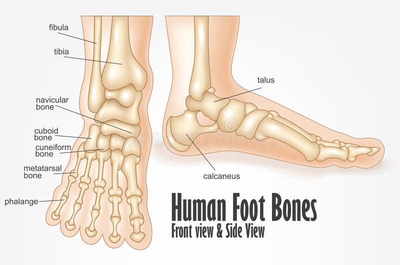
Free with trial Human foot bones front and side view anatomy. Vector Illustration Of Human foot bones front and side view anatomy. Tarsal bones vectors Human foot bones front and side view anatomy. Vector Illustration Of Human foot bones front and side view anatomy
Free with trial Human foot bones front and side view anatomy. Vector Illustration Of Human foot bones front and side view anatomy. Tarsal bones vectors Human foot bones front and side view anatomy. Vector Illustration Of Human foot bones front and side view anatomy
Free with trial Bones of the human foot, hand drawn vector illustration isolated on a white background, orthopedics medicine anatomy sketch. Tarsal bones vectors Bones of the human foot, hand drawn vector illustration isolated on a white background. Orthopedics medicine anatomy sketch
Free with trial Bones of the foot. Vector drawing. Tarsal bones vectors Bones of the foot. Vector drawing
Free with trial Healthy limb posture section element diagnostic concept on white text space. Black line hand drawn hip medial care logo pictogram in doodle art vintage style. Analysis abstract frontal above top view. Tarsal bones vectors Bones of the foot. Vector drawing. Healthy limb posture section element diagnostic concept on white text space. Black line hand drawn hip medial care logo. Healthy limb posture section element diagnostic concept on white text space. Black line hand drawn hip medial care logo pictogram in doodle art vintage style. Analysis abstract frontal above top view
Free with trial Human foot bones anatomy. Colored foot parts structure. Human leg base diagram vector illustration. Educational content for medical university. Tarsal bones vectors Human foot bones anatomy. Colored foot parts structure. Human leg base diagram vector illustration. Educational content for medical university
Free with trial Human foot bones anatomy with descriptions. Foot parts structure. Human internal organ illustration. Talus, calcaneus, tarsals, cuboid, metatarsals leg base parts. Tarsal bones vectors Human foot bones anatomy with descriptions. Foot parts structure. Human internal organ illustration. Talus, calcaneus, tarsals, cuboid, metatarsals leg base
Free with trial Human foot bones anatomy drawing with a pen on notebook. Foot parts structure diagram with bones description. Human internal organ illustration. Talus, calcaneus, cuboid, metatarsals leg base parts. Tarsal bones vectors Human foot bones anatomy drawing with a pen on notebook. Foot parts structure diagram with bones description. Human. Internal organ illustration. Talus. Human foot bones anatomy drawing with a pen on notebook. Foot parts structure diagram with bones description. Human internal organ illustration. Talus, calcaneus, cuboid, metatarsals leg base parts.
Free with trial Foot and ankle Bones Skeleton Human front back view with partly transparent bones position. Set of realistic flat natural color concept Vector illustration of anatomy isolated on white background. Tarsal bones vectors Foot and ankle Bones Skeleton Human front back view with partly transparent bones position. Set of realistic flat. Natural color concept Vector illustration of. Foot and ankle Bones Skeleton Human front back view with partly transparent bones position. Set of realistic flat natural color concept Vector illustration of anatomy isolated on white background
Free with trial Foot and ankle Bones Human Skeleton Anatomically correct side lateral view joints, toe phalanges, cuboid, metatarsal, navicular, cuneiform. Realistic flat natural color concept Vector illustration. Tarsal bones vectors Foot and ankle Bones Human Skeleton Anatomically correct side lateral view joints, toe phalanges, cuboid, metatarsal. Navicular, cuneiform. Realistic flat. Foot and ankle Bones Human Skeleton Anatomically correct side lateral view joints, toe phalanges, cuboid, metatarsal, navicular, cuneiform. Realistic flat natural color concept Vector illustration
Free with trial Foot and ankle Bones Human Skeleton back Posterior dorsal view. Set of Anatomically correct joints, toe phalanges, cuboid, metatarsal, navicular, cuneiform realistic flat concept Vector illustration. Tarsal bones vectors Foot and ankle Bones Human Skeleton back Posterior dorsal view. Set of Anatomically correct joints, toe phalange, cuboid. Foot and ankle Bones Human Skeleton. Foot and ankle Bones Human Skeleton back Posterior dorsal view. Set of Anatomically correct joints, toe phalanges, cuboid, metatarsal, navicular, cuneiform realistic flat concept Vector illustration
Free with trial Above view foot bone. Hand drawn illustration of the foot bones on white, artistic anatomy graphic study. Tarsal bones vectors Above view foot bone. Hand drawn illustration of the foot bones on white, artistic anatomy graphic study
Free with trial Healthy limb posture section element diagnostic concept on white text space. Black line hand drawn hip medial care logo pictogram emblem chart in doodle art vintage style. Analysis abstract side view. Tarsal bones vectors Bones of the foot. Vector drawing. Healthy limb posture section element diagnostic concept on white text space. Black line hand drawn hip medial care logo. Healthy limb posture section element diagnostic concept on white text space. Black line hand drawn hip medial care logo pictogram emblem chart in doodle art vintage style. Analysis abstract side view
 Sign in