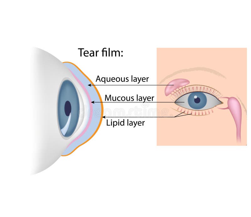
Free with trial Tear film formation. The lacrimal gland, meibomian glands and conjunctival goblet cells secretion. Tarsal vectors Tear film formation. The lacrimal gland, meibomian glands and conjunctival goblet cells secretion
Free with trial Tears chemical composition. The lacrimal gland, meibomian glands and conjunctival goblet cells secretion, anterior and lateral view. Tarsal vectors Tears chemical composition. The lacrimal gland, meibomian glands and conjunctival goblet cells secretion, anterior and lateral view
Free with trial Eye conjunctiva. And other accessory structures of the orbit, eps10. Tarsal vectors Eye conjunctiva. And other accessory structures of the orbit, eps10
Free with trial Vector illustration of healthy human foot and a medial ankle injury. Tarsal tunnel syndrome. EPS 10. Tarsal vectors Medial ankle injury_Tarsal tunnel syndrome. Vector illustration of healthy human foot and a medial ankle injury. Tarsal tunnel syndrome. EPS 10. Vector illustration of healthy human foot and a medial ankle injury. Tarsal tunnel syndrome. EPS 10.
Free with trial Chalazion & sty. Medical illustration of symptoms of chalazion and sty. Tarsal vectors Chalazion & sty. Medical illustration of symptoms of chalazion and sty
Free with trial Nerves of the foot. Vector illustration diagram of the nerves and cutaneous innervation of the human foot with palmar and dorsal view. Used transparency. Tarsal vectors Nerves of the foot. Vector illustration diagram of the nerves and cutaneous innervation of the human foot with palmar and dorsal view. Used transparency
Free with trial Anatomy of bee educational labeled body structure scheme vector illustration. Biological description with insect head, thorax and abdomen detailed parts names. Examination for zoology handout purpose. Tarsal vectors Anatomy of bee educational labeled body structure scheme vector illustration. Biological description with insect head, thorax and abdomen detailed parts names
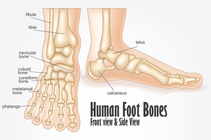
Free with trial Human foot bones front and side view anatomy. Illustration Of Human foot bones front and side view anatomy. Tarsal vectors Human foot bones front and side view anatomy. Illustration Of Human foot bones front and side view anatomy
Free with trial Foot fat pad atrophy. Vector illustration, diagram of a healthy foot and a foot with a fat pad atrophy. Medial and plantar view of a human foot. Tarsal vectors Foot fat pad atrophy. Vector illustration, diagram of a healthy foot and a foot with a fat pad atrophy. Medial and plantar view of a human foot
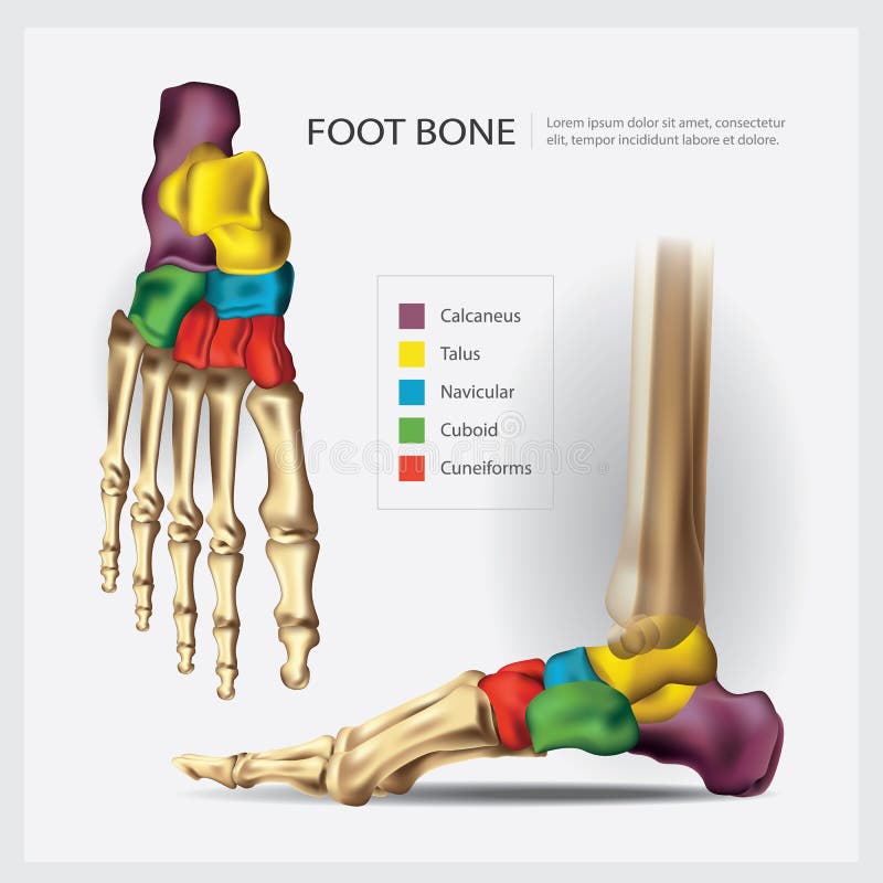
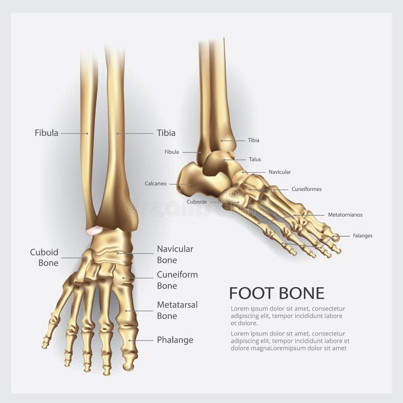
Free with trial Human Anatomy Foot Bone. Vector Illustration. Tarsal vectors Human Anatomy Foot Bone. Vector Illustration
Free with trial In the dorsal (posterior) view of the bones of the feet, the tarsal bones are visible, including the talus, calcaneus, navicular, cuboid, and the three cuneiform bones. The metatarsal bones, numbering from one to five, can also be observed, extending towards the toes. Additionally, the phalanges, consisting of proximal, middle, and distal phalanges, complete the skeletal structure of the feet in the dorsal view. Tarsal vectors Bones of feet, dorsal (posterior) view. In the dorsal (posterior) view of the bones of the feet, the tarsal bones are visible, including the talus, calcaneus. In the dorsal (posterior) view of the bones of the feet, the tarsal bones are visible, including the talus, calcaneus, navicular, cuboid, and the three cuneiform bones. The metatarsal bones, numbering from one to five, can also be observed, extending towards the toes. Additionally, the phalanges, consisting of proximal, middle, and distal phalanges, complete the skeletal structure of the feet in the dorsal view.
Free with trial Vector illustration, diagram of the Baxter`s neuropathy problem, inflammation of the inferior calcaneal nerve. Medial and plantar view of a human foot. Tarsal vectors Nerves of the foot-02. Vector illustration, diagram of the Baxter`s neuropathy problem, inflammation of the inferior calcaneal nerve. Medial and plantar view of. Vector illustration, diagram of the Baxter`s neuropathy problem, inflammation of the inferior calcaneal nerve. Medial and plantar view of a human foot
Free with trial Too many toes. Vector illustration of the normal human foot and the foot with pronation or flatfoot, with hindfoot deformity. Too many toes sign. Tarsal vectors Too many toes. Vector illustration of the normal human foot and the foot with pronation or flatfoot, with hindfoot deformity. Too many toes sign
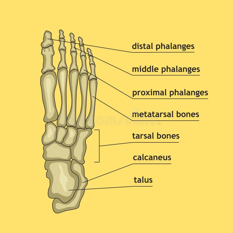
Free with trial Vector illustration of foot bones with explanation. Skeleton part for medical or health care design. Anatomy guide skeletal human foot. Tarsal vectors Foot bones with explanation. Vector illustration of foot bones with explanation. Skeleton part for medical or health care design. Anatomy guide skeletal human. Vector illustration of foot bones with explanation. Skeleton part for medical or health care design. Anatomy guide skeletal human foot.
Free with trial Vector illustration of a human leg with denominations of the bones of the foot. Anatomy of lateral or side view of the foot. For advertising or medical publications. EPS 10. Tarsal vectors Anatomy_bones of the human foot lateral view. Vector illustration of a human leg with denominations of the bones of the foot. Anatomy of lateral or side view of. Vector illustration of a human leg with denominations of the bones of the foot. Anatomy of lateral or side view of the foot. For advertising or medical publications. EPS 10.
Free with trial Vector illustration of a human leg with denominations of the bones of the foot. Anatomy of medial or side view of the foot. For advertising or medical publications. EPS 10. Tarsal vectors Anatomy_bones of the human foot medial view. Vector illustration of a human leg with denominations of the bones of the foot. Anatomy of medial or side view of. Vector illustration of a human leg with denominations of the bones of the foot. Anatomy of medial or side view of the foot. For advertising or medical publications. EPS 10.
Free with trial Vector illustration of a human leg with denominations of the bones of the foot. Anatomy of dorsal and plantar views of the foot. For advertising or medical publications. EPS 10. Tarsal vectors Anatomy_bones of the human foot dorsal and plantar view. Vector illustration of a human leg with denominations of the bones of the foot. Anatomy of dorsal and. Vector illustration of a human leg with denominations of the bones of the foot. Anatomy of dorsal and plantar views of the foot. For advertising or medical publications. EPS 10.
Free with trial Anatomy Bones of the Feet. Superior view. Orthotics for foot Superior view. Tarsal vectors Anatomy Bones of the Feet. Superior view. Anatomy Bones of the Feet. Orthotics for foot Superior view
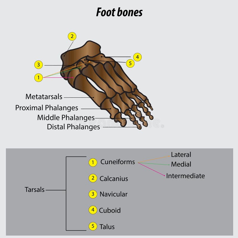
Free with trial Bones of the Foot. Tarsals or tarsus, Metatarsals, Phalanges. Tarsal vectors Bones of the Foot .Tarsals or tarsus, Metatarsals, Phalanges. Bones of the Foot.Tarsals or tarsus, Metatarsals, Phalanges Anatomy
Free with trial Human Anatomy Foot Bone. Vector Illustration. Tarsal vectors Human Anatomy Foot Bone. Vector Illustration
Free with trial Foot pain causes from zones diagnosis and painful spots areas outline diagram. Labeled educational scheme with medical illness, disease or trauma diagnostics vector illustration. Tendonitis, bursitis. Tarsal vectors Foot pain causes from zones diagnosis and painful spots areas outline diagram. Labeled educational scheme with medical illness, disease or trauma diagnostics
Free with trial The external structure of the eye. Vector illustration. Tarsal vectors The external structure of the eye. Vector. Illustration
Free with trial The external structure of the eye. Vector illustration. Tarsal vectors The external structure of the eye. Vector. Illustration
Free with trial Severs disease. calcaneus apophysitis. inflammation at the back of the heel growth plate. Foot anatomy. side view of a Human foot with the name of bones. Arches of the feet. skeleton anatomy. realistic Vector poster. Tarsal vectors Severs disease. calcaneus apophysitis. inflammation at the back of the heel growth plate. Foot anatomy. side view of a Human foot with the name of bones
Free with trial A striking, high-resolution illustration of a Common Wasp (Vespula vulgaris), showcasing the intricate details of this well-known social insect. The asset features the characteristic bright yellow and black aposematic patterning across the abdomen and thorax, serving as a natural warning signal. The rendering captures the delicate veining of the translucent wings, the segmented antennae, and the sharp, hooked tarsal claws on the legs. Detailed compound eyes and mandibles are visible on the head, emphasizing its role as a specialized predator. Isolated on a transparent grid background, this entomological specimen is an ideal resource for biodiversity education, pest management guides, and high-end digital media focusing on insect anatomy and ecology. Tarsal vectors Common Wasp Specimen, High-Fidelity Hymenoptera Asset, Transparent Background. A striking, high-resolution illustration of a Common Wasp (Vespula vulgaris). A striking, high-resolution illustration of a Common Wasp (Vespula vulgaris), showcasing the intricate details of this well-known social insect. The asset features the characteristic bright yellow and black aposematic patterning across the abdomen and thorax, serving as a natural warning signal. The rendering captures the delicate veining of the translucent wings, the segmented antennae, and the sharp, hooked tarsal claws on the legs. Detailed compound eyes and mandibles are visible on the head, emphasizing its role as a specialized predator. Isolated on a transparent grid background, this entomological specimen is an ideal resource for biodiversity education, pest management guides, and high-end digital media focusing on insect anatomy and ecology.
Free with trial Front view foot bones. Hand drawn illustration of the foot bones on white, artistic anatomy graphic study. Tarsal vectors Front view foot bones. Hand drawn illustration of the foot bones on white, artistic anatomy graphic study
Free with trial Human foot bones and joints skeleton vector sketch body anatomy icon. Isolated symbol of feet and toes limbs structure of leg organ for anatomical orthopedic or medical surgery design element. Tarsal vectors Vector sketch icon of human foot bones or joints. Human foot bones and joints skeleton vector sketch body anatomy icon. Isolated symbol of feet and toes limbs. Human foot bones and joints skeleton vector sketch body anatomy icon. Isolated symbol of feet and toes limbs structure of leg organ for anatomical orthopedic or medical surgery design element
Free with trial Vector illustration of a mans soles. feet, sole, body part. Tarsal vectors Vector illustration of a mans soles. Feet, sole, body part
Free with trial Dislocation of the foot forward, vintage engraved illustration. Usual Medicine Dictionary - Paul Labarthe - 1885. Tarsal vectors Dislocation of the foot forward, vintage engraving. Dislocation of the foot forward, vintage engraved illustration. Usual Medicine Dictionary - Paul Labarthe. Dislocation of the foot forward, vintage engraved illustration. Usual Medicine Dictionary - Paul Labarthe - 1885
Free with trial Vector drawing of human leg human leg anatomy drawing labeled. Drawing with layers reedit able. Tarsal vectors Vector drawing of human leg human leg anatomy drawing labeled. Drawing with layers reedit able
Free with trial Healthy posture section diagnostic concept on white space for text. Outline black hand drawn hip leg logo emblem pictogram in doodle art vintage vector style. Analysis coccyx cure abstract side view. Tarsal vectors Spine. Vector drawing. Healthy posture section diagnostic concept on white space for text. Outline black hand drawn hip leg logo emblem pictogram in doodle art. Healthy posture section diagnostic concept on white space for text. Outline black hand drawn hip leg logo emblem pictogram in doodle art vintage vector style. Analysis coccyx cure abstract side view
Free with trial Vector drawing of human foot human foot anatomy drawing labeled. Drawing with layers reedit able. Tarsal vectors Vector drawing of human foot human foot anatomy drawing labeled. Drawing with layers reedit able
Free with trial Vector illustration of a mans feet. Legs, sole, body part. Tarsal vectors Vector illustration of a mans feet. Legs, sole, body part
Free with trial Fracture of the metatarsal bones in the foot. Anatomical structure of the foot. Skeleton. Broken bones. Vector illustration on isolated background. Tarsal vectors Fracture of the metatarsal bones in the foot. Anatomical structure of the foot. Skeleton. Broken bones. Vector. Illustration on isolated background
Free with trial Structure and functions of the eye. The main parts that make up For basic medical education. Signatures and text. Also for clinics set. Tarsal vectors Structure and functions of the eye. The main parts that make up For basic medical education. Signatures and text. Also for clinics set
Free with trial Vector illustration of a womans soles. feet, sole, body part. Tarsal vectors Vector illustration of a womans soles. Feet, sole, body part
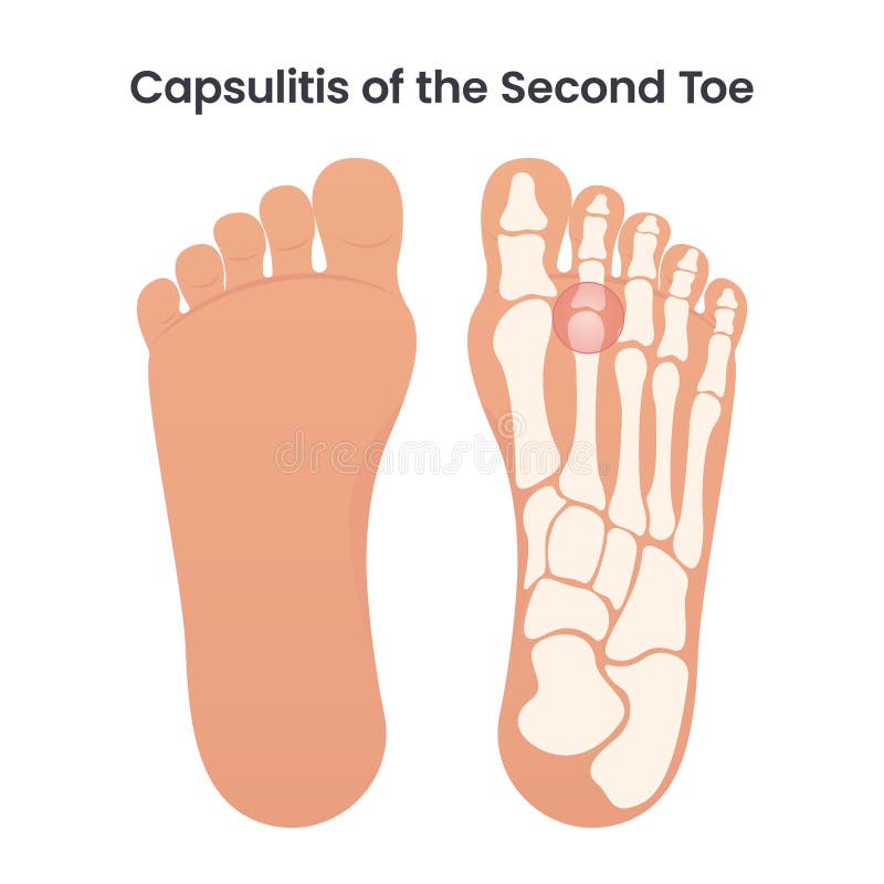
Free with trial Capsulitis of the second toe occurs in the ligaments surrounding the joint at the base of the second toe. These ligaments form a capsule, which helps the joint to function properly. Capsulitis is a condition in which these ligaments have become inflamed. Tarsal vectors Capsulitis of the Second Bone educational medical vector illustration. Capsulitis of the second toe occurs in the ligaments surrounding the joint at the base of. Capsulitis of the second toe occurs in the ligaments surrounding the joint at the base of the second toe. These ligaments form a capsule, which helps the joint to function properly. Capsulitis is a condition in which these ligaments have become inflamed
Free with trial Structure and functions of the eye. The main parts that make up For basic medical education. Signatures and text. Also for clinics set. Tarsal vectors Structure and functions of the eye. The main parts that make up For basic medical education. Signatures and text. Also for clinics set
Free with trial Illustration human foot bone. on white background. vector about science and medical. Tarsal vectors Illustration human foot bone. on white background. Illustration of human foot bone. on white background. vector about science and medical
Free with trial Meibomian gland. Front and side view of human eye with eyelids, Eyelashes and openings of tarsal glands. Eye sebaceous glands anatomy. Medical illustration. Infographic diagram. Vector. Tarsal vectors Meibomian gland. Front and side view of human eye with eyelids, Eyelashes and openings of tarsal glands. Eye sebaceous glands. Anatomy. Medical illustration. Meibomian gland. Front and side view of human eye with eyelids, Eyelashes and openings of tarsal glands. Eye sebaceous glands anatomy. Medical illustration. Infographic diagram. Vector
Free with trial Tarsal glands anatomy. Front view of human eye with Meibomian, Lacrimal glands. Close-up of lower eyelid with Openings of palpebral or tarsoconjunctival glands and meibum oil droplets can be seen on tarsal plate. Eye's tear film. Medical illustration. Infographic diagram. Vector. Tarsal vectors Tarsal glands anatomy. Front view of human eye with Meibomian, Lacrimal glands. Close-up of lower eyelid with Openings of palpebral or tarsoconjunctival glands
Free with trial Realistic 3D render of human tarsal bones slightly rotated showing talus calcaneus navicular cuboid and cuneiforms isolated on white background with ultra sharp professional focus. Tarsal vectors Realistic 3D Render Human Tarsal Bones Detailed Ankle Anatomy. Realistic 3D render of human tarsal bones slightly rotated showing talus calcaneus navicular. Realistic 3D render of human tarsal bones slightly rotated showing talus calcaneus navicular cuboid and cuneiforms isolated on white background with ultra sharp professional focus
Free with trial Educational podiatry and orthopedics detailing tarsal tunnel syndrome compared to a normal tarsal tunnel in full color. The labeled cross-sectional anatomical diagram illustrates the medial ankle region beneath the flexor retinaculum, identifying the medial malleolus, tendons, and posterior tibial nerve. It clearly contrasts an uncompressed normal tarsal tunnel against tarsal tunnel syndrome characterized by localized swelling, inflamed surrounding tendons, and compression of the posterior tibial nerve. High-quality infographic graphic ideal for podiatry clinic posters, orthopedic textbooks, physical therapy study guides, sports medicine manuals, medical school lecture slides, and patient healthcare education presentations. Tarsal vectors Tarsal Tunnel Syndrome vs Normal Foot Anatomy Posterior Tibial Nerve Compression Color Vector Illustration. Educational podiatry and orthopedics detailing tarsal. Educational podiatry and orthopedics detailing tarsal tunnel syndrome compared to a normal tarsal tunnel in full color. The labeled cross-sectional anatomical diagram illustrates the medial ankle region beneath the flexor retinaculum, identifying the medial malleolus, tendons, and posterior tibial nerve. It clearly contrasts an uncompressed normal tarsal tunnel against tarsal tunnel syndrome characterized by localized swelling, inflamed surrounding tendons, and compression of the posterior tibial nerve. High-quality infographic graphic ideal for podiatry clinic posters, orthopedic textbooks, physical therapy study guides, sports medicine manuals, medical school lecture slides, and patient healthcare education presentations.
Free with trial This image illustrates the intricate structure of the human foot, highlighting the bones and muscles. The foot is composed of numerous bones that work together to provide support, balance, and mobility. The image shows the bones in the toes, the metatarsals, the tarsal bones, and the ankle joint. The muscles are depicted in a way that shows their attachment points and how they contribute to the. Tarsal vectors Detailed anatomy of the human foot. This image illustrates the intricate structure of the human foot, highlighting the bones and muscles. The foot is composed of. This image illustrates the intricate structure of the human foot, highlighting the bones and muscles. The foot is composed of numerous bones that work together to provide support, balance, and mobility. The image shows the bones in the toes, the metatarsals, the tarsal bones, and the ankle joint. The muscles are depicted in a way that shows their attachment points and how they contribute to the
Free with trial This is a detailed illustration of a human foot skeleton, showcasing the intricate structure of the skeletal system. The image features a gray foot skeleton with a transparent background, allowing for clear visibility of the bones. The skeleton is composed of multiple bones, including the phalanges, metatarsal, tarsal, calcaneus, talus, cuboid, navicular, and cuneiform. This image is ideal for medical, scientific, and educational purposes, providing a comprehensive view of the human foot anatomy. Tarsal vectors A detailed illustration of a human foot skeleton on transparent background. This is a detailed illustration of a human foot skeleton, showcasing the intricate. This is a detailed illustration of a human foot skeleton, showcasing the intricate structure of the skeletal system. The image features a gray foot skeleton with a transparent background, allowing for clear visibility of the bones. The skeleton is composed of multiple bones, including the phalanges, metatarsal, tarsal, calcaneus, talus, cuboid, navicular, and cuneiform. This image is ideal for medical, scientific, and educational purposes, providing a comprehensive view of the human foot anatomy.
Free with trial Human foot and ankle joint skeleton vector illustration isolated on black background. X-ray style side view of foot bones, anatomy tarsal tibia fibula for medical poster, sprain injury design. Tarsal vectors Human foot and ankle joint skeleton vector illustration isolated on black background. X-ray style side view of foot bones, anatomy. Tarsal tibia fibula for. Human foot and ankle joint skeleton vector illustration isolated on black background. X-ray style side view of foot bones, anatomy tarsal tibia fibula for medical poster, sprain injury design
Free with trial This image is an anatomical diagram of the human foot, showcasing the intricate structure of bones and muscles. The diagram highlights the skeletal framework and the detailed arrangement of muscles that support the foot. It provides a clear view of the bones in the toes, the metatarsals, and the tarsal bones, as well as the muscles that control movement and provide stability. Tarsal vectors Anatomical diagram of the human foot. This image is an anatomical diagram of the human foot, showcasing the intricate structure of bones and muscles. The diagram. This image is an anatomical diagram of the human foot, showcasing the intricate structure of bones and muscles. The diagram highlights the skeletal framework and the detailed arrangement of muscles that support the foot. It provides a clear view of the bones in the toes, the metatarsals, and the tarsal bones, as well as the muscles that control movement and provide stability
Free with trial Illustration of a horses lower leg anatomy detailing key bones and joints. It includes the cannon bone (third metacarpal tarsal bone), the splint bone (second and fourth metacarpal tarsal bones), the fetlock joint (metacarpophalangeal metatarsophalangeal joint), the pastern bones (proximal and middle phalanges), and the coffin bone (distal phalanx). The image serves as an educational tool for understanding the structure and function of a horses limb. Tarsal vectors Illustration of a horses lower leg anatomy detailing key bones and joints. It includes the cannon bone (third metacarpal tarsal bone), the splint bone (second
Free with trial Detailed silhouette icon of a human foot skeleton ankle bones anatomy on transparent background. Tarsal vectors Human foot skeleton ankle tarsal bones anatomy silhouette. AI generated detailed silhouette icon of a human foot skeleton ankle bones anatomy on transparent. detailed silhouette icon of a human foot skeleton ankle bones anatomy on transparent background
Free with trial A black-and-white anatomical diagram presents eight labeled bones of the foot: the calcaneus, talus, navicular, cuboid, medial, intermediate and lateral cuneiforms, and first metatarsal. Each bone is shown separately as a simplified, outlined with shaded areas suggesting its shape and contours; the first metatarsal appears elongated, while the tarsal bones are more compact and irregular. The clean layout on a white background emphasizes identification and comparison, with bold labels beneath each drawing. Tarsal vectors A black-and-white anatomical diagram presents eight labeled bones of the foot: the calcaneus, talus, navicular, cuboid, medial. Intermediate and lateral. A black-and-white anatomical diagram presents eight labeled bones of the foot: the calcaneus, talus, navicular, cuboid, medial, intermediate and lateral cuneiforms, and first metatarsal. Each bone is shown separately as a simplified, outlined with shaded areas suggesting its shape and contours; the first metatarsal appears elongated, while the tarsal bones are more compact and irregular. The clean layout on a white background emphasizes identification and comparison, with bold labels beneath each drawing.
Free with trial Vector illustration, diagram. Normal human foot and a foot with talocalcaneal coalition, area of pain. Medial view of the foot, section of the ankle and subtalar joint bones. For medical publications. Tarsal vectors Normal foot and a foot with talocalcaneal coalition. Vector illustration, diagram. Normal human foot and a foot with talocalcaneal coalition, area of pain. Vector illustration, diagram. Normal human foot and a foot with talocalcaneal coalition, area of pain. Medial view of the foot, section of the ankle and subtalar joint bones. For medical publications
Free with trial Tear film of the eye. Cross section of a human eye with Conjunctiva, Meibomian gland, Lacrimal gland and Cornea. Layers of Tear film. Mucin, Lipid, and Aqueous layers. Medical illustration. Infographic diagram. Vector. Tarsal vectors Tear film. eye with Conjunctiva, Meibomian gland, Lacrimal gland and Cornea. Layers of Tear film. Tear film of the eye. Cross section of a human eye with. Tear film of the eye. Cross section of a human eye with Conjunctiva, Meibomian gland, Lacrimal gland and Cornea. Layers of Tear film. Mucin, Lipid, and Aqueous layers. Medical illustration. Infographic diagram. Vector
Free with trial Calcaneus Lateral View Anatomy. Detailed Medical Illustration of Heel Bone. Human Calcaneal Bone Anatomy Labeled. Lateral Surface. 3D Render. Tarsal vectors Calcaneus Lateral View Anatomy. Detailed Medical Illustration of Heel Bone. Human Calcaneal Bone Anatomy Labeled. Lateral Surface. 3D Render
Free with trial High-detail medical illustration of the human calcaneus shown in superior view, alongside a contextual foot skeleton. Calcaneus. Bone Superior View. Tarsal vectors High-detail medical illustration of the human calcaneus shown in superior view, alongside a contextual foot skeleton. Calcaneus. Bone Superior View
Free with trial Plantar fasciitis is inflammation in the plantar fascia is one of the most common orthopedic complaints. The plantar fascia is a thick weblike ligament that connects heel to the front of foot. Medical. Tarsal vectors Plantar fasciitis is inflammation in the plantar fascia. Is one of the most common orthopedic complaints.The plantar fascia is a thick weblike ligament that. Plantar fasciitis is inflammation in the plantar fascia is one of the most common orthopedic complaints.The plantar fascia is a thick weblike ligament that connects heel to the front of foot.Medical
Free with trial Plantar fasciitis is one of the most common orthopedic complaints. causes pain in the bottom of the heel. The plantar fascia is a thick weblike ligament that connects heel to the front of foot. Medical. Tarsal vectors Plantar fasciitis is one of the most common orthopedic. Complaints. causes pain in the bottom of the heel.The plantar fascia is a thick weblike ligament that. Plantar fasciitis is one of the most common orthopedic complaints. causes pain in the bottom of the heel.The plantar fascia is a thick weblike ligament that connects heel to the front of foot.Medical
Free with trial Clean silhouette of a human foot skeleton in black and white, isolated on white background. Perfect for medical, anatomical, or design projects. Tarsal vectors Human foot skeleton silhouette isolated. Clean silhouette of a human foot skeleton in black and white, isolated on white background. Perfect for medical. Clean silhouette of a human foot skeleton in black and white, isolated on white background. Perfect for medical, anatomical, or design projects
Free with trial Human Ankle Joint Bones Skeleton Icons. Presented in outline and solid black styles for medical or anatomical representation. Tarsal vectors Human Ankle Joint Bones Skeleton Icons. Presented in outline and solid black styles for medical or anatomical representation
Free with trial Human heel bone icon outline and solid black fill for medical and anatomical use. Tarsal vectors Human Heel Bone Icon Outline and Solid Black Fill. For medical and anatomical use. Human heel bone icon outline and solid black fill for medical and anatomical use
Free with trial Forefoot pain caused by increased pressure or stress on the metatarsal heads, commonly affecting the ball of the foot. Tarsal vectors Ball of Foot Pain (Metatarsalgia) flashcard. Forefoot pain caused by increased pressure or stress on the metatarsal heads, commonly affecting the ball of the. Forefoot pain caused by increased pressure or stress on the metatarsal heads, commonly affecting the ball of the foot.
Free with trial Anatomy of bone ankle toe talus, concept Vector illustration of anatomy isolated on white background. Tarsal vectors Anatomy of bone ankle toe talus. Concept Vector illustration of anatomy isolated on white background
Free with trial This high-quality, professional digital illustration features a side profile of a large grasshopper (Acrididae) in an alert, active pose. The artwork meticulously captures the intricate anatomical details, including the segmented abdomen with its vibrant yellow and black striped patterns and the textured, leathery forewings. Rendered with high fidelity, the asset highlights the long filiform antennae, large compound eye, and the powerful, spiny hind jumping legs characteristic of the species. Every detail, from the delicate tarsal segments to the fine structural lines on the thorax, is clearly defined. Perfectly isolated on a transparent background, this premium entomology cutout is an ideal resource for commercial graphic design, biology-related educational materials, agricultural pest management marketing, and professional nature-themed illustrations. Tarsal vectors Side View of a Large Grasshopper Isolated on Transparent Background. This high-quality, professional digital illustration features a side profile of a large. This high-quality, professional digital illustration features a side profile of a large grasshopper (Acrididae) in an alert, active pose. The artwork meticulously captures the intricate anatomical details, including the segmented abdomen with its vibrant yellow and black striped patterns and the textured, leathery forewings. Rendered with high fidelity, the asset highlights the long filiform antennae, large compound eye, and the powerful, spiny hind jumping legs characteristic of the species. Every detail, from the delicate tarsal segments to the fine structural lines on the thorax, is clearly defined. Perfectly isolated on a transparent background, this premium entomology cutout is an ideal resource for commercial graphic design, biology-related educational materials, agricultural pest management marketing, and professional nature-themed illustrations.
Free with trial Human foot bones illustration. Detailed anatomical illustration of a human foot showcasing its bones and structure, ideal for medical or educational purposes. Tarsal vectors Human foot bones illustration. Detailed anatomical illustration of a human foot showcasing its bones and structure, ideal for medical or educational purposes
Free with trial Detailed anatomical illustration of human feet bones in a vintage style, set against a dark background, showcasing skeletal structure and medical detail. Tarsal vectors Anatomical Illustration of Human Feet Bones on Dark Background. Detailed anatomical illustration of human feet bones in a vintage style, set against a dark. Detailed anatomical illustration of human feet bones in a vintage style, set against a dark background, showcasing skeletal structure and medical detail
Free with trial This high-quality, professional digital illustration features a cluster of five European hornets ($Vespa$ $crabro$) in a dynamic, social group. The artwork meticulously details the insects' striking yellow and black abdominal banding, reddish-brown thorax, and large, compound eyes. Each hornet is rendered with high fidelity, showcasing complex anatomical features such as the delicate wing venation, jointed legs with fine tarsal claws, and segmented antennae. This premium entomological asset is perfectly isolated on a transparent background, making it an ideal resource for commercial graphic design, educational biology materials, pest control informational guides, and professional nature study posters. Tarsal vectors Full Body Group of European Hornets Nesting and Crawling Isolated on Transparent Background. This high-quality, professional digital illustration features a. This high-quality, professional digital illustration features a cluster of five European hornets ($Vespa$ $crabro$) in a dynamic, social group. The artwork meticulously details the insects' striking yellow and black abdominal banding, reddish-brown thorax, and large, compound eyes. Each hornet is rendered with high fidelity, showcasing complex anatomical features such as the delicate wing venation, jointed legs with fine tarsal claws, and segmented antennae. This premium entomological asset is perfectly isolated on a transparent background, making it an ideal resource for commercial graphic design, educational biology materials, pest control informational guides, and professional nature study posters.
Free with trial Detailed anatomical illustration of a human foot skeleton, showcasing the bones and structure, set against a stark black background for emphasis. Tarsal vectors Anatomical Illustration of a Human Foot Skeleton on Black Background. Detailed anatomical illustration of a human foot skeleton, showcasing the bones and. Detailed anatomical illustration of a human foot skeleton, showcasing the bones and structure, set against a stark black background for emphasis
Free with trial Visually striking 3D rendering of a fractured foot bone highlights injury and the potential for healing. Ideal for medical education, patient communication, and health awareness campaigns. Tarsal vectors Detailed 3D rendering of a fractured foot bone showing injury and healing. Visually striking 3D rendering of a fractured foot bone highlights injury and the. Visually striking 3D rendering of a fractured foot bone highlights injury and the potential for healing. Ideal for medical education, patient communication, and health awareness campaigns
Free with trial Explore the intricate details of the human foot skeleton. Essential for medical, educational, and health science applications, offering clear anatomical representation. Tarsal vectors Detailed human foot bone structure for medical and educational presentations. Explore the intricate details of the human foot skeleton. Essential for medical. Explore the intricate details of the human foot skeleton. Essential for medical, educational, and health science applications, offering clear anatomical representation
Free with trial Stylized cartoon illustration of a skeletal foot and ankle on a pink sparkly background. AI generated. Tarsal vectors Stylized cartoon illustration of a skeletal foot and ankle on a pink sparkly background. AI generated
Free with trial Heel pain, specifically consistent with Sever's disease, calcaneal apophysitis. Tarsal vectors Heel pain, specifically consistent with Sever\'s disease. Heel pain, specifically consistent with Sever's disease, calcaneal apophysitis. Heel pain, specifically consistent with Sever's disease, calcaneal apophysitis.
Free with trial Detailed diagram of a human foot showcasing its skeletal structure and individual bones, perfect for educational materials or medical illustrations. This image has a transparent background. Tarsal vectors Human foot bones diagram with transparent background. Detailed diagram of a human foot showcasing its skeletal structure and individual bones, perfect for. Detailed diagram of a human foot showcasing its skeletal structure and individual bones, perfect for educational materials or medical illustrations. This image has a transparent background
Free with trial A clear black silhouette icon depicting the intricate human foot skeleton, isolated on a clean white background. This anatomical illustration showcases the structure of the tarsal bones, metatarsals, and phalanges. Ideal for medical, educational, and healthcare-related projects, it can be used in presentations, textbooks, websites, or apps to represent anatomy, orthopedics, podiatry, bone health, or skeletal system concepts. The minimalist design ensures versatility and readability for various applications. Tarsal vectors Human Foot Skeleton Anatomy Icon. A clear black silhouette icon depicting the intricate human foot skeleton, isolated on a clean white background. This. A clear black silhouette icon depicting the intricate human foot skeleton, isolated on a clean white background. This anatomical illustration showcases the structure of the tarsal bones, metatarsals, and phalanges. Ideal for medical, educational, and healthcare-related projects, it can be used in presentations, textbooks, websites, or apps to represent anatomy, orthopedics, podiatry, bone health, or skeletal system concepts. The minimalist design ensures versatility and readability for various applications.
Free with trial 3D Isometric Flat Vector Illustration of Comparison of Plantigrade, Digitigrade and Unguligrade, Walking Methods of Human and Animals. Item 1. Tarsal vectors 3D Isometric Flat Vector Illustration of Comparison of Plantigrade, Digitigrade and Unguligrade. Item 1. 3D Isometric Flat Vector Illustration of Comparison of. 3D Isometric Flat Vector Illustration of Comparison of Plantigrade, Digitigrade and Unguligrade, Walking Methods of Human and Animals. Item 1
Free with trial 3D Isometric Flat Vector Illustration of Comparison of Plantigrade, Digitigrade and Unguligrade, Walking Methods of Human and Animals. Item 2. Tarsal vectors 3D Isometric Flat Vector Illustration of Comparison of Plantigrade, Digitigrade and Unguligrade. Item 2. 3D Isometric Flat Vector Illustration of Comparison of. 3D Isometric Flat Vector Illustration of Comparison of Plantigrade, Digitigrade and Unguligrade, Walking Methods of Human and Animals. Item 2
Free with trial 3D Isometric Flat Vector Illustration of Comparison of Plantigrade, Digitigrade and Unguligrade, Walking Methods of Human and Animals. Item 3. Tarsal vectors 3D Isometric Flat Vector Illustration of Comparison of Plantigrade, Digitigrade and Unguligrade. Item 3. 3D Isometric Flat Vector Illustration of Comparison of. 3D Isometric Flat Vector Illustration of Comparison of Plantigrade, Digitigrade and Unguligrade, Walking Methods of Human and Animals. Item 3
Free with trial The structure of the working bee. Monochrome illustration in gray color. Tarsal vectors The structure of the working bee. Monochrome illustration in gray color
Free with trial Human foot skeleton anatomy with captions, medical scientific chart. Vector illustration. Tarsal vectors Human foot skeleton anatomy with captions, medical scientific chart. Vector illustration
Free with trial Precise anatomical rendering of the human foot skeleton, highlighting bone structure and a distinct fracture. Ideal for medical education, research, and diagnostic visualization. Tarsal vectors Detailed human foot skeleton with visible bone fracture illustration. Precise anatomical rendering of the human foot skeleton, highlighting bone structure and a. Precise anatomical rendering of the human foot skeleton, highlighting bone structure and a distinct fracture. Ideal for medical education, research, and diagnostic visualization
Free with trial A clear, line-art illustration depicting the skeletal structure of the human foot and ankle, showing the tibia, fibula, tarsals, metatarsals, and phalanges. Generated using AI. Tarsal vectors Anatomical Illustration of the Human Foot and Ankle Bones. A clear, line-art illustration depicting the skeletal structure of the human foot and ankle, showing. A clear, line-art illustration depicting the skeletal structure of the human foot and ankle, showing the tibia, fibula, tarsals, metatarsals, and phalanges. Generated using AI.
Free with trial Detailed anatomical illustration of human feet showcasing the skeletal structure, perfect for medical or educational resources. This image has a transparent background. Tarsal vectors Foot bones diagram with transparent background. Detailed anatomical illustration of human feet showcasing the skeletal structure, perfect for medical or. Detailed anatomical illustration of human feet showcasing the skeletal structure, perfect for medical or educational resources. This image has a transparent background
 Sign in