Free with trial Human bones icon set, cartoon style. Human bones icon set. Cartoon set of human bones vector icons for web design isolated on white background. Tibia vectors Human bones icon set, cartoon style. Human bones icon set. Cartoon set of human bones vector icons for web design isolated on white background
Free with trial Human bones icon set, cartoon style. Human bones icon set. Cartoon set of human bones vector icons for web design isolated on white background. Tibia vectors Human bones icon set, cartoon style. Human bones icon set. Cartoon set of human bones vector icons for web design isolated on white background
Free with trial Cat skeleton veterinary vector illustration, cat osteology, bones. vector. Tibia vectors Cat skeleton veterinary vector illustration, cat osteology, bones. Vector
Free with trial Bones joint icon. Thin linear bones joint outline icon isolated on white background from human body parts collection. Line vector bones joint sign, symbol for web and mobile. Tibia vectors Bones joint icon. Thin linear bones joint outline icon isolated on white background from human body parts collection. Line vector. Bones joint sign, symbol for
Free with trial Ankle joint anatomy diagram shows how tibia, fibula and talus connect with key ligaments for stability, illustrated in lateral and medial views. Outline diagram. Tibia vectors Ankle joint anatomy diagram shows how tibia, fibula and talus ... Ankle joint anatomy diagram shows how tibia, fibula and talus connect with key ligaments for. Ankle joint anatomy diagram shows how tibia, fibula and talus connect with key ligaments for stability, illustrated in lateral and medial views. Outline diagram
Free with trial Tibia. Drawing of tibia. Medical illustration. Tibia vectors Tibia. Drawing of tibia. Medical illustration
Free with trial A vector image illustration (humerus,tibia,femur,fibula,clavicle,sternum,scapula,mandible,axial skeleton detailed medical illustrations. Latin medical terms. Isolated on a white background. ). Tibia vectors Humerus,tibia,femur,fibula,clavicle,sternum,scapula,mandible,axial skeleton detailed medical illustrations . A vector image illustration (humerus,tibia,femur. A vector image illustration (humerus,tibia,femur,fibula,clavicle,sternum,scapula,mandible,axial skeleton detailed medical illustrations .Latin medical terms. Isolated on a white background.)
Free with trial Foot bones vector illustration with labels. Medical diagram with tibia, fibula, malleous, talus and navicular. Educational scheme with labeled cuneiforms, cuboid, lateral, calcanius and phalanges. Tibia vectors Vector illustration with foot bones. Medical diagram and educational scheme with tibia, fibula, malleous, talus and navicular. Foot bones vector illustration. Foot bones vector illustration with labels. Medical diagram with tibia, fibula, malleous, talus and navicular. Educational scheme with labeled cuneiforms, cuboid, lateral, calcanius and phalanges.
Free with trial Monkey holding tibia. Vintage black engraving. Illustration for poster and t-shirt design. Isolated on white background. Hand drawn design element. Tibia vectors Monkey holding tibia. Vintage black engraving. Illustration for poster and t-shirt design. Isolated on white background. Hand drawn design element
Free with trial Knee ligaments structure anatomy diagram joint knee disease. Labeled tendon, ligament, femur, patella, fibula, tibia. Synovial joint anatomy. Frontal view of human knee joint. Vector illustration. Tibia vectors Knee ligaments structure anatomy diagram joint knee disease. Labeled tendon, ligament, femur, patella, fibula, tibia. Synovial. Joint anatomy. Frontal view of. Knee ligaments structure anatomy diagram joint knee disease. Labeled tendon, ligament, femur, patella, fibula, tibia. Synovial joint anatomy. Frontal view of human knee joint. Vector illustration
Free with trial Anatomy Of Knee Joint Medical Infographic Poster. Patella, Femur, Articular Cartilage. External or Internal Meniscus, Tibia, Patellar, Tibial, Collagen Ligament. Anterior Ligament of the Fibular Head. Tibia vectors Anatomy Of Knee Joint Medical Infographics. Patella, Femur, Articular Cartilage. External Or Internal Meniscus, Tibia. Anatomy Of Knee Joint Medical Infographic. Anatomy Of Knee Joint Medical Infographic Poster. Patella, Femur, Articular Cartilage. External or Internal Meniscus, Tibia, Patellar, Tibial, Collagen Ligament. Anterior Ligament of the Fibular Head
Free with trial Hamstring muscle injury. . Leg muscle sport trauma. Isolated femur, fibula, tibia shown injury location. Orthopedic, sport medicine, diagnostics or corrective therapy. Vector illustration. Tibia vectors Hamstring muscle injury. . Leg muscle sport trauma. Isolated femur, fibula, tibia shown injury location. Orthopedic, sport medicine, diagnostics or corrective
Free with trial Leg tibia, fibula, Foot, ankle Skeleton Human front Anterior ventral view. Set of Anatomically correct 3D realistic flat natural color concept Vector illustration isolated on white background. Tibia vectors Leg tibia, fibula, Foot, ankle Skeleton Human front Anterior ventral view. Set of Anatomically correct 3D realistic flat. Natural color concept Vector. Leg tibia, fibula, Foot, ankle Skeleton Human front Anterior ventral view. Set of Anatomically correct 3D realistic flat natural color concept Vector illustration isolated on white background
Free with trial Vector illustration anatomy of a healthy human male and female knee joint with Q angle between femur and tibia. Anterior or front view of the leg. For advertising or medical publications. EPS 10. Tibia vectors Male and female Q angle. Vector illustration anatomy of a healthy human male and female knee joint with Q angle between femur and tibia. Anterior or front view. Vector illustration anatomy of a healthy human male and female knee joint with Q angle between femur and tibia. Anterior or front view of the leg. For advertising or medical publications. EPS 10.
Free with trial The ankle joint anatomy. Talocrural region or the jumping bone. Part of human body where the foot and the leg meet. Plantar fascia and Achilles tendon. Lateral collateral ligaments of ankle joint Anterior talofibular, Posterior talofibular, and Calcaneofibular ligament. Bones of foot Talus, Fibula, Tibia, and Calcaneus. Vector illustration. Tibia vectors The ankle joint anatomy. Talocrural region or the jumping bone. Part of human body where the foot and the leg meet. Plantar fascia and Achilles tendon. Lateral
Free with trial Vector illustration of a proximal surface of tibia in a healthy human knee joint and unhealthy knee with horizontal tear of meniscus and meniscal cyst. For advertising and medical publications. EPS 10. Tibia vectors Meniscus _Meniscal cyst. Vector illustration of a proximal surface of tibia in a healthy human knee joint and unhealthy knee with horizontal tear of meniscus and. Vector illustration of a proximal surface of tibia in a healthy human knee joint and unhealthy knee with horizontal tear of meniscus and meniscal cyst. For advertising and medical publications. EPS 10
Free with trial Popliteus muscle as leg and knee muscular joint anatomy outline diagram. Labeled educational scheme with femur and tibia bones that is connected with popliteal muscular system vector illustration. Tibia vectors Popliteus muscle as leg and knee muscular joint anatomy outline diagram. Labeled educational scheme with femur and tibia bones that is connected with popliteal
Free with trial Vastus lateralis muscle location and hip or leg skeletal bone outline diagram. Labeled educational human hip scheme with anatomical ilium, femur, patella and tibia body parts vector illustration. Tibia vectors Vastus lateralis muscle location and hip or leg skeletal bone outline diagram. Labeled educational human hip scheme with anatomical ilium, femur, patella and
Free with trial The knee joint joins the thigh with the leg and consists of two articulations: one between the femur and tibia tibiofemoral joint, and one between the femur and patella. It is the largest joint in the human body. Health care education infographic. Vector design. Tibia vectors Anatomy of the Knee Joint. Eeducation infographic. Vector design. The knee joint joins the thigh with the leg and consists of two articulations: one between the. The knee joint joins the thigh with the leg and consists of two articulations: one between the femur and tibia tibiofemoral joint, and one between the femur and patella. It is the largest joint in the human body. Health care education infographic. Vector design.
Free with trial Vector illustration. Anatomy of a proximal surface of the tibia with meniscus in the healthy human knee joint. Front view of a human knee. For advertising, medical publications. EPS 10. Tibia vectors Meniscus _Human knee. Vector illustration. Anatomy of a proximal surface of the tibia with meniscus in the healthy human knee joint. Front view of a human knee. Vector illustration. Anatomy of a proximal surface of the tibia with meniscus in the healthy human knee joint. Front view of a human knee. For advertising, medical publications. EPS 10
Free with trial Plantaris muscle as leg superficial posterior compartment part outline diagram. Labeled educational scheme with calcaneus, tendon of plantaris, fibula and tibia skeletal bones vector illustration. Tibia vectors Plantaris muscle as leg superficial posterior compartment outline diagram. Plantaris muscle as leg superficial posterior compartment part outline diagram. Plantaris muscle as leg superficial posterior compartment part outline diagram. Labeled educational scheme with calcaneus, tendon of plantaris, fibula and tibia skeletal bones vector illustration.
Free with trial Bone marrow transplant. The preferred sites for the transplantation procedure Sternum, iliac crest, tibia or femur. man silhouette with highlighted sites of the skeleton. Vector illustration for medical and educational use. Tibia vectors Bone marrow transplant. The preferred sites for the transplantation procedure. Sternum, iliac crest, tibia or femur. man silhouette with highlighted sites of. Bone marrow transplant. The preferred sites for the transplantation procedure Sternum, iliac crest, tibia or femur. man silhouette with highlighted sites of the skeleton. Vector illustration for medical and educational use
Free with trial Knee joint capsule anatomy infographic diagram. structure and components including bone femur tibia patella muscle ligament cartilage cavity and bursa. lateral view vector drawing. illustration for medical science. Tibia vectors Knee joint capsule anatomy infographic diagram. Structure and components including bone femur tibia patella muscle ligament cartilage cavity and bursa. lateral
Free with trial Knee joint structure and anatomy infographic diagram including parts femur tibia fibulae and patella bones also synovia and cartilage for medical science education clinics and healthcare vector. Tibia vectors Knee joint structure and anatomy infographic diagram. Including parts femur tibia fibulae and patella bones also synovia and cartilage for medical science. Knee joint structure and anatomy infographic diagram including parts femur tibia fibulae and patella bones also synovia and cartilage for medical science education clinics and healthcare vector
Free with trial Ankle joint vector illustration. Labeled educational leg structure scheme. Physiological orthopedics explanation with isolated toe closeup. Cross section with phalanges, tibia, tarsals, ligament graph. Tibia vectors Ankle joint vector illustration. Labeled educational leg structure scheme. Physiological orthopedics explanation with isolated toe closeup. Cross section with
Free with trial The knee joint joins the thigh with the leg and consists of two articulations: one between the femur and tibia and one between the femur and patella. It is the largest joint in the human body. Illustration anatomy body. Tibia vectors Knee Joint symptom. The knee joint joins the thigh with the leg and consists of two articulations: one between the femur and tibia and one between the femur and. The knee joint joins the thigh with the leg and consists of two articulations: one between the femur and tibia and one between the femur and patella. It is the largest joint in the human body. Illustration anatomy body.
Free with trial Black vector floral seamless horror background with white tibia bone, turqouise rose and leaf, graphic pattern in grunge design style. Tibia vectors Black floral seamless horror background. Black vector floral seamless horror background with white tibia bone, turqouise rose and leaf, graphic pattern in grunge. Black vector floral seamless horror background with white tibia bone, turqouise rose and leaf, graphic pattern in grunge design style
Free with trial The knee joint joins the thigh with the leg and consists of two articulations: one between the femur and tibia and one between the femur and patella. It is the largest joint in the human body. Illustration anatomy body. Tibia vectors Anatomy of the Knee Joint. The knee joint joins the thigh with the leg and consists of two articulations: one between the femur and tibia and one between the. The knee joint joins the thigh with the leg and consists of two articulations: one between the femur and tibia and one between the femur and patella. It is the largest joint in the human body. Illustration anatomy body.
Free with trial Floral bones background. Floral vector bones background in grunge style, textured seamless pattern woth tibia bones and pink rose flowers. Tibia vectors Floral bones background. Floral vector bones background in grunge style, textured seamless pattern woth tibia bones and pink rose flowers
Free with trial Osgood–Schlatter disease. inflammation of the patellar ligament at the tibial tuberosity. Anatomy of human knee femur, patella, patellar ligament, tibia and fibula. vector illustration. leg bones and joint. Side and front anterior view of the knee. Tibia vectors Osgood–Schlatter disease. inflammation of the patellar ligament in knee. Osgood–Schlatter disease. inflammation of the patellar ligament at the tibial. Osgood–Schlatter disease. inflammation of the patellar ligament at the tibial tuberosity. Anatomy of human knee femur, patella, patellar ligament, tibia and fibula. vector illustration. leg bones and joint. Side and front anterior view of the knee
Free with trial Vector illustration. Anatomy of a proximal surface of the tibia with soft tissues in the healthy human knee joint. For advertising and medical publications. EPS 10. Tibia vectors Superior aspect of the tibial plateau. Vector illustration. Anatomy of a proximal surface of the tibia with soft tissues in the healthy human knee joint. For. Vector illustration. Anatomy of a proximal surface of the tibia with soft tissues in the healthy human knee joint. For advertising and medical publications. EPS 10.
Free with trial Vector Stick man with leg fracture on wheelchair and text bubble ( Disability , Physical Therapy concept ) ( Flat design ) ( Tibia and Fibular fracture ). Tibia vectors Vector Stick man with leg fracture on wheelchair and text bubble ( Disability , Physical Therapy concept ) ( Flat design ) ( Tibia. And Fibular fracture
Free with trial Ankle fracture and right foot anterior trauma medical view outline diagram. Labeled educational scheme with anatomical comparison for healthy and fractured bones vector illustration. Leg tibia injury. Tibia vectors Ankle fracture and right foot anterior trauma medical view outline diagram. Labeled educational scheme with anatomical comparison for healthy and fractured
Free with trial Set of Limb injury ( stick man with wheel chair , axillary crutch , tripod cane ) ( Fracture tibia and fibula , ankle sprain , knee arthralgia )( Vector Medical stick man , physical therapy concept ). Tibia vectors Set of Limb injury ( stick man with wheel chair , axillary crutch , tripod cane ) ( Fracture tibia and fibula , ankle sprain. Knee arthralgia )( Vector. Set of Limb injury ( stick man with wheel chair , axillary crutch , tripod cane ) ( Fracture tibia and fibula , ankle sprain , knee arthralgia )( Vector Medical stick man , physical therapy concept )
Free with trial Knee joint anatomy. Tibiofemoral joint. Joint capsule with Femur, Tibia and Fibula bone. Vector poster. Tibia vectors Knee joint anatomy. Tibiofemoral joint. Joint capsule with Femur, Tibia and Fibula bone. Vector poster
Free with trial Articular cartilage structure and location in knee joint outline diagram. Labeled educational scheme with femur, tibia and patella parts vector illustration. Healthy leg skeleton movement explanation. Tibia vectors Articular cartilage structure and location in knee joint outline diagram. Labeled educational scheme with femur, tibia and patella parts vector illustration
Free with trial This image is a detailed anatomical illustration of a human knee joint. It shows the bone structure, including the femur, tibia, and patella, as well as the cartilage and meniscus components. The image highlights the complex structure and interaction between the bones and soft tissues that enable the knee's function and movement. Tibia vectors Detailed illustration of a human knee joint. This image is a detailed anatomical illustration of a human knee joint. It shows the bone structure, including the. This image is a detailed anatomical illustration of a human knee joint. It shows the bone structure, including the femur, tibia, and patella, as well as the cartilage and meniscus components. The image highlights the complex structure and interaction between the bones and soft tissues that enable the knee's function and movement
Free with trial Knee joint anatomy. Meniscus and ligaments. Of human knee. Joint structure. Head of tibia seen from above. Front aspect of knee. Poster. Vector illustration. Tibia vectors Knee joint anatomy. Meniscus and ligaments. Of human knee. Joint structure. Head of tibia seen from above. Front aspect of knee. Poster. Vector illustration
Free with trial Knee joint anatomy infographic diagram. medical science education. structure parts. bones femur patella tibia fibula. anterior lateral posterior view. cartoon vector drawing chart. illustration scheme. Tibia vectors Knee joint anatomy infographic diagram medical science education. Knee joint anatomy infographic diagram. medical science education. structure parts. bones femur. Knee joint anatomy infographic diagram. medical science education. structure parts. bones femur patella tibia fibula. anterior lateral posterior view. cartoon vector drawing chart. illustration scheme
Free with trial Anatomy of human knee vector sketch of leg bones and joint, medicine design. Side and front view of knee bones, hand drawn femur, patella, tibia and fibula, tibial plateau and lateral condyle. Tibia vectors Knee bones and joint, vector sketch human anatomy. Anatomy of human knee vector sketch of leg bones and joint, medicine design. Side and front view of knee bones. Anatomy of human knee vector sketch of leg bones and joint, medicine design. Side and front view of knee bones, hand drawn femur, patella, tibia and fibula, tibial plateau and lateral condyle
Free with trial Leg Bones, vintage engraving. Leg Bones, Tibia and Fibula, vintage engraved illustration. Usual Medicine Dictionary by Dr Labarthe - 1885. Tibia vectors Leg Bones, vintage engraving. Leg Bones, Tibia and Fibula, vintage engraved illustration. Usual Medicine Dictionary by Dr Labarthe - 1885
Free with trial Realistic 3d Detailed Human Knee Joint Anatomy Concept Card Include of Femur, Tibia, Patella and Fibula. Vector illustration. Tibia vectors Realistic 3d Detailed Human Knee Joint Anatomy Concept Card. Vector. Realistic 3d Detailed Human Knee Joint Anatomy Concept Card Include of Femur, Tibia, Patella. Realistic 3d Detailed Human Knee Joint Anatomy Concept Card Include of Femur, Tibia, Patella and Fibula. Vector illustration
Free with trial Skeleton Human leg with pelvis hip silhouette body bones - Thighs, femur, patella, tibia, fibula front view flat black color concept Vector illustration of anatomy isolated on white background. Tibia vectors Skeleton Human leg with pelvis hip silhouette body bones - Thighs, femur, patella, tibia, fibula front view flat black. Color concept Vector illustration of. Skeleton Human leg with pelvis hip silhouette body bones - Thighs, femur, patella, tibia, fibula front view flat black color concept Vector illustration of anatomy isolated on white background
Free with trial Set of human joints, knee joint, elbow joint, ankle joint, wrist, skeletal spinal bone structure of Human Spine, emblem or sign of medical diagnostic center or clinic, flat vector illustration. Tibia vectors Set of human joints. Set of human joints, knee joint, elbow joint, ankle joint, wrist, skeletal spinal bone structure of Human Spine, emblem or sign of medical. Set of human joints, knee joint, elbow joint, ankle joint, wrist, skeletal spinal bone structure of Human Spine, emblem or sign of medical diagnostic center or clinic, flat vector illustration.
Free with trial Knee. Art illustration of leg with a highlight for the kneecap (patella. Tibia vectors Knee. Art illustration of leg with a highlight for the kneecap (patella
Free with trial Knee. Art illustration of leg with a highlight for the kneecap (patella. Tibia vectors Knee. Art illustration of leg with a highlight for the kneecap (patella
Free with trial Vector illustration of a Ultrasound Scan of the Knee. Tibia vectors Vector illustration of a Ultrasound Scan of the Knee.
Free with trial Knee Joint Diagram. A knee joint with high levels of detailed. Tibia vectors Knee Joint Diagram. A knee joint with high levels of detailed
Free with trial Knee anatomy. Structure of knee joint. Major parts of human leg. Bones, muscles, cartilage, tendon and Ligaments of knee. Side view. Vector poster. Tibia vectors Knee anatomy. Structure of knee joint. Major parts of human leg. Bones, muscles, cartilage, tendon and Ligaments of knee. Side view. Vector poster
Free with trial Vector illustration anatomy of a healthy human knee joint isolated on white background. Front, back and side or lateral view of the knee joint. For advertising and other medical publications. EPS 10. Tibia vectors Anatomy1_Knee. Vector illustration anatomy of a healthy human knee joint isolated on white background. Front, back and side or lateral view of the knee joint. Vector illustration anatomy of a healthy human knee joint isolated on white background. Front, back and side or lateral view of the knee joint. For advertising and other medical publications. EPS 10
Free with trial Ankle, foot bones. Vector illustration of the ankle bones of the foot. Tibia vectors Ankle, foot bones. Vector illustration of the ankle bones of the foot
Free with trial Anatomy of a fly. Анатомия мухи. Управляйте внешней анатомией. Морфология мухи боковое представление. Зеленая обыкновенная муха. Насекомое - реалистическая муха, управляйте силуэтом. Диаграмма показывая части мухи. Внешняя структура обобщенного насекомого / Anatomy of a fly. Fly external anatomy. Morphology of a fly lateral view. A green common fly. Insect - a realistic fly, fly silhouette. Diagram showing parts of fly. External structure of a generalized insect. Tibia vectors Anatomy of a fly. Анатомия мухи. Управляйте внешней анатомией. Морфология мухи боковое представление. Зеленая обыкновенная муха. Насекомое - реалистическая муха, управляйте силуэтом. Диаграмма показывая части мухи. Внешняя структура обобщенного насекомого / Anatomy of a fly. Fly external anatomy. Morphology of a fly lateral view. A green common fly. Insect - a realistic fly, fly silhouette. Diagram showing parts of fly. External structure of a generalized insect
Free with trial Vector illustration. Anatomy of a meniscus in the healthy human knee joint. Types of meniscal tear with cross section of the menisci. For advertising, medical publications. EPS 10. Tibia vectors Meniscus _Tear types. Vector illustration. Anatomy of a meniscus in the healthy human knee joint. Types of meniscal tear with cross section of the menisci. For. Vector illustration. Anatomy of a meniscus in the healthy human knee joint. Types of meniscal tear with cross section of the menisci. For advertising, medical publications. EPS 10
Free with trial Medial knee injuries. joint anatomy. Vector illustration for biological, medical, science and educational use. Tibia vectors Medial knee injuries. Joint anatomy. Vector illustration for biological, medical, science and educational use
Free with trial Meniscus composition. Top view of a human knee joint. Meniscus anatomy. Types of collagen in White zone and Red zone. Vector poster. Tibia vectors Meniscus composition. knee joint. Meniscus composition. Top view of a human knee joint. Meniscus anatomy. Types of collagen in White zone and Red zone. Vector. Meniscus composition. Top view of a human knee joint. Meniscus anatomy. Types of collagen in White zone and Red zone. Vector poster
Free with trial Diagram of leg have bone fracture. This illustration have description of inner leg composition. Tibia vectors Diagram of leg fracture. Diagram of leg have bone fracture. This illustration have description of inner leg composition. Diagram of leg have bone fracture. This illustration have description of inner leg composition.
Free with trial Vector illustration. Anatomy of a meniscus in the healthy human knee joint. Oblique meniscal tears with cross section of the menisci. For advertising, medical publications. EPS 10. Tibia vectors Meniscus _Oblique tears. Vector illustration. Anatomy of a meniscus in the healthy human knee joint. Oblique meniscal tears with cross section of the menisci. Vector illustration. Anatomy of a meniscus in the healthy human knee joint. Oblique meniscal tears with cross section of the menisci. For advertising, medical publications. EPS 10.
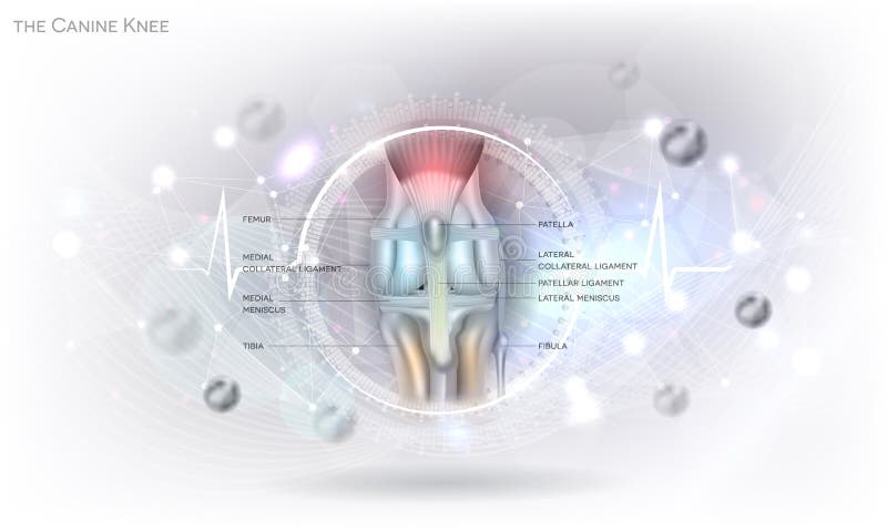
Free with trial Dog knee. Anatomy of the canine dog`s knee joint colorful design on an abstract glowing light grey scientific background. Tibia vectors Dog knee. Anatomy of the canine dog`s knee joint colorful design on an abstract glowing light grey scientific background
Free with trial Luxating patella in dogs, dislocated knee cap detailed info graphic poster, the severity of the condition, grades, diagnostic and treatment etc. beautiful aesthetic design. Tibia vectors Luxating patella in dogs. Dislocated knee cap detailed info graphic poster, the severity of the condition, grades, diagnostic and treatment etc. beautiful
Free with trial Extensor digitorum brevis muscle for foot phalanges movement outline diagram. Labeled educational scheme with feet anatomy and skeleton structure vector illustration. Physiological anterior view. Tibia vectors Extensor digitorum brevis muscle for foot phalanges movement outline diagram. Labeled educational scheme with feet anatomy and skeleton structure vector
Free with trial Vector illustration of Arthritis of an ankle joint. Front view of the human foot with pain. EPS 10. Tibia vectors Arthritis of the ankle joint. Vector illustration of Arthritis of an ankle joint. Front view of the human foot with pain. EPS 10. Vector illustration of Arthritis of an ankle joint. Front view of the human foot with pain. EPS 10.
Free with trial Krater `Centaur and Maenad`, Ancient Greek vase. For watering down wine, red figure style amphora vase painting vector illustration. Tibia vectors Krater `Centaur and Maenad`, Ancient Greek vase. For watering down wine, red figure style amphora vase painting vector illustration
Free with trial Vector illustration of a healthy human knee joint and unhealthy knee with horizontal tear of meniscus and meniscal cyst. Front, anterior view of knee. For advertising and medical publications. EPS 10. Tibia vectors Meniscus _Meniscal cyst front view. Vector illustration of a healthy human knee joint and unhealthy knee with horizontal tear of meniscus and meniscal cyst. Vector illustration of a healthy human knee joint and unhealthy knee with horizontal tear of meniscus and meniscal cyst. Front, anterior view of knee. For advertising and medical publications. EPS 10
Free with trial A medical illustrations showing the symptoms of patellar dislocation. Tibia vectors Patellar dislocation. A medical illustrations showing the symptoms of patellar dislocation. A medical illustrations showing the symptoms of patellar dislocation.
Free with trial Arteries of the foot dorsal and plantar view of an ankle. diagram. Tibia vectors Arteries of the foot. Dorsal and plantar view of an ankle. diagram
Free with trial Calcaneal spur. orthopedics. Vector illustration of a heel spur. Tibia vectors Calcaneal spur. orthopedics. Vector illustration of a heel spur. orthopedics
Free with trial Vector illustration. Anatomy of a meniscus in the healthy human knee joint. Peripheral meniscal tears with cross section of the menisci. For advertising, medical publications. EPS 10. Tibia vectors Meniscus _Peripheral tears. Vector illustration. Anatomy of a meniscus in the healthy human knee joint. Peripheral meniscal tears with cross section of the. Vector illustration. Anatomy of a meniscus in the healthy human knee joint. Peripheral meniscal tears with cross section of the menisci. For advertising, medical publications. EPS 10.
Free with trial Knee. Art illustration of leg with a highlight for the kneecap (patella. Tibia vectors Knee. Art illustration of leg with a highlight for the kneecap (patella
Free with trial Set of human knee and elbow, ankle and shoulder joints, wrist and spine and tooth, anatomy icons, emblem or sign of medical diagnostic center or clinic, flat design, vector. Tibia vectors Set of humane joints. Set of human knee and elbow, ankle and shoulder joints, wrist and spine and tooth, anatomy icons, emblem or sign of medical diagnostic. Set of human knee and elbow, ankle and shoulder joints, wrist and spine and tooth, anatomy icons, emblem or sign of medical diagnostic center or clinic, flat design, vector
Free with trial Vector illustration of a human leg with denominations of the bones of the foot. Anatomy of lateral or side view of the foot. For advertising or medical publications. EPS 10. Tibia vectors Anatomy_bones of the human foot lateral view. Vector illustration of a human leg with denominations of the bones of the foot. Anatomy of lateral or side view of. Vector illustration of a human leg with denominations of the bones of the foot. Anatomy of lateral or side view of the foot. For advertising or medical publications. EPS 10.
Free with trial Structure of a human knee joint. Knee anatomy. structure of a human knee joint: Articular cartilage, Bones, ligaments, tendons, and Meniscus. Vector poster. Tibia vectors Structure of a human knee joint. Knee anatomy. structure of a human knee joint: Articular cartilage, Bones, ligaments, tendons, and Meniscus. Vector poster
Free with trial Joint. Human skeleton, medicine. 3d vector icon. On white background. Tibia vectors Joint. Human skeleton, medicine. 3d vector icon. On white background
Free with trial Vector illustration anatomy of a healthy knee joint. Anterior or front view of flexed knee and posterior or back view of straight knee. For advertising or medical publications. EPS 10. Tibia vectors Ligaments of the knee_Anatomy. Vector illustration anatomy of a healthy knee joint. Anterior or front view of flexed knee and posterior or back view of straight. Vector illustration anatomy of a healthy knee joint. Anterior or front view of flexed knee and posterior or back view of straight knee. For advertising or medical publications. EPS 10.
Free with trial The canine knee blue background. Anatomy of the healthy canine dog`s knee joint health care concept design on an abstract blue scientific background. Tibia vectors The canine knee blue background. Anatomy of the healthy canine dog`s knee joint health care concept design on an abstract blue scientific background
Free with trial Disease of the joints and bones,human joints, knee joint, elbow joint, ankle joint, wrist, skeletal spinal bone structure of Human Spine, medical healthcare flat vector illustration. Tibia vectors Disease of the joints and bones. Disease of the joints and bones,human joints, knee joint, elbow joint, ankle joint, wrist, skeletal spinal bone structure of. Disease of the joints and bones,human joints, knee joint, elbow joint, ankle joint, wrist, skeletal spinal bone structure of Human Spine, medical healthcare flat vector illustration.
Free with trial Disease of the joints and bones, medical health care flat vector illustration. Tibia vectors Disease of the joints and bones. Disease of the joints and bones, medical health care flat vector illustration. Disease of the joints and bones, medical health care flat vector illustration.
Free with trial Arthrosis medical vector illustration diagram with damaged knee structure and healthy knee comparison. Bone exposure, osteophytes, cartilage fragments, erosion of cartilage and inflamed joint capsule. Tibia vectors Arthrosis medical vector illustration diagram with damaged knee structure and healthy knee comparison. Bone exposure, osteophytes, cartilage fragments, erosion
Free with trial Arthrosis medical vector illustration diagram with damaged knee structure and healthy knee comparison. Bone exposure, osteophytes, cartilage fragments, erosion of cartilage and inflamed joint capsule. Tibia vectors Arthrosis medical vector illustration diagram with damaged knee structure and healthy knee comparison. Bone exposure, osteophytes, cartilage fragments, erosion
Free with trial Vector illustration of Peroneal Tendon Injuries. Longitudinal tear of the peroneus brevis tendon. Lateral ankle injury. Tibia vectors Peroneal Tendon Injuries_Longitudinal tear of the peroneus brevis tendon. Vector illustration of Peroneal Tendon Injuries. Longitudinal tear of the peroneus. Vector illustration of Peroneal Tendon Injuries. Longitudinal tear of the peroneus brevis tendon. Lateral ankle injury.
Free with trial Knee anatomy. Human joint structure. Lateral and Front aspects of right knee. Vector poster. Tibia vectors Knee anatomy. Human joint structure. Lateral and Front aspects of right knee. Vector poster
 Sign in