Free with trial Nervous System. Vector illustration of diagram of nervous system. Tibial vectors Nervous System. Vector illustration of diagram of nervous system
Free with trial Front view of the right knee in flexion. Anatomy of the front of the right knee in flexion. Tibial vectors Front view of the right knee in flexion. Anatomy of the front of the right knee in flexion
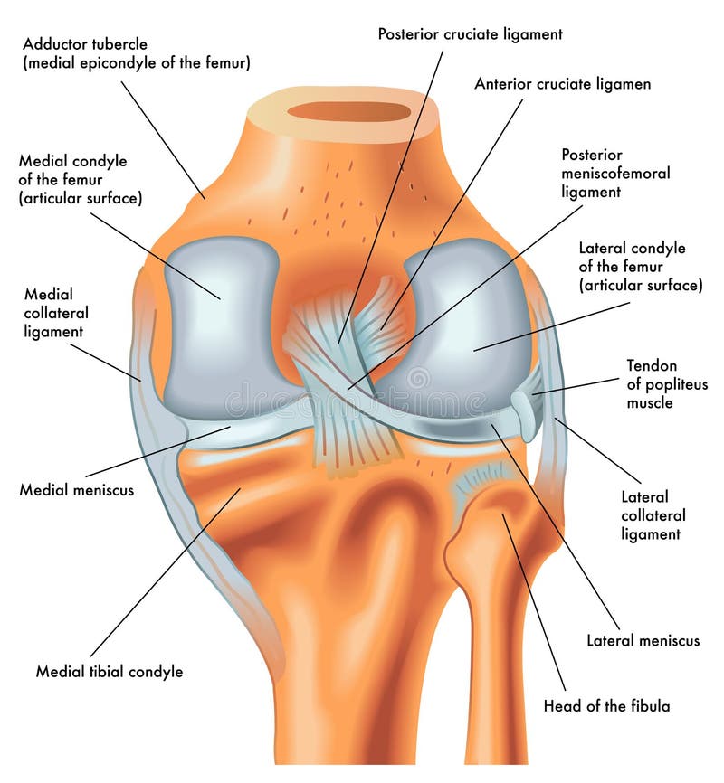
Free with trial Posterior view of the right knee in extension. Anatomy of the posterior of the right knee in extension. Tibial vectors Posterior view of the right knee in extension. Anatomy of the posterior of the right knee in extension
Free with trial Peripheral nervous system, medical vector illustration diagram with brain, spinal cord and full body nerve scheme. Tibial vectors Peripheral nervous system, medical vector illustration diagram with full body nerve scheme. Peripheral nervous system, medical vector illustration diagram with. Peripheral nervous system, medical vector illustration diagram with brain, spinal cord and full body nerve scheme.
Free with trial Components of knee replacement surgery joint. Human body parts of Anatomy. Health care education infographic. Vector design. Tibial vectors Components of knee replacement surgery joint. Human body parts of Anatomy. Health care education infographic. Vector design
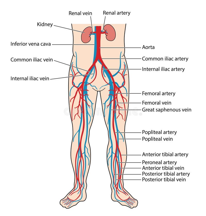
Free with trial The main veins and arteries of the lower body, including the abdominal aorta, inferior vena cava, femoral artery and vein to the anterior and posterior tibial artery and vein of the lower leg. Tibial vectors Blood vessels of the lower body. The main veins and arteries of the lower body, including the abdominal aorta, inferior vena cava, femoral artery and vein to the. The main veins and arteries of the lower body, including the abdominal aorta, inferior vena cava, femoral artery and vein to the anterior and posterior tibial artery and vein of the lower leg
Free with trial The major arteries and pulse points of the head, arms and legs. Created in Adobe Illustrator. Tibial vectors Major pulse points. The major arteries and pulse points of the head, arms and legs. Created in Adobe Illustrator. The major arteries and pulse points of the head, arms and legs. Created in Adobe Illustrator.
Free with trial Components of a Knee Replacement. Total Knee Replacement. Knee implant. Vector. Tibial vectors Components of a Knee Replacement. Total Knee Replacement. Knee implant. Vector
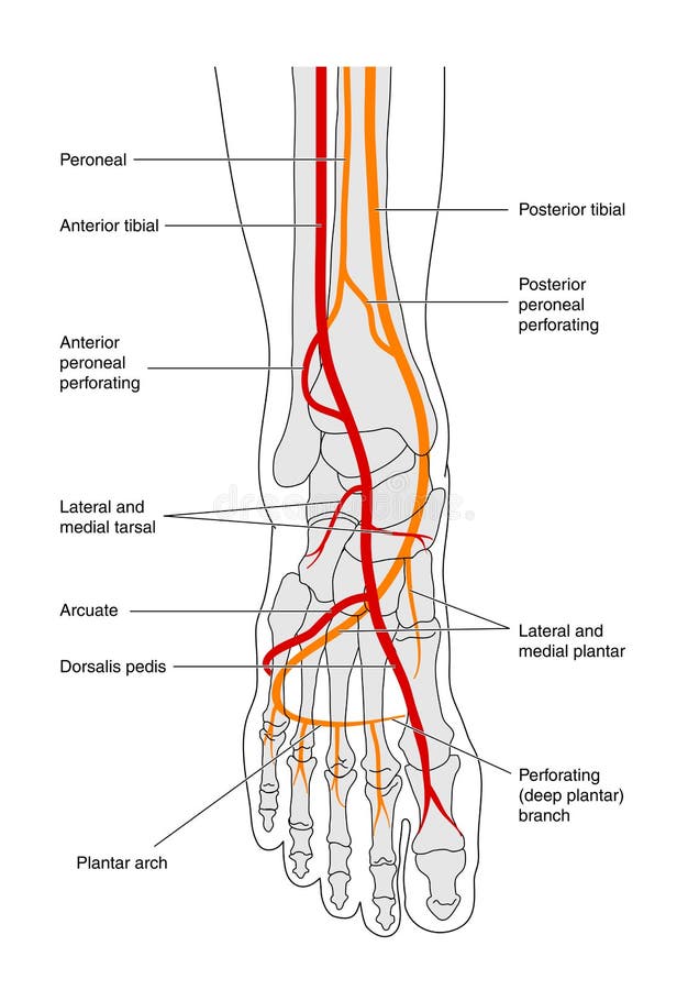
Free with trial Circulation of the foot - labelled. Blood supply to foot, showing bones and major blood vessels, labelled. Tibial vectors Circulation of the foot - labelled. Blood supply to foot, showing bones and major blood vessels, labelled
Free with trial Nerves of the foot. Vector illustration diagram of the nerves and cutaneous innervation of the human foot with palmar and dorsal view. Used transparency. Tibial vectors Nerves of the foot. Vector illustration diagram of the nerves and cutaneous innervation of the human foot with palmar and dorsal view. Used transparency
Free with trial Medial collateral ligament rupture. Medical Illustration of medial collateral ligament rupture. Tibial vectors Medial collateral ligament rupture. Medical Illustration of medial collateral ligament rupture
Free with trial Shin splints vector illustration. Leg muscle sport trauma and bone pain labeled diagram. Isolated femur, patella, fibula, tibia and foot bones with shown injury location. Tibial vectors Shin splints vector illustration. Leg muscle sport trauma and bone pain. Shin splints vector illustration. Leg muscle sport trauma and bone pain labeled diagram. Shin splints vector illustration. Leg muscle sport trauma and bone pain labeled diagram. Isolated femur, patella, fibula, tibia and foot bones with shown injury location.
Free with trial Vector illustration of healthy human foot and a medial ankle injury. Tarsal tunnel syndrome. EPS 10. Tibial vectors Medial ankle injury_Tarsal tunnel syndrome. Vector illustration of healthy human foot and a medial ankle injury. Tarsal tunnel syndrome. EPS 10. Vector illustration of healthy human foot and a medial ankle injury. Tarsal tunnel syndrome. EPS 10.
Free with trial Medically accurate vector illustration of human feet, includes nervous system, veins, arteries, etc. Tibial vectors Medically accurate vector illustration of human feet, includes nervous system, veins, arteries, etc.
Free with trial Foot circulation. Blood supply to foot, showing bones and major blood vessels. Tibial vectors Foot circulation. Blood supply to foot, showing bones and major blood vessels
Free with trial Vector illustration of healthy human foot and a medial ankle injury. Posterior tibial tendon dysfunction. EPS 10. Tibial vectors Medial ankle injury_Posterior tibial tendon dysfunction. Vector illustration of healthy human foot and a medial ankle injury. Posterior tibial tendon dysfunction. Vector illustration of healthy human foot and a medial ankle injury. Posterior tibial tendon dysfunction. EPS 10.
Free with trial Osgood schlatter disease condition with leg and knee joint pain outline diagram. Labeled educational medical scheme with patella tendon inflammation and tibial tuberosity anatomy vector illustration. Tibial vectors Osgood schlatter disease condition with knee joint pain outline diagram. Osgood schlatter disease condition with leg and knee joint pain outline diagram. Labeled. Osgood schlatter disease condition with leg and knee joint pain outline diagram. Labeled educational medical scheme with patella tendon inflammation and tibial tuberosity anatomy vector illustration.
Free with trial Vector illustration of healthy human foot and a medial ankle injury. Posterior tibial tendonitis. EPS 10. Tibial vectors Medial ankle injury_Posterior tibial tendonitis. Vector illustration of healthy human foot and a medial ankle injury. Posterior tibial tendonitis. EPS 10. Vector illustration of healthy human foot and a medial ankle injury. Posterior tibial tendonitis. EPS 10.
Free with trial Vector illustration of healthy human foot and a medial ankle injury. Posterior tibial tendon rupture. EPS 10. Tibial vectors Medial ankle injury_Posterior tibial tendon rupture. Vector illustration of healthy human foot and a medial ankle injury. Posterior tibial tendon rupture. EPS 10. Vector illustration of healthy human foot and a medial ankle injury. Posterior tibial tendon rupture. EPS 10.
Free with trial Partial knee replacement. unicompartmental knee arthroplasty and uni knee implant. Tibial vectors Partial knee replacement. Unicompartmental knee arthroplasty and uni knee implant
Free with trial Major veins of the body. Venous network, showing the major veins from the periphery to the heart. Created in Adobe Illustrator. EPS 10. Tibial vectors Major veins of the body. Venous network, showing the major veins from the periphery to the heart. Created in Adobe Illustrator. EPS 10
Free with trial Doppler probe measurement of pedal pulse, showing the major pedal pulse points with named arteries. Created in Adobe Illustrator. Contains transparencies. EPS 10. Tibial vectors Doppler probe measurement of pedal pulse. Showing the major pedal pulse points with named arteries. Created in Adobe Illustrator. Contains transparencies. EPS
Free with trial A medical illustration with a normal knee and a dislocated knee. Tibial vectors Dislocation of knee. A medical illustration with a normal knee and a dislocated knee. A medical illustration with a normal knee and a dislocated knee.
Free with trial Vector illustration of a healthy bones of human leg and a leg with tibial shaft fracture. Twisting, blunt trauma injury. Front view of the foot with knee. For advertising, medical publications. EPS 10. Tibial vectors Bone fracture_Tibial shaft fracture. Vector illustration of a healthy bones of human leg and a leg with tibial shaft fracture. Twisting, blunt trauma injury. Vector illustration of a healthy bones of human leg and a leg with tibial shaft fracture. Twisting, blunt trauma injury. Front view of the foot with knee. For advertising, medical publications. EPS 10
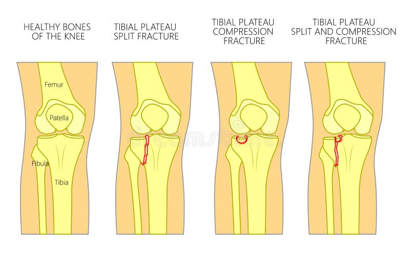
Free with trial Vector illustration of a healthy bones of human knee and a knee with tibial plateau split, compression or depression fractures. Front view of the knee. For advertising, medical publications. EPS 10. Tibial vectors Bone fracture_Tibial plateau fracture. Vector illustration of a healthy bones of human knee and a knee with tibial plateau split, compression or depression. Vector illustration of a healthy bones of human knee and a knee with tibial plateau split, compression or depression fractures. Front view of the knee. For advertising, medical publications. EPS 10.
Free with trial Vector illustration. Front view of a healthy human knee, anatomy of a proximal surface of tibial plateau with soft tissues and blood supply of meniscus. For advertising, medical publications. EPS 8. Tibial vectors Meniscus _Blood supply_Superior view. Vector illustration. Front view of a healthy human knee, anatomy of a proximal surface of tibial plateau with soft tissues. Vector illustration. Front view of a healthy human knee, anatomy of a proximal surface of tibial plateau with soft tissues and blood supply of meniscus. For advertising, medical publications. EPS 8
Free with trial Medical illustration showing arthrosis of the knee with annotations next to total knee replacement diagram. Tibial vectors Arthrosis medical illustration. Medical illustration showing arthrosis of the knee with annotations next to total knee replacement diagram. Medical illustration showing arthrosis of the knee with annotations next to total knee replacement diagram.
Free with trial Osgood–Schlatter disease. Healthy Joint and Apophysitis of the tibial tubercle. Vector illustration. Poster for medical use. Tibial vectors Osgood–Schlatter disease. Healthy Joint and Apophysitis of the tibial tubercle. Vector illustration. Poster for medical use
Free with trial Vector illustration of healthy adolescent knee joint and unhealthy knee with avulsion of tibial tuberosity. Lateral or side view of the leg. For advertising and other medical publications. EPS 10. Tibial vectors Knee problem_Avulsion of tibial tuberosity. Vector illustration of healthy adolescent knee joint and unhealthy knee with avulsion of tibial tuberosity. Lateral. Vector illustration of healthy adolescent knee joint and unhealthy knee with avulsion of tibial tuberosity. Lateral or side view of the leg. For advertising and other medical publications. EPS 10.
Free with trial Osgood–Schlatter disease. inflammation of the patellar ligament at the tibial tuberosity. Anatomy of human knee femur, patella, patellar ligament, tibia and fibula. vector illustration. leg bones and joint. Side and front anterior view of the knee. Tibial vectors Osgood–Schlatter disease. inflammation of the patellar ligament in knee. Osgood–Schlatter disease. inflammation of the patellar ligament at the tibial. Osgood–Schlatter disease. inflammation of the patellar ligament at the tibial tuberosity. Anatomy of human knee femur, patella, patellar ligament, tibia and fibula. vector illustration. leg bones and joint. Side and front anterior view of the knee
Free with trial Anatomy of human knee vector sketch of leg bones and joint, medicine design. Side and front view of knee bones, hand drawn femur, patella, tibia and fibula, tibial plateau and lateral condyle. Tibial vectors Knee bones and joint, vector sketch human anatomy. Anatomy of human knee vector sketch of leg bones and joint, medicine design. Side and front view of knee bones. Anatomy of human knee vector sketch of leg bones and joint, medicine design. Side and front view of knee bones, hand drawn femur, patella, tibia and fibula, tibial plateau and lateral condyle
Free with trial Anatomy Of Knee Joint Medical Infographic Poster. Patella, Femur, Articular Cartilage. External or Internal Meniscus, Tibia, Patellar, Tibial, Collagen Ligament. Anterior Ligament of the Fibular Head. Tibial vectors Anatomy Of Knee Joint Medical Infographics. Patella, Femur, Articular Cartilage. External Or Internal Meniscus, Tibia. Anatomy Of Knee Joint Medical Infographic. Anatomy Of Knee Joint Medical Infographic Poster. Patella, Femur, Articular Cartilage. External or Internal Meniscus, Tibia, Patellar, Tibial, Collagen Ligament. Anterior Ligament of the Fibular Head
Free with trial Human Knee Implant illustration. Human Knee Implant vector diagram illustration. Tibial vectors Human Knee Implant illustration. Human Knee Implant vector diagram illustration
Free with trial Pulses. A diagram depicting the pulses of the body. Tibial vectors Pulses. A diagram depicting the pulses of the body
Free with trial Too many toes. Vector illustration of the normal human foot and the foot with pronation or flatfoot, with hindfoot deformity. Too many toes sign. Tibial vectors Too many toes. Vector illustration of the normal human foot and the foot with pronation or flatfoot, with hindfoot deformity. Too many toes sign
Free with trial Arteries of the foot dorsal and plantar view of an ankle. diagram. Tibial vectors Arteries of the foot. Dorsal and plantar view of an ankle. diagram
Free with trial The main veins of the leg, from the foot to the inferior vena cava, including the femoral and iliac veins. Tibial vectors Main veins of the leg. The main veins of the leg, from the foot to the inferior vena cava, including the femoral and iliac veins. The main veins of the leg, from the foot to the inferior vena cava, including the femoral and iliac veins.
Free with trial Nervous system of human body with nerve network anatomy outline diagram. Labeled educational medical scheme with CNS brain structure and peripheral inner cord, plexus and nerves vector illustration. Tibial vectors Nervous system of human body with nerve network anatomy outline diagram. Labeled educational medical scheme with CNS brain structure and peripheral inner cord
Free with trial Total knee implant. Close-up of a Healthy knee joint and joint with implant. Vector poster. Tibial vectors Total knee implant. Close-up of a Healthy knee joint and joint with implant. Vector poster
Free with trial Vector illustration of a human legs anterior and medial view with shin pain. Vector illustration for advertising, medical health care publications. EPS 10. Tibial vectors Bone fracture_Shin pain-09. Vector illustration of a human legs anterior and medial view with shin pain. Vector illustration for advertising, medical health care. Vector illustration of a human legs anterior and medial view with shin pain. Vector illustration for advertising, medical health care publications. EPS 10
Free with trial Chondromalacia patella knee breakdown compared with healthy outline diagram. Labeled educational kneecap tissue damage with cartilage problem and anatomical leg joint structure vector illustration. Tibial vectors Chondromalacia patella knee breakdown compared with healthy outline diagram. Labeled educational kneecap tissue damage with cartilage problem and anatomical
Free with trial Vector illustration. Anatomy of a proximal surface of the tibia with soft tissues in the healthy human knee joint. For advertising and medical publications. EPS 10. Tibial vectors Superior aspect of the tibial plateau. Vector illustration. Anatomy of a proximal surface of the tibia with soft tissues in the healthy human knee joint. For. Vector illustration. Anatomy of a proximal surface of the tibia with soft tissues in the healthy human knee joint. For advertising and medical publications. EPS 10.
Free with trial Medical illustration of the cutaneous innervation of the foot, with annotations. Tibial vectors Cutaneous innervation of the foot. Medical illustration of the cutaneous innervation of the foot, with annotations. Medical illustration of the cutaneous innervation of the foot, with annotations.
Free with trial A vector medical illustration of the symptoms of a bone edema on a knee. Tibial vectors Medical illustration of bone edema on knee. A vector medical illustration of the symptoms of a bone edema on a knee. A vector medical illustration of the symptoms of a bone edema on a knee.
Free with trial The major arteries and pulse points on human body vector illustration. Tibial vectors The major arteries and pulse points on human body vector illustration.
Free with trial Medically accurate anatomy illustration of human feet legs with nervous and blood system. Educational x-ray style illustration. Tibial vectors Medically accurate anatomy illustration of human feet legs with nervous and blood system. Educational x-ray style illustration
Free with trial Medically accurate vector illustration of human feet, includes nervous system, veins, arteries, etc. Tibial vectors Medically accurate vector illustration of human back chest, includes nervous system, veins, arteries, heart, etc. Medically accurate vector illustration of. Medically accurate vector illustration of human feet, includes nervous system, veins, arteries, etc.
Free with trial The Sciatic Nerve. Sensory innervation of the sciatic nerve. Sciatica. Medical Illustration. Tibial vectors The Sciatic Nerve. Sensory innervation of the sciatic nerve. Sciatica. Medical Illustration
Free with trial Female and male skeleton differences poster. Q angle in comparison. Major gender nuances. Vector illustration on a dark grey background. Tibial vectors Female and male skeleton differences. Poster. Q angle in comparison. Major gender nuances. Vector illustration on a dark grey background
Free with trial Central nervous system anatomical diagram. Nerves send electrical signals to and from brain and spinal cord in human body. CNS and PNS concept. Medical poster for neurology clinic vector illustration. Tibial vectors Central nervous system. Anatomical diagram. Nerves send electrical signals to and from brain and spinal cord in human body. CNS and PNS concept. Medical poster. Central nervous system anatomical diagram. Nerves send electrical signals to and from brain and spinal cord in human body. CNS and PNS concept. Medical poster for neurology clinic vector illustration.
Free with trial Osteochondroma knee problem as medical bone tumor overgrowth outline diagram. Labeled educational scheme with leg pathology explanation vector illustration. Femur cross section anatomical structure. Tibial vectors Osteochondroma knee problem as medical bone tumor overgrowth outline diagram. Labeled educational scheme with leg pathology explanation vector illustration
Free with trial Central nervous system anatomical diagram. Nerves send electrical signals to and from brain and spinal cord in human body. CNS and PNS concept. Medical poster for neurology clinic vector illustration. Tibial vectors Central nervous system. Anatomical diagram. Nerves send electrical signals to and from brain and spinal cord in human body. CNS and PNS concept. Medical poster. Central nervous system anatomical diagram. Nerves send electrical signals to and from brain and spinal cord in human body. CNS and PNS concept. Medical poster for neurology clinic vector illustration.
Free with trial Central nervous system anatomical diagram. Nerves send electrical signals to and from brain and spinal cord in human body. CNS and PNS concept. Medical poster for neurology clinic vector illustration. Tibial vectors Central nervous system. Anatomical diagram. Nerves send electrical signals to and from brain and spinal cord in human body. CNS and PNS concept. Medical poster. Central nervous system anatomical diagram. Nerves send electrical signals to and from brain and spinal cord in human body. CNS and PNS concept. Medical poster for neurology clinic vector illustration.
Free with trial Medical Vector Infographics Depicting The Knee Joint Anatomy, Including Bones, Ligaments, And Tendons And Cartilage, Illustrating Its Structure And Function For Medical Education Purposes. Tibial vectors Medical Vector Infographics Depicting The Knee Joint Anatomy, Including Bones, Ligaments, And Tendons And Cartilage. Illustrating Its Structure And Function. Medical Vector Infographics Depicting The Knee Joint Anatomy, Including Bones, Ligaments, And Tendons And Cartilage, Illustrating Its Structure And Function For Medical Education Purposes
Free with trial Deep plantar region, vintage engraving. Deep plantar region, vintage engraved illustration. Usual Medicine Dictionary - Paul Labarthe - 1885. Tibial vectors Deep plantar region, vintage engraving. Deep plantar region, vintage engraved illustration. Usual Medicine Dictionary - Paul Labarthe - 1885
Free with trial The major arteries and pulse points on human body. Heartbeat evaluation. Placements. Cardiovascular system. Flat vector illustration. Tibial vectors The major arteries and pulse points on human body. Heartbeat evaluation. Placements. Cardiovascular system. Flat vector illustration
Free with trial Central nervous system 3d realistic vector illustration. Nerves send electrical signals to and from brain and spinal cord in human body. CNS and PNS concept. X ray medical poster for neurology clinic. Tibial vectors Central nervous system. 3d realistic vector illustration. Nerves send electrical signals to and from brain and spinal cord in human body. CNS and PNS concept. X. Central nervous system 3d realistic vector illustration. Nerves send electrical signals to and from brain and spinal cord in human body. CNS and PNS concept. X ray medical poster for neurology clinic.
Free with trial Educational medical illustration of leg bones. And knee. Tibial vectors Educational medical illustration of leg bones. And knee
Free with trial 3d illustration of posterior collateral ligament of human knee bone on dark blue background. Used in medicine, education, science and industry. Tibial vectors 3d illustration of posterior collateral ligament of human knee bone on dark blue background. Used in medicine, education, science and industry
Free with trial Tibial Nerve Schwannoma Illustration in Human Leg Anatomy. Medical Diagram of Schwannoma on Tibial Nerve Peripheral Nerve Tumor. Visualization. Tibial vectors Tibial Nerve Schwannoma Illustration in Human Leg Anatomy. Medical Diagram of Schwannoma on Tibial Nerve Peripheral Nerve Tumor. Visualization
Free with trial Educational podiatry and orthopedics detailing tarsal tunnel syndrome compared to a normal tarsal tunnel in full color. The labeled cross-sectional anatomical diagram illustrates the medial ankle region beneath the flexor retinaculum, identifying the medial malleolus, tendons, and posterior tibial nerve. It clearly contrasts an uncompressed normal tarsal tunnel against tarsal tunnel syndrome characterized by localized swelling, inflamed surrounding tendons, and compression of the posterior tibial nerve. High-quality infographic graphic ideal for podiatry clinic posters, orthopedic textbooks, physical therapy study guides, sports medicine manuals, medical school lecture slides, and patient healthcare education presentations. Tibial vectors Tarsal Tunnel Syndrome vs Normal Foot Anatomy Posterior Tibial Nerve Compression Color Vector Illustration. Educational podiatry and orthopedics detailing tarsal. Educational podiatry and orthopedics detailing tarsal tunnel syndrome compared to a normal tarsal tunnel in full color. The labeled cross-sectional anatomical diagram illustrates the medial ankle region beneath the flexor retinaculum, identifying the medial malleolus, tendons, and posterior tibial nerve. It clearly contrasts an uncompressed normal tarsal tunnel against tarsal tunnel syndrome characterized by localized swelling, inflamed surrounding tendons, and compression of the posterior tibial nerve. High-quality infographic graphic ideal for podiatry clinic posters, orthopedic textbooks, physical therapy study guides, sports medicine manuals, medical school lecture slides, and patient healthcare education presentations.
Free with trial Knee osgood schlatter joint knee disease. Labeled inflamed patellar ligament Tibial tuberosity inflammation and traction apophysitis Synovial joint anatomy. Frontal and side view. Vector illustration. Tibial vectors Knee osgood schlatter joint knee disease. Labeled inflamed patellar ligament Tibial tuberosity inflammation and traction. Apophysitis Synovial joint anatomy. Knee osgood schlatter joint knee disease. Labeled inflamed patellar ligament Tibial tuberosity inflammation and traction apophysitis Synovial joint anatomy. Frontal and side view. Vector illustration
Free with trial Tibial Nerve Tumor Anatomy. Peripheral Nerve Sheath Tumor in Leg. Schwannoma Medical Visualization. Detailed medical illustration showing the surgical removal of a benign tumor from a tibial nerve. Tibial vectors Tibial Nerve Tumor Anatomy. Peripheral Nerve Sheath Tumor in Leg. Schwannoma Medical Visualization. Detailed medical illustration. Showing the surgical removal. Tibial Nerve Tumor Anatomy. Peripheral Nerve Sheath Tumor in Leg. Schwannoma Medical Visualization. Detailed medical illustration showing the surgical removal of a benign tumor from a tibial nerve.
Free with trial Exploded anatomical view showing femoral component, tibial implant, polyethylene insert and patella replacement for orthopedic. Education and surgery. Tibial vectors Exploded anatomical view showing femoral component, tibial implant, polyethylene insert and patella replacement for orthopedic. Education and surgery
Free with trial Skeletal traction sites on the human body, including proximal tibial, calcaneal, and metacarpal areas diagram hand drawn schematic vector illustration. Medical science educational illustration. Tibial vectors Skeletal Traction Sites diagram medical science. Skeletal traction sites on the human body, including proximal tibial, calcaneal, and metacarpal areas diagram. skeletal traction sites on the human body, including proximal tibial, calcaneal, and metacarpal areas diagram hand drawn schematic vector illustration. Medical science educational illustration
Free with trial Healthy foot and a foot with Sesamoid boneS fracture. Vector illustration for medical publiatios. Tibial vectors Healthy foot and a foot with Sesamoid boneS fracture. Vector illustration for medical publiatios
Free with trial Compartment syndrome brief anatomy illustrates a leg cross-section with normal vs swollen muscle and fasciotomy relief, focusing on muscles, nerves, and vessels. Outline diagram. Tibial vectors Compartment syndrome brief anatomy illustrates a leg cross-section with. Normal vs swollen muscle and fasciotomy relief, focusing on muscles, nerves, and. Compartment syndrome brief anatomy illustrates a leg cross-section with normal vs swollen muscle and fasciotomy relief, focusing on muscles, nerves, and vessels. Outline diagram
Free with trial Medical displaying an anatomical comparison between normal leg alignment and bowed lower limbs, highlighting genu varum, rickets, osteomalacia, knock knees (genu valgum), achondroplasia, and tibial torsion for orthopedic diagnosis, pediatric skeletal development, and physiotherapy awareness concepts. Tibial vectors Genu Varum Bow Legs Alignment Vector. Medical displaying an anatomical comparison between normal leg alignment and bowed lower limbs, highlighting genu varum. Medical displaying an anatomical comparison between normal leg alignment and bowed lower limbs, highlighting genu varum, rickets, osteomalacia, knock knees (genu valgum), achondroplasia, and tibial torsion for orthopedic diagnosis, pediatric skeletal development, and physiotherapy awareness concepts.
Free with trial Diagram illustrating skeletal traction, featuring two views of a human knee joint. The left side shows a pin inserted laterally through the femur, indicated by a hand drill and text noting "2-3 fingers" for spacing. The right side labels "femoral entry point" and "tibial entry point," depicting entry points on the femur and tibia. This is used in orthopedic procedures to align and stabilize fractures by applying tension via an external device. The bones are simplified and labeled for clarity. Tibial vectors Skeletal Traction Illustration. Diagram illustrating skeletal traction, featuring two views of a human knee joint. The left side shows a pin inserted laterally. Diagram illustrating skeletal traction, featuring two views of a human knee joint. The left side shows a pin inserted laterally through the femur, indicated by a hand drill and text noting "2-3 fingers" for spacing. The right side labels "femoral entry point" and "tibial entry point," depicting entry points on the femur and tibia. This is used in orthopedic procedures to align and stabilize fractures by applying tension via an external device. The bones are simplified and labeled for clarity.
Free with trial Educational vector illustration of the common peroneal nerve anatomy, highlighting its pathway through the lower leg and foot. Ideal for neurology, orthopaedics, anatomy education, rehabilitation, medical textbooks, and healthcare presentations. Tibial vectors Common Peroneal Nerve Anatomy Lower Leg and Foot Medical Vector Illustration. Educational vector illustration of the common peroneal nerve anatomy, highlighting. Educational vector illustration of the common peroneal nerve anatomy, highlighting its pathway through the lower leg and foot. Ideal for neurology, orthopaedics, anatomy education, rehabilitation, medical textbooks, and healthcare presentations.
Free with trial Nerve Conduction Study (NCS). Medical diagnostic procedure illustration. Tibial vectors Nerve Conduction Study (NCS). Medical diagnostic procedure illustration
Free with trial This vector shows a detailed human knee joint with highlighted bones and ligaments in orange, illustrating anatomy clearly and educationally. Informative mood. Generative AI. Tibial vectors Human Knee Joint Anatomy Medical Illustration with Highlighted Bones and Ligaments Vector. This vector shows a detailed human knee joint with highlighted bones. This vector shows a detailed human knee joint with highlighted bones and ligaments in orange, illustrating anatomy clearly and educationally. Informative mood. Generative AI
Free with trial Highly detailed 3D illustration of a total knee replacement prosthesis isolated on a white background for orthopedic surgery, medical education and healthcare. Tibial vectors 3D illustration of a total knee replacement prosthesis isolated on a white background for orthopedic surgery,. Highly detailed 3D illustration of a total knee. Highly detailed 3D illustration of a total knee replacement prosthesis isolated on a white background for orthopedic surgery, medical education and healthcare
Free with trial This detailed anatomical shows the human foot and ankle skeletal structure, highlighting the complex arrangement of bones and joints in a clear side profile view. Tibial vectors Anatomical of the human foot and ankle skeletal structure showing bones and joints in a side profile view for medical. This detailed anatomical shows the human. This detailed anatomical shows the human foot and ankle skeletal structure, highlighting the complex arrangement of bones and joints in a clear side profile view
Free with trial Osgood Schlatter disease or OSD is inflammation of the patellar ligament at the tibial tuberosity. Apophysitis. Tibial vectors Osgood Schlatter disease or OSD is inflammation of the patellar ligament at the tibial tuberosity. Apophysitis
Free with trial Fracture tibial plateau and knee icon. Vector flat design for radiology orthopedic research hospital for body joints, orthopedic, sport medicine, diagnostics or corrective therapy. Vector illustration. Tibial vectors Fracture tibial plateau and knee icon. Vector flat design for radiology orthopedic research hospital. For body joints, orthopedic, sport medicine, diagnostics. Fracture tibial plateau and knee icon. Vector flat design for radiology orthopedic research hospital for body joints, orthopedic, sport medicine, diagnostics or corrective therapy. Vector illustration
Free with trial Large tibial white bone. 3D vector accessory for Halloween. Part of skeleton. Isolated object with shadow. Element for scary night concept. Vector design. Tibial vectors Large tibial white bone. 3D vector accessory for Halloween. Part of skeleton. Isolated object with shadow. Element for scary night concept. Vector design
Free with trial Dog foodstuff icon, concept label hound plaything stuff toy bone, two skeleton tibial item isolated on white, flat vector illustration. Doggie food product sticker, animal care different item. Tibial vectors Dog foodstuff icon, concept label hound plaything stuff toy bone, two skeleton tibial item isolated on white, flat. Vector illustration. Doggie food product. Dog foodstuff icon, concept label hound plaything stuff toy bone, two skeleton tibial item isolated on white, flat vector illustration. Doggie food product sticker, animal care different item.
Free with trial Osgood-Schlatter disease. Destruction of the tibial nucleus. Human leg on the side. Medical poster, infographics. Vector illustration. Tibial vectors Osgood-Schlatter disease. Destruction of the tibial nucleus. Human leg on the side. Medical poster, infographics. Vector illustration
Free with trial Stifle Joint Ligaments where internal lateral femoro tibial ligament is present, vintage line drawing or engraving illustration. Tibial vectors Stifle Joint Ligaments, vintage illustration. Stifle Joint Ligaments where internal lateral femoro tibial ligament is present, vintage line drawing or engraving. Stifle Joint Ligaments where internal lateral femoro tibial ligament is present, vintage line drawing or engraving illustration
 Sign in