Free with trial Vector illustration of Peroneal Tendon Injuries. Peroneal tendonitis. Inflammation of peroneal tendons. Lateral ankle injury. Tibialis vectors Peroneal Tendon Injuries_Peroneal tendonitis. Vector illustration of Peroneal Tendon Injuries. Peroneal tendonitis. Inflammation of peroneal tendons. Lateral. Vector illustration of Peroneal Tendon Injuries. Peroneal tendonitis. Inflammation of peroneal tendons. Lateral ankle injury.
Free with trial Vector illustration of healthy human foot and a medial ankle injury. Tibialis anterior tendonitis. EPS 10. Tibialis vectors Medial ankle injury_Tibialis anterior tendonitis. Vector illustration of healthy human foot and a medial ankle injury. Tibialis anterior tendonitis. EPS 10. Vector illustration of healthy human foot and a medial ankle injury. Tibialis anterior tendonitis. EPS 10.
Free with trial Vector illustration of Peroneal Tendon Injuries. Longitudinal tear of the peroneus brevis tendon. Lateral ankle injury. Tibialis vectors Peroneal Tendon Injuries_Longitudinal tear of the peroneus brevis tendon. Vector illustration of Peroneal Tendon Injuries. Longitudinal tear of the peroneus. Vector illustration of Peroneal Tendon Injuries. Longitudinal tear of the peroneus brevis tendon. Lateral ankle injury.
Free with trial Man doing foam roller anterior tibialis or shin stretch. Flat vector illustration isolated on white background. Tibialis vectors Man doing foam roller anterior tibialis or shin stretch. Flat vector illustration isolated on white background
Free with trial Woman doing foam roller anterior tibialis or shin stretch. Flat vector illustration isolated on white background. Tibialis vectors Woman doing foam roller anterior tibialis or shin stretch. Flat vector illustration isolated on white background
Free with trial Lower limb muscles diagram showing anterior and posterior views with labeled quadriceps, hamstrings, and gastrocnemius for study and anatomy reference. Outline diagram. Tibialis vectors Lower limb muscles diagram showing anterior and posterior views with ... Lower limb muscles diagram showing anterior and posterior views with labeled quadriceps. Lower limb muscles diagram showing anterior and posterior views with labeled quadriceps, hamstrings, and gastrocnemius for study and anatomy reference. Outline diagram
Free with trial Vector illustration of Peroneal Tendon Injuries. Peroneal tendon dislocation or subluxation when the superior peroneal retinaculum or retinacula is torn. Lateral ankle injury. Tibialis vectors Peroneal Tendon Injuries_Peroneal tendon dislocation. Vector illustration of Peroneal Tendon Injuries. Peroneal tendon dislocation or subluxation when the. Vector illustration of Peroneal Tendon Injuries. Peroneal tendon dislocation or subluxation when the superior peroneal retinaculum or retinacula is torn. Lateral ankle injury.
Free with trial Vector illustration of Peroneal Tendon Injuries. Rupture of the peroneus longus tendon. Lateral ankle injury. Tibialis vectors Peroneal Tendon Injuries_Rupture of the peroneus longus tendon. Vector illustration of Peroneal Tendon Injuries. Rupture of the peroneus longus tendon. Lateral. Vector illustration of Peroneal Tendon Injuries. Rupture of the peroneus longus tendon. Lateral ankle injury.
Free with trial Vector illustration of Peroneal Tendon Injuries. Degeneration of the peroneus longus and brevis tendons. Tendinosis or tendinopathy. Lateral ankle injury. Tibialis vectors Peroneal Tendon Injuries_Degeneration of the peroneus longus and. Vector illustration of Peroneal Tendon Injuries. Degeneration of the peroneus longus and brevis. Vector illustration of Peroneal Tendon Injuries. Degeneration of the peroneus longus and brevis tendons. Tendinosis or tendinopathy. Lateral ankle injury.
Free with trial Foot, Fig 1. Muscles, Fig 2. Skeleton, vintage engraved illustration. Dictionary of words and things - Larive and Fleury - 1895. Tibialis vectors Foot, Fig 1. Muscles, Fig 2. Skeleton, vintage engraving. Foot, Fig 1. Muscles, Fig 2. Skeleton, vintage engraved illustration. Dictionary of words and things. Foot, Fig 1. Muscles, Fig 2. Skeleton, vintage engraved illustration. Dictionary of words and things - Larive and Fleury - 1895
Free with trial Vector illustration. Front view of a human foot. Anterior ankle injury. Tibialis anterior tendonitis. For medical publications. Tibialis vectors Anterior ankle injury. Tibialis anterior tendonitis problem. Front view. Vector illustration. Front view of a human foot. Anterior ankle injury. Tibialis. Vector illustration. Front view of a human foot. Anterior ankle injury. Tibialis anterior tendonitis. For medical publications
Free with trial This detailed medical depicts the posterior and lateral views of the human lower leg and foot anatomy. It features labeled muscle groups including the gastrocnemius, soleus, and tibialis anterior, along with skeletal structures like the tibia and calcaneus, and connective tissues such as the Achilles tendon and fascia. Tibialis vectors Anatomical Diagram of the Human Lower Leg and Foot Muscles and Bones. This detailed medical depicts the posterior and lateral views of the human lower leg and. This detailed medical depicts the posterior and lateral views of the human lower leg and foot anatomy. It features labeled muscle groups including the gastrocnemius, soleus, and tibialis anterior, along with skeletal structures like the tibia and calcaneus, and connective tissues such as the Achilles tendon and fascia.
Free with trial A comprehensive anatomical diagram illustrating the muscular system of the human lower body. The graphic features four different perspectives: anterior, posterior, and lateral views. It highlights key muscle groups including the gluteus maximus, quadriceps femoris, hamstrings, gastrocnemius, and tibialis anterior, along with tendons and ligaments. This clear, detailed medical is ideal for educational materials, fitness training, and healthcare presentations. Tibialis vectors Detailed anatomical of the human leg muscles from front, back, and side views. A comprehensive anatomical diagram illustrating the muscular system of the human. A comprehensive anatomical diagram illustrating the muscular system of the human lower body. The graphic features four different perspectives: anterior, posterior, and lateral views. It highlights key muscle groups including the gluteus maximus, quadriceps femoris, hamstrings, gastrocnemius, and tibialis anterior, along with tendons and ligaments. This clear, detailed medical is ideal for educational materials, fitness training, and healthcare presentations.
Free with trial Body anatomy muscle set featuring biceps triceps brachioradialis deltoid pectoralis latissimus trapezius teres rhomboid abdominis oblique erector gluteus quadriceps hamstring. Tibialis vectors Body anatomy muscle set. Featuring biceps triceps brachioradialis deltoid pectoralis latissimus trapezius teres rhomboid abdominis oblique erector gluteus
Free with trial Body anatomy muscle set featuring biceps triceps brachioradialis deltoid pectoralis latissimus trapezius teres rhomboid abdominis oblique erector gluteus quadriceps hamstring. Tibialis vectors Body anatomy muscle set. Featuring biceps triceps brachioradialis deltoid pectoralis latissimus trapezius teres rhomboid abdominis oblique erector gluteus
Free with trial Body anatomy muscle set featuring biceps triceps brachioradialis deltoid pectoralis latissimus trapezius teres rhomboid abdominis oblique erector gluteus quadriceps hamstring. Tibialis vectors Body anatomy muscle set. Featuring biceps triceps brachioradialis deltoid pectoralis latissimus trapezius teres rhomboid abdominis oblique erector gluteus
Free with trial The image features a black and white illustration of an athletic male figure demonstrating a running motion. The figure is segmented into different sections, each labeled with the name of a specific muscle group, such as the deltoid, biceps, pectorals, quadriceps, and hamstrings. The labels highlight the muscles engaged during the activity and their anatomical locations. Tibialis vectors Athletic figure showcasing various muscle groups in motion. The image features a black and white illustration of an athletic male figure demonstrating a running. The image features a black and white illustration of an athletic male figure demonstrating a running motion. The figure is segmented into different sections, each labeled with the name of a specific muscle group, such as the deltoid, biceps, pectorals, quadriceps, and hamstrings. The labels highlight the muscles engaged during the activity and their anatomical locations
Free with trial Timed icon, communication, vector icon, perfect for scheduling, timing, or communication-related designs. Tibialis vectors Thigh and Calf Muscle Anatomy Outline Icon Vector Illustration. Timed icon, communication, vector icon, perfect for scheduling, timing, or communication-related. Timed icon, communication, vector icon, perfect for scheduling, timing, or communication-related designs.
Free with trial Thigh and calf muscle anatomy outline icon, a vector illustration showing the muscles of the thigh and calf in an anatomical diagram. Tibialis vectors Thigh and Calf Muscle Anatomy Outline Icon Vector Illustration. Thigh and calf muscle anatomy outline icon, a vector illustration showing the muscles of the. Thigh and calf muscle anatomy outline icon, a vector illustration showing the muscles of the thigh and calf in an anatomical diagram.
Free with trial Thigh and calf muscle anatomy outline icon, a vector illustration showing the muscles of the thigh and calf in an anatomical diagram. Tibialis vectors Thigh and Calf Muscle Anatomy Outline Icon Vector Illustration. Thigh and calf muscle anatomy outline icon, a vector illustration showing the muscles of the. Thigh and calf muscle anatomy outline icon, a vector illustration showing the muscles of the thigh and calf in an anatomical diagram.
Free with trial Thigh and calf muscle anatomy outline icon, a vector illustration showing the muscles of the thigh and calf in an anatomical diagram. Tibialis vectors Thigh and Calf Muscle Anatomy Outline Icon Vector Illustration. Thigh and calf muscle anatomy outline icon, a vector illustration showing the muscles of the. Thigh and calf muscle anatomy outline icon, a vector illustration showing the muscles of the thigh and calf in an anatomical diagram.
Free with trial Timed icon, communication, vector icon, perfect for scheduling, timing, or communication-related designs. Tibialis vectors Thigh and Calf Muscle Anatomy Outline Icon Vector Illustration. Timed icon, communication, vector icon, perfect for scheduling, timing, or communication-related. Timed icon, communication, vector icon, perfect for scheduling, timing, or communication-related designs.
Free with trial Human muscular system of legs in front view. Quadriceps and adductor magnus muscles. Intermedius, medialis and lateralis vastus. Anatomical poster. Bodybuilding and strong body vector illustration. Tibialis vectors Muscular system legs. Human muscular system of legs in front view. Quadriceps and adductor magnus muscles. Intermedius, medialis and lateralis vastus. Anatomical. Human muscular system of legs in front view. Quadriceps and adductor magnus muscles. Intermedius, medialis and lateralis vastus. Anatomical poster. Bodybuilding and strong body vector illustration
Free with trial 3D Isometric Flat Vector Illustration of Major Glute Muscles , Anatomical Structure. Tibialis vectors 3D Isometric Flat Vector Illustration of Major Glute Muscles. Anatomical Structure
Free with trial 3D Isometric Flat Vector Conceptual Illustration of Talus Fracture, Ankle Trauma. Tibialis vectors 3D Isometric Flat Vector Conceptual Illustration of Talus Fracture. Ankle Trauma
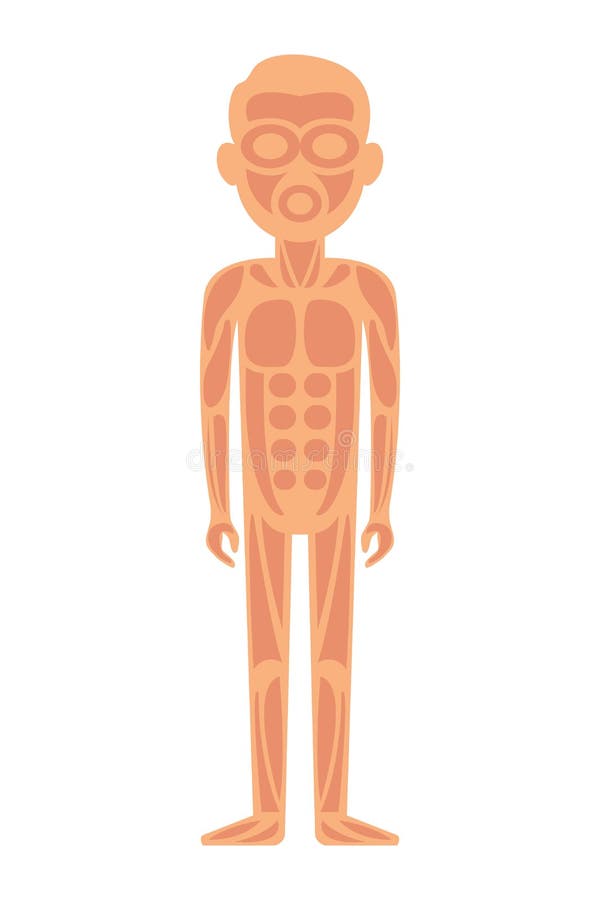
Free with trial Anatomy of man muscular system - anterior view. Tibialis vectors Anatomy of man muscular system - anterior view.
Free with trial 3D Isometric Flat Vector Conceptual Illustration of Extensor Hallucis Longus Muscle, Labeled Educational Anatomical Scheme. Tibialis vectors 3D Isometric Flat Vector Conceptual Illustration of Extensor Hallucis Longus Muscle. Labeled Educational Anatomical Scheme
Free with trial Leg, hip and knee muscle anatomical structure set. Front, side and back view scheme with anatomical captions. Vastus lateralis, medialis, intermedius and rectus femoris. Flat vector illustration. Tibialis vectors Leg, hip and knee muscle anatomical structure set. Front, side. And back view scheme with anatomical captions. Vastus lateralis, medialis, intermedius and. Leg, hip and knee muscle anatomical structure set. Front, side and back view scheme with anatomical captions. Vastus lateralis, medialis, intermedius and rectus femoris. Flat vector illustration.
Free with trial Human muscular system of legs in front view. Quadriceps and adductor magnus muscles. Intermedius, medialis and lateralis vastus. Anatomical poster. Bodybuilding and strong body vector illustration. Tibialis vectors Muscular system legs. Human muscular system of legs in front view. Quadriceps and adductor magnus muscles. Intermedius, medialis and lateralis vastus. Anatomical. Human muscular system of legs in front view. Quadriceps and adductor magnus muscles. Intermedius, medialis and lateralis vastus. Anatomical poster. Bodybuilding and strong body vector illustration
Free with trial Male muscle anatomy. Body with tibialis anterior and peroneal muscles highlighted in red. Training and Workout. Design element for sports poster. Cartoon flat vector illustration isolated on white. Tibialis vectors Male muscle anatomy concept. Male muscle anatomy. Body with tibialis anterior and peroneal muscles highlighted in red. Training and Workout. Design element for. Male muscle anatomy. Body with tibialis anterior and peroneal muscles highlighted in red. Training and Workout. Design element for sports poster. Cartoon flat vector illustration isolated on white
 Sign in