Free with trial Uterus & types of fibroids. Uterine fibroids are benign tumors. They are many times denser than normal myometrium. Vector illustration. Tumors vectors Uterus & types of fibroids. Uterine fibroids are benign tumors. They are many times denser than normal myometrium. Vector illustration
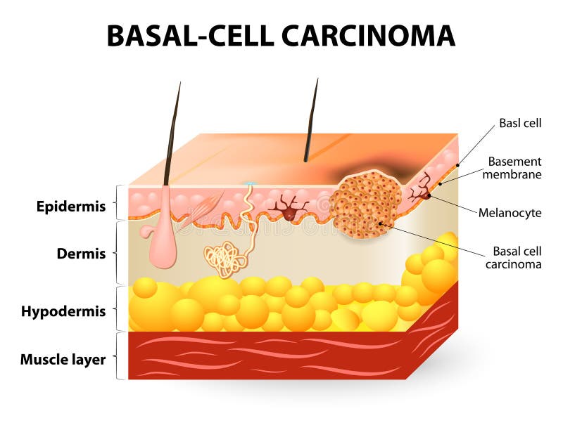
Free with trial Skin cancer. Basal-cell carcinoma or basal cell cancer (BCC). Schematic representation of skin. The separation between epidermis and dermis occurs at the basement membrane zone. Tumors vectors Basal-cell carcinoma or basal cell cancer. Skin cancer. Basal-cell carcinoma or basal cell cancer (BCC). Schematic representation of skin. The separation between. Skin cancer. Basal-cell carcinoma or basal cell cancer (BCC). Schematic representation of skin. The separation between epidermis and dermis occurs at the basement membrane zone.
Free with trial Bowel cancer. Old man suffered colon cancer and charts the growth of tumors in the colon infographic. Vector illustration. Tumors vectors Bowel cancer. Old man suffered colon cancer and charts the growth of tumors in the colon infographic.Vector illustration
Free with trial Types sarcoma. A sarcoma is a cancer. Sarcoma - malignant tumors made of cancellous bone, cartilage, fat, muscle, vascular, and tissues. Tumors vectors Types sarcoma. A sarcoma is a cancer. Sarcoma - malignant tumors made of cancellous bone, cartilage, fat, muscle, vascular, and tissues. Types sarcoma
Free with trial Different types of brain tumors. They categorized by the type of cell where the tumor begins. The most common primary brain cancer. Medical Illustration. human Anatomy. Tumors vectors The most common primary brain tumors. Different types of brain tumors. They categorized by the type of cell where the tumor begins. The most common primary brain. Different types of brain tumors. They categorized by the type of cell where the tumor begins. The most common primary brain cancer. Medical Illustration. human Anatomy
Free with trial Sarcoidosis signs and symptoms. Sarcoidosis it causes the development of granulomas. Granulomas it is a clumps of cells from the immune system resembling little tumors. Human silhouette with internal organs. Tumors vectors Sarcoidosis signs and symptoms. Sarcoidosis it causes the development of granulomas. Granulomas it is a clumps of cells from the immune system resembling
Free with trial Vertebral compression fracture. Medical illustration of the symptoms of vertebral compression fracture. Tumors vectors Vertebral compression fracture. Medical illustration of the symptoms of vertebral compression fracture
Free with trial Headache disorders. Info Graphics in circle. Vector illustrations. Tumors vectors Headache disorders. Info Graphics in circle.Vector illustrations
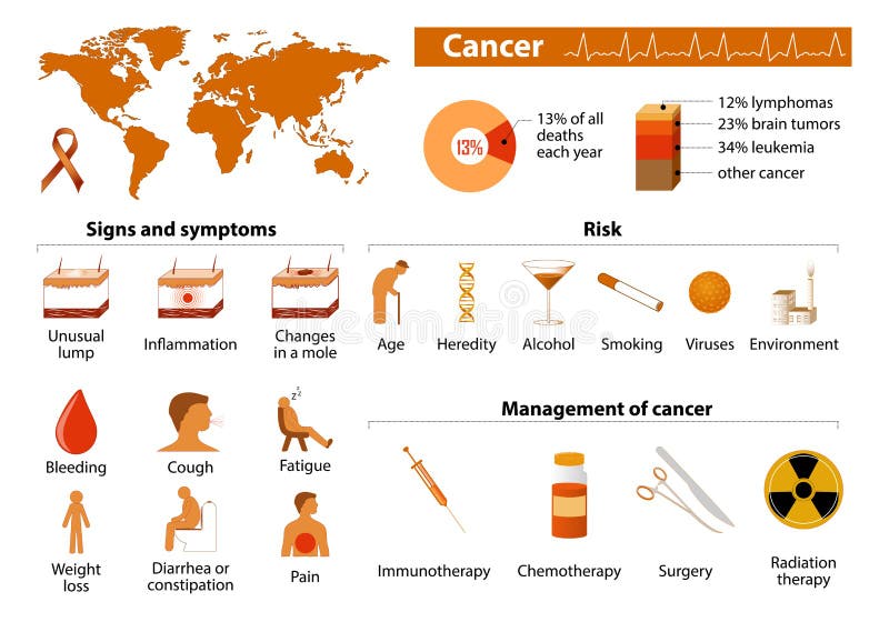
Free with trial Cancer infographic. Cancer signs, symptoms and management. malignant tumor. Medical infographic. Set elements and symbols for design. Tumors vectors Cancer infographic. Cancer signs, symptoms and management. malignant tumor. Medical infographic. Set elements and symbols for design
Free with trial Lipoma are adipose tumors located in the subcutaneous tissues. Tumors vectors Lipoma. Are adipose tumors located in the subcutaneous tissues
Free with trial Hematuria. Blood in Urine Signs Causes and Treatment isolated on white background. Vector illustrations. Tumors vectors Hematuria. Blood in Urine Signs Causes and Treatment isolated on white background. Vector illustrations
Free with trial Endometrial polyp or uterine polyp. Sessile polyp and pedunculated polyp. The polyps consist of dense, fibrous tissue, blood vessels and endometrial epithelium. They are attached by a thin stalk or sessile. Vector diagram. Tumors vectors Endometrial polyp or uterine polyp. Sessile polyp and pedunculated polyp. The polyps consist of dense, fibrous tissue, blood vessels and endometrial epithelium
Free with trial Vocal cord diseases. Medical illustration of the vocal cord diseases. Tumors vectors Vocal cord diseases. Medical illustration of the vocal cord diseases
Free with trial Eye diseases. Sick Eye Old Man diseases Circle, illustration. Tumors vectors Eye diseases. Sick Eye Old Man diseases Circle, illustration
Free with trial Fertilisation in the Uterus. A diagram of the action of fertilisation. Tumors vectors Fertilisation in the Uterus. A diagram of the action of fertilisation
Free with trial Sarcoidosis. Human`s lungs with granulomas. And Enlarged lymph nodes. Tumors vectors Sarcoidosis. Human`s lungs with granulomas. And Enlarged lymph nodes
Free with trial Stages of liver damage from healthy, fatty liver, fibrosis, cirrhosis to liver cancer. Medical infographic, liver diseases icons in flat design isolated on white background. Tumors vectors Stages of liver damage from healthy, fatty liver, fibrosis, cirrhosis to liver cancer. Medical infographic, liver. Diseases icons in flat design isolated on
Free with trial Trephine biopsy cancer tumor. Vector illustration of a trephine biopsy of cancer tumors. Tumors vectors Trephine biopsy cancer tumor. Vector illustration of a trephine biopsy of cancer tumors
Free with trial Illustration explain the origin of exosomes (also called microvesicles, epididimosomes, argosomes, exosome-like vesicles, microparticles, promininosomes, prostasomes, dexosomes, texosomes, dex, tex, archeosomes and oncosomes). Tumors vectors Exosomes. Illustration explain the origin of exosomes (also called microvesicles, epididimosomes, argosomes, exosome-like vesicles, microparticles. Illustration explain the origin of exosomes (also called microvesicles, epididimosomes, argosomes, exosome-like vesicles, microparticles, promininosomes, prostasomes, dexosomes, texosomes, dex, tex, archeosomes and oncosomes)
Free with trial Colon cancer. Doctor and team is helping patient from colon cancer. Tumors vectors Colon cancer. Doctor and team is helping patient from colon cancer
Free with trial Illustration of formation of tumor cells through abnormal cell division, and cancer associated other processes like angiogenesis and metastasis. Tumors vectors Angiogenesis of blood vessels in tumor. Illustration of formation of tumor cells through abnormal cell division, and cancer associated other processes like. Illustration of formation of tumor cells through abnormal cell division, and cancer associated other processes like angiogenesis and metastasis.
Free with trial Osteosarcoma. Osteogenic sarcoma is a cancerous tumor in a bone. malignant neoplasm. Two human bones: healthy femur and bone with osteosarcoma. Vector illustration for biological, science, medical use. Tumors vectors Two human bones: healthy femur and bone with osteosarcoma. Osteosarcoma. Osteogenic sarcoma is a cancerous tumor in a bone. malignant neoplasm. Two human bones. Osteosarcoma. Osteogenic sarcoma is a cancerous tumor in a bone. malignant neoplasm. Two human bones: healthy femur and bone with osteosarcoma. Vector illustration for biological, science, medical use
Free with trial Prostatitis pathology poster. Prostatitis can cause cancer tumors without urgent treatment. Tumors vectors Prostatitis pathology poster. Prostatitis can cause cancer tumors without urgent treatment
Free with trial Infographics of prostate cancer illustration. Prostate cancer symptoms, risk factors. Prostate cancer symptoms. Tumors vectors Infographics of prostate cancer illustration. Prostate cancer symptoms, risk factors. Infographics of prostate cancer illustration. Prostate cancer symptoms
Free with trial Set of Cancer and Chemotherapy Line Icons. Oncology, Chest Tumors, Sarcoma, Leukemia, Dna Molecule, Stethoscope, Microscope, MRI Scanner and more. Pack of 48x48 Pixel Icons. Tumors vectors Set of Cancer and Chemotherapy Line Icons. Oncology, Sarcoma, Leukemia and more. Set of Cancer and Chemotherapy Line Icons. Oncology, Chest Tumors, Sarcoma. Set of Cancer and Chemotherapy Line Icons. Oncology, Chest Tumors, Sarcoma, Leukemia, Dna Molecule, Stethoscope, Microscope, MRI Scanner and more. Pack of 48x48 Pixel Icons
Free with trial Fallopian tube obstruction or Blocked fallopian tubes. cause of female infertility. Uterus and uterine tubes. Human anatomy. female reproductive system. Vector diagram. Gynecological diseases. Tumors vectors Fallopian tube obstruction or Blocked fallopian tubes. Cause of female infertility. Uterus and uterine tubes. Human anatomy. female reproductive system. Vector
Free with trial Surgery of benign tumors of atheroma. Vector illustration of surgery of benign tumors of atheroma. Tumors vectors Surgery of benign tumors of atheroma. Vector illustration of surgery of benign tumors of atheroma
Free with trial Mutation of cells into tumors. Vector illustration diagram of the formation of tumors from mutated cells caused by radiation, mutagens, or infectious agents. Tumors vectors Mutation of cells into tumors. Vector illustration diagram of the formation of tumors from mutated cells caused by radiation, mutagens, or infectious agents
Free with trial Lung cancer stages presented in a brief medical graphic, lungs, tumors, and lymph nodes show progression from Stage I to IV with metastasis. Outline diagram. Tumors vectors Lung cancer stages presented in a brief medical graphic, lungs, ... Lung cancer stages presented in a brief medical graphic, lungs, tumors, and lymph nodes show. Lung cancer stages presented in a brief medical graphic, lungs, tumors, and lymph nodes show progression from Stage I to IV with metastasis. Outline diagram
Free with trial Sarcoidosis signs and symptoms. Development of granulomas. Little tumors. vector illustration. Labeled medical scheme. Tumors vectors Sarcoidosis signs and symptoms. Development of granulomas. Little tumors. vector illustration. Labeled medical scheme
Free with trial Lipoma. adipose tumors. Skin layers. adipose tumors located into the subcutaneous tissues in the skin. Vector illustration. Tumors vectors Lipoma. adipose tumors. Skin layers. Lipoma. adipose tumors located into the subcutaneous tissues in the skin. Vector illustration. Skin layers
Free with trial Brain cancer. different types of primary brain tumors. They categorized by the type of cell where the tumor begins. Tumors vectors Brain cancer. different types of primary brain tumors. They categorized by the type of cell where the tumor begins
Free with trial Epithelial tumors of the bladder. Vector illustration of bladder epithelial tumors. urology. Tumors vectors Epithelial tumors of the bladder. Vector illustration of bladder epithelial tumors. urology
Free with trial Brain tumors in children can develop in different parts of the brain, including the upper brain (supratentorial), cerebellum, brainstem, and lower brain regions. The location of the tumor affects symptoms, treatment, and outcomes. Tumors vectors Brain Tumors in Children Illustration. Brain tumors in children can develop in different parts of the brain, including the upper brain (supratentorial). Brain tumors in children can develop in different parts of the brain, including the upper brain (supratentorial), cerebellum, brainstem, and lower brain regions. The location of the tumor affects symptoms, treatment, and outcomes.
Free with trial Lipoma are adipose tumors located in the subcutaneous tissues. Lipoma are adipose tumors located in the subcutaneous tissues, vector illustration. Tumors vectors Lipoma are adipose tumors located in the subcutaneous tissues. Lipoma are adipose tumors located in the subcutaneous tissues, vector illustration
Free with trial Human eye with tumors line icon. Eye cancer, disease visual organ, retinoblastoma symbol. And sign illustration design. Isolated on white background. Tumors vectors Human eye with tumors line icon. Eye cancer, disease visual organ, retinoblastoma symbol. And sign illustration design. Isolated on white background
Free with trial Brain cancer concept. Primary brain tumors types. Malignant cells develop in the different parts of human brain. Human brain structure anatomy. Flat vector illustration. Tumors vectors Brain cancer concept. Primary brain tumors types. Malignant cells develop. In the different parts of human brain. Human brain structure anatomy. Flat vector
Free with trial How mobile phone radiation penetrates the brain infographic,vector illustration. Tumors vectors How mobile phone radiation penetrates the brain infographic. Vector illustration
Free with trial A medical illustration compares a healthy eye with one affected by lymphoma of the conjunctiva. Tumors vectors Lymphoma of conjunctiva. A medical illustration compares a healthy eye with one affected by lymphoma of the conjunctiva. A medical illustration compares a healthy eye with one affected by lymphoma of the conjunctiva.
Free with trial Biopsy imprint. diagnosis of cancer. Vector illustration of a biopsy imprint. Tumors vectors Biopsy imprint.diagnosis of cancer. Vector illustration of a biopsy imprint.diagnosis of cancer
Free with trial Nose cancer. Nasal Infections. Cancer stage. Nasal Cavity and Paranasal Sinus Cancer. Nasal cancer - tumors of the nasal cavity and sinuses. Tumors vectors Nose cancer. Nasal Infections. Cancer stage. Nasal Cavity and Paranasal Sinus Cancer. Nasal cancer - tumors of the nasal cavity and sinuses
Free with trial Lipoma. adipose tumors located into the subcutaneous tissues in the skin. Cross section of a human skin. Vector illustration. Tumors vectors Lipoma. Cross section of a human skin. Lipoma. adipose tumors located into the subcutaneous tissues in the skin. Cross section of a human skin. Vector. Lipoma. adipose tumors located into the subcutaneous tissues in the skin. Cross section of a human skin. Vector illustration
Free with trial Tumor angiogenesis. transition of tumors from a benign state to a malignant. tumor grow. Cancer cells and Spread of metastasis. Vector poster. Tumors vectors Tumor angiogenesis. Transition of tumors from a benign state to a malignant. tumor grow. Cancer cells and Spread of metastasis. Vector poster
Free with trial Skin redness appear on facial and neck of kid from sunburn. Illustration about danger of Ultraviolet radiation. Tumors vectors Skin redness appear on facial and neck of kid from sunburn. Illustration about danger of Ultraviolet radiation
Free with trial A medical illustration of a person with a nasal tumor and a magnified detail. Tumors vectors Medical illustration of nasal tumor. A medical illustration of a person with a nasal tumor and a magnified detail. A medical illustration of a person with a nasal tumor and a magnified detail.
Free with trial Woman with thyroid cancer. Businesswoman with thyroid cancer on the blue background. Tumors vectors Woman with thyroid cancer. Businesswoman with thyroid cancer on the blue background
Free with trial Sad picture of everyday life in polluted town. Tumors vectors Pulluted city. Sad picture of everyday life in polluted town. Sad picture of everyday life in polluted town.
Free with trial Beautiful vector illustration of medical asthma world day logotype. Editable image in green, orange and dark blue colors useful for a poster, icon, placard, sign, ad and web banner creative design. Tumors vectors World Asthma Day. Beautiful vector illustration of medical asthma world day logotype. Editable image in green, orange and dark blue colors useful for a poster. Beautiful vector illustration of medical asthma world day logotype. Editable image in green, orange and dark blue colors useful for a poster, icon, placard, sign, ad and web banner creative design.
Free with trial The word LYMPHOMA written in colorful fragmented word art on a vibrant background. Vector EPS 10 available. Tumors vectors Lymphoma Concept Colorful Word Art Illustration. The word LYMPHOMA written in colorful fragmented word art on a vibrant background. Vector EPS 10 available. The word LYMPHOMA written in colorful fragmented word art on a vibrant background. Vector EPS 10 available.
Free with trial Interventional radiology as minimally invasive procedures outline diagram. Labeled educational scheme with medical biopsy treatment process for tumors, stenting and angioplasty vector illustration. Tumors vectors Interventional radiology as minimally invasive procedures outline diagram. Labeled educational scheme with medical biopsy treatment process for tumors
Free with trial The injection syringe to the brain, the concept of direct treatment of brain tumors and other diseases. The concept of treating diseases of the brain. Tumors vectors The injection syringe to the brain, the concept of direct treatment of brain tumors and other diseases. The concept of treating diseases of the brain
Free with trial Illustration depicting the progression of fibrous dysplasia in the femur. The stages are shown left to right: a normal femur, early stage with slight deformity, progression with fibrous tissue replacement and visible swelling, and finally, a complication with a fracture. Each stage includes text explaining changes. The deformed areas are highlighted in pink, and arrows indicate the progression from normal to complication. Tumors vectors Fibrous Dysplasia of Bone medical flashcard illustration. Illustration depicting the progression of fibrous dysplasia in the femur. The stages are shown left to. Illustration depicting the progression of fibrous dysplasia in the femur. The stages are shown left to right: a normal femur, early stage with slight deformity, progression with fibrous tissue replacement and visible swelling, and finally, a complication with a fracture. Each stage includes text explaining changes. The deformed areas are highlighted in pink, and arrows indicate the progression from normal to complication.
Free with trial Medical illustration of Ewing Sarcoma, highlighting the femur with a marked area indicating the presence of a tumor. The diagram includes a silhouette of a human body to show the location on the thigh. Labels point to the "Ewing tumor" on the femur and a "warm lump. " The highlighted area on the bone is colored red to denote the tumor's presence, providing a clear visual reference for educational purposes. Tumors vectors Ewing Sarcoma, ewing tumor flashcard medical illustration. Medical illustration of Ewing Sarcoma, highlighting the femur with a marked area indicating the. Medical illustration of Ewing Sarcoma, highlighting the femur with a marked area indicating the presence of a tumor. The diagram includes a silhouette of a human body to show the location on the thigh. Labels point to the "Ewing tumor" on the femur and a "warm lump." The highlighted area on the bone is colored red to denote the tumor's presence, providing a clear visual reference for educational purposes.
Free with trial People using different electronic devices such as smartphone, laptop, tablet. Radio fields, electromagnetic pollution, radiation concept, violet palette. Vector illustration on white background. Tumors vectors Radio fields influence concept vector illustration. People using different electronic devices such as smartphone, laptop, tablet. Radio fields, electromagnetic. People using different electronic devices such as smartphone, laptop, tablet. Radio fields, electromagnetic pollution, radiation concept, violet palette. Vector illustration on white background.
Free with trial Liver damage infographic diagram cancer disease anatomy. Liver damage organ tissue hepatic carcinoma fibrosis stages. Liver damage infographic diagram cancer. Tumors vectors Liver damage infographic diagram cancer disease anatomy. Liver damage organ tissue hepatic carcinoma fibrosis stages. Liver damage infographic diagram cancer
Free with trial Cross-section diagram comparing Craniopharyngioma and Pituitary Adenoma in the brain. Both illustrations show the hypothalamus, pituitary gland, and sphenoid sinus. The left image highlights a Craniopharyngioma, a benign tumor near the pituitary, common in children and young adults, leading to hormone deficiencies. The right image depicts a Pituitary Adenoma, a noncancerous tumor of the pituitary gland, often causing hormone imbalances and vision problems. Both tumors are indicated in red against the internal anatomy of the brain. Tumors vectors Craniopharyngioma vs Pituitary Adenoma. Cross-section diagram comparing Craniopharyngioma and Pituitary Adenoma in the brain. Both illustrations show the. Cross-section diagram comparing Craniopharyngioma and Pituitary Adenoma in the brain. Both illustrations show the hypothalamus, pituitary gland, and sphenoid sinus. The left image highlights a Craniopharyngioma, a benign tumor near the pituitary, common in children and young adults, leading to hormone deficiencies. The right image depicts a Pituitary Adenoma, a noncancerous tumor of the pituitary gland, often causing hormone imbalances and vision problems. Both tumors are indicated in red against the internal anatomy of the brain.
Free with trial The pancreas is affected dense and papillary epithelial tumors. Vector illustration on background. Tumors vectors The pancreas is affected dense and papillary. Epithelial tumors. Vector illustration on background
Free with trial People using different electronic devices such as smartphone, laptop, tablet. Radio fields, electromagnetic pollution, radiation concept, violet palette. Vector illustration on white background. Tumors vectors Radio fields influence concept vector illustration. People using different electronic devices such as smartphone, laptop, tablet. Radio fields, electromagnetic. People using different electronic devices such as smartphone, laptop, tablet. Radio fields, electromagnetic pollution, radiation concept, violet palette. Vector illustration on white background.
Free with trial People using different electronic devices such as smartphone, laptop, tablet. Radio fields, electromagnetic pollution, radiation concept, violet palette. Vector illustration on white background. Tumors vectors Radio fields influence concept vector illustration. People using different electronic devices such as smartphone, laptop, tablet. Radio fields, electromagnetic. People using different electronic devices such as smartphone, laptop, tablet. Radio fields, electromagnetic pollution, radiation concept, violet palette. Vector illustration on white background.
Free with trial Healthy Liver and Liver diseases - fatty liver, hepatitis, fibrosis, cirrhosis, alcoholic hepatitis, liver cancer - medical infographic elements isolated on white background. Tumors vectors Healthy Liver and Liver diseases - fatty liver, hepatitis, fibrosis, cirrhosis, alcoholic hepatitis, liver cancer -. Medical infographic elements isolated on
Free with trial Drops from the dropper to the brain, the concept of a direct treatment of tumors and other diseases of the brain. The concept of treating diseases of the brain. Tumors vectors Drops from the dropper to the brain, the concept of a direct treatment of tumors and other diseases of the brain. The concept of treating diseases of the brain
Free with trial Chemo Brain word cloud on a white background. Tumors vectors Chemo Brain Word Cloud. On a white background. Chemo Brain word cloud on a white background.
Free with trial Chemotherapy word cloud on a white background. Tumors vectors Chemotherapy Word Cloud. On a white background. Chemotherapy word cloud on a white background.
Free with trial Cancer word cloud on a white background. Tumors vectors Cancer Word Cloud. On a white background. Cancer word cloud on a white background
Free with trial Drops from the eyedropper into the brain, the concept of direct treatment of brain tumors and other diseases. The concept of treating diseases of the brain. Tumors vectors Drops from the eyedropper into the brain, the concept of direct treatment of brain tumors and other diseases. The concept of treating diseases of the brain
Free with trial Cancer word cloud on a white background. Tumors vectors Cancer Word Cloud. On a white background. Cancer word cloud on a white background
Free with trial Cancer Treatment Breakthrough concept. CAR-T cell immunotherapy revolutionizing oncology with targeted cancer cell destruction. Flat vector illustration. Tumors vectors Cancer Treatment Breakthrough concept. Flat. Cancer Treatment Breakthrough concept. CAR-T cell immunotherapy revolutionizing oncology with targeted cancer cell. Cancer Treatment Breakthrough concept. CAR-T cell immunotherapy revolutionizing oncology with targeted cancer cell destruction. Flat vector illustration.
Free with trial Skin cancer word concepts banner. Skin tumors and growths. Melanoma. Infographics with linear icons on blue background. Isolated typography. Vector outline RGB color illustration. Tumors vectors Skin cancer word concepts banner. Skin tumors and growths. Melanoma. Infographics with linear icons on blue background. Isolated typography. Vector outline RGB
Free with trial Cancer word cloud on a white background. Tumors vectors Cancer Word Cloud. On a white background. Cancer word cloud on a white background
Free with trial Cancer word cloud on a white background. Tumors vectors Cancer Word Cloud. On a white background. Cancer word cloud on a white background
Free with trial Cancer word cloud on a white background. Tumors vectors Cancer Word Cloud. On a white background. Cancer word cloud on a white background
Free with trial Mesenchymal stem cells (MSCs) are a type of multipotent adult stem cell found in various tissues throughout the body, including bone marrow, adipose tissue (fat), umbilical cord tissue, and dental pulp. They have the capacity to differentiate into a variety of cell types, including osteoblasts (bone cells), chondrocytes (cartilage cells), and adipocytes (fat cells), among others. Tumors vectors Mesenchymal stem cells. (MSCs) are a type of multipotent adult stem cell found in various tissues throughout the body, including bone marrow, adipose tissue (. Mesenchymal stem cells (MSCs) are a type of multipotent adult stem cell found in various tissues throughout the body, including bone marrow, adipose tissue (fat), umbilical cord tissue, and dental pulp. They have the capacity to differentiate into a variety of cell types, including osteoblasts (bone cells), chondrocytes (cartilage cells), and adipocytes (fat cells), among others.
Free with trial Fibroids color icon. Abnormal growth. Develop in the uterus. Cancerous or noncancerous tumors. Health protection. Women health concept. Isolated vector illustration. Tumors vectors Fibroids color icon. Abnormal growth. Develop in the uterus. Cancerous or noncancerous tumors.Health protection. Women health concept. Isolated vector
Free with trial Metastasis RGB color icon. Cancer cells spread. Pathogenic tumors. Oncology. Malignant tumor. Disease development. Isolated vector illustration. Simple filled line drawing. Editable stroke. Tumors vectors Metastasis RGB color icon. Cancer cells spread. Pathogenic tumors. Oncology. Malignant tumor. Disease development. Isolated vector illustration. Simple filled
Free with trial Types of fatty liver. Human liver diseases. Alcoholic fatty liver disease. Hepatitis, liver cirrhosis, fibrosis, steatosis. Medical infographic banner. Vector. Tumors vectors Types of fatty liver. Human liver diseases. Alcoholic fatty liver disease. Medical infographic banner. Vector. Types of fatty liver. Human liver diseases. Types of fatty liver. Human liver diseases. Alcoholic fatty liver disease. Hepatitis, liver cirrhosis, fibrosis, steatosis. Medical infographic banner. Vector
Free with trial National Young Adult Cancer awareness week - first week in April. Lavender or violet color ribbon Cancer Awareness Products. Vector illustration. Tumors vectors National Young Adult Cancer awareness week - first week in April. Lavender or violet color of the ribbon Cancer. National Young Adult Cancer awareness week. National Young Adult Cancer awareness week - first week in April. Lavender or violet color ribbon Cancer Awareness Products. Vector illustration.
Free with trial Here we are offering you a World Cancer Day icons pack that can be used in project related marketing and consist of a love and romance hope you will enjoy it!. Tumors vectors Doctors analyzing brain for tumors or problems in the laboratory Illustration. Here we are offering you a World Cancer Day icons pack that can be used in. Here we are offering you a World Cancer Day icons pack that can be used in project related marketing and consist of a love and romance hope you will enjoy it !
Free with trial Vector illustration of a girl with moles all over her body. Changes in a mole are symptoms of a hormonal imbalance, an. Infection in the body. Tumors vectors Vector illustration of a girl with moles all over her body. Changes in a mole are symptoms of a hormonal imbalance, an. Infection in the body
Free with trial Vector illustration of ovarian cancer awareness tapes isolated on a white background. Realistic vector teal silk ribbon with loop. Design for the poster. Tumors vectors Vector illustration of ovarian cancer awareness tapes isolated on a white background. Realistic vector teal silk ribbon. With loop.Design for the poster
Free with trial Vector banner template for International Neuroendocrine Tumors Awareness Month in November featuring zebra print ribbon, dark background, and world map silhouette. Tumors vectors International Neuroendocrine Tumors Awareness Month Vector With Zebra Ribbon. Vector banner template for International Neuroendocrine Tumors Awareness Month in. Vector banner template for International Neuroendocrine Tumors Awareness Month in November featuring zebra print ribbon, dark background, and world map silhouette
 Sign in