Free with trial ECG set. Normal and diseased ecg collection, eps8. Ventricular vectors ECG set. Normal and diseased ecg collection, eps8
Free with trial Pulmonary hypertension. Artwork depicting a pulmonary hypertension. Ventricular vectors Pulmonary hypertension. Artwork depicting a pulmonary hypertension
Free with trial Diseases of heart muscle. 3 different diseases of heart muscle, eps8. Ventricular vectors Diseases of heart muscle. 3 different diseases of heart muscle, eps8
Free with trial Ventricular septal defect. Congenital heart disease: ventricular septal defect, eps8. Ventricular vectors Ventricular septal defect. Congenital heart disease: ventricular septal defect, eps8
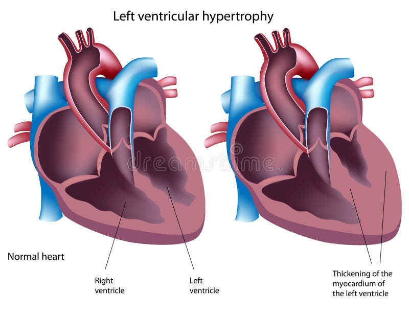
Free with trial Left ventricular hypertrophy. Normal heart and heart with left ventricular hypertrophy, eps8. Ventricular vectors Left ventricular hypertrophy. Normal heart and heart with left ventricular hypertrophy, eps8
Free with trial Heart failure or congestive heart failure. Ventricular vectors Heart failure. Or congestive. Heart failure or congestive heart failure
Free with trial Heart disease: valve disease, aneurysm, coronary artery disease, cardiac arrhythmia, heart failture, cardiomyopathy and pericarditis. Ventricular vectors Heart disease. Valve disease, aneurysm, coronary artery disease, cardiac arrhythmia, heart failture, cardiomyopathy and pericarditis
Free with trial Cardiac arrhythmia. cardiac dysrhythmia or irregular heartbeat. Medical illustration. Ventricular vectors Cardiac arrhythmia. Cardiac dysrhythmia or irregular heartbeat. Medical illustration
Free with trial Heart sounds. The mechanism of the production of the typical lub dub heart sounds. Ventricular vectors Heart sounds. The mechanism of the production of the typical lub dub heart sounds
Free with trial Congestive heart. Medical illustration of the symptoms and consequences of congestive heart. Ventricular vectors Congestive heart. Medical illustration of the symptoms and consequences of congestive heart
Free with trial Tetralogy of Fallot. Congenital heart disease: Tetralogy of Fallot, eps8. Ventricular vectors Tetralogy of Fallot. Congenital heart disease: Tetralogy of Fallot, eps8
Free with trial Diabetic nephropathy. Medical illustration of the effects of the diabetic nephropathy. Ventricular vectors Diabetic nephropathy. Medical illustration of the effects of the diabetic nephropathy
Free with trial Dilated cardiomyopathy. Medical Illustration of the effects of dilated cardiomyopathy. Ventricular vectors Dilated cardiomyopathy. Medical Illustration of the effects of dilated cardiomyopathy
Free with trial Congenital heart defect. Comparison of normal heart and the congenital heart defect Tetralogy of Fallot. Ventricular vectors Congenital heart defect. Comparison of normal heart and the congenital heart defect Tetralogy of Fallot
Free with trial Detailed medical illustration of human heart anatomy showing chambers and blood flow directions. Suitable for anatomy education and healthcare materials. Ventricular vectors Human Heart Anatomy Diagram with Blood Flow Direction Illustration. Detailed medical illustration of human heart anatomy showing chambers and blood flow. Detailed medical illustration of human heart anatomy showing chambers and blood flow directions. Suitable for anatomy education and healthcare materials.
Free with trial Drawing of the heart electrical conduction system showing normal activity and erratic impulses in atrial fibrillation. Ventricular vectors Normal heart electrical conduction and atrial fibrillation. Drawing of the heart electrical conduction system showing normal activity and erratic impulses in. Drawing of the heart electrical conduction system showing normal activity and erratic impulses in atrial fibrillation.
Free with trial Heart anatomy and types of heart disease vector illustration. Isolated on white. Ventricular vectors Heart anatomy and types of heart disease vector illustration. Isolated on white
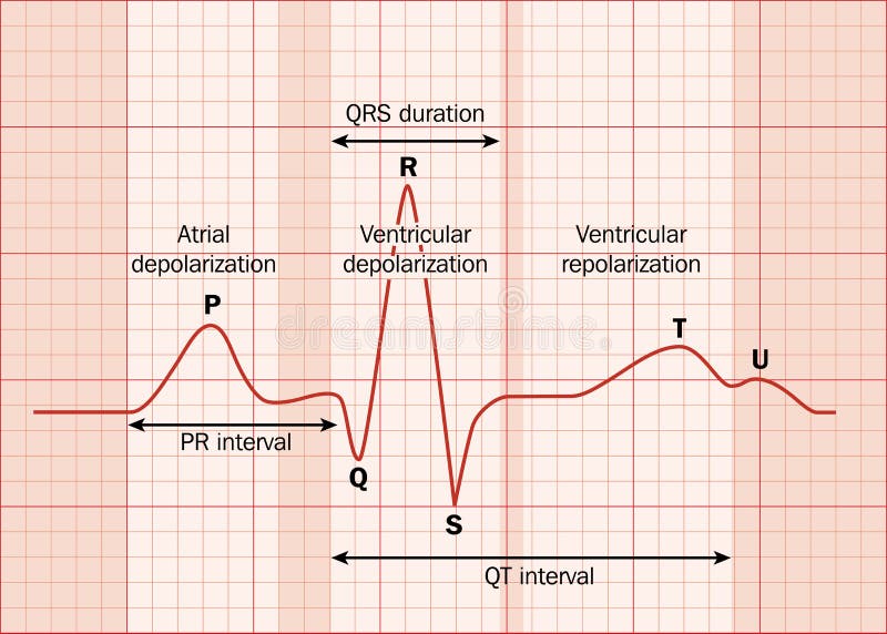
Free with trial Heart ECG (EKG) showing the major intervals. Created in Adobe Illustrator. EPS 10. Ventricular vectors Heart ECG. (EKG) showing the major intervals. Created in Adobe Illustrator. EPS 10
Free with trial Heart. Anatomy of the Heart. The Cardiac Cycle. Diastole Ventricular Relaxation and Filling. Systole Ventricular Contraction and Ejection. Ventricular vectors Heart. Anatomy of the . The Cardiac Cycle. Diastole Ventricular Relaxation and Filling. Systole Ventricular Contraction and Ejection
Free with trial Medical diagram illustrating human circulatory system with heart, lungs, arteries and veins. Ideal for anatomy education and healthcare materials. Ventricular vectors Human Circulatory System Diagram Showing Heart Lungs and Blood Flow. Medical diagram illustrating human circulatory system with heart, lungs, arteries and veins. Medical diagram illustrating human circulatory system with heart, lungs, arteries and veins. Ideal for anatomy education and healthcare materials.
Free with trial Types of cardiomyopathy: the walls of the ventricles thicken and become stiff; the left ventricle is enlarged and weakened; the walls of the ventricles become stiff, but not necessarily thickened. Ventricular vectors Diseases of the Heart Muscle. Types of cardiomyopathy: the walls of the ventricles thicken and become stiff; the left ventricle is enlarged and weakened; the. Types of cardiomyopathy: the walls of the ventricles thicken and become stiff; the left ventricle is enlarged and weakened; the walls of the ventricles become stiff, but not necessarily thickened
Free with trial Types of heart diseases: hypertrophic cardiomyopathy and dilated cardiomyopathy. healthy heart and heart with Pathology. vector illustration for medical use. Ventricular vectors Hypertrophic cardiomyopathy, dilated cardiomyopathy and healthy. Types of heart diseases: hypertrophic cardiomyopathy and dilated cardiomyopathy. healthy heart. Types of heart diseases: hypertrophic cardiomyopathy and dilated cardiomyopathy. healthy heart and heart with Pathology. vector illustration for medical use
Free with trial Ventricular septal defect VSD. Congenital heart defect. Ventricular vectors Ventricular septal defect VSD. Congenital heart defect
Free with trial Ventricular system. Cross Section of a Human brain with ventricles and Cerebrospinal fluid. Ventricular vectors Cross Section of a Human brain with ventricles and Cerebrospinal fluid. Ventricular system. Cross Section of a Human brain with ventricles and Cerebrospinal. Ventricular system. Cross Section of a Human brain with ventricles and Cerebrospinal fluid
Free with trial Normal Atrial And Ventricular Depolarization With. Electrocardiogram Description Vector Illustration. Ventricular vectors Normal Atrial And Ventricular Depolarization With. Electrocardiogram Description Vector Illustration
Free with trial Heart checkup. Magnifying glass on an ECG diagram, eps8. Ventricular vectors Heart checkup. Magnifying glass on an ECG diagram, eps8
Free with trial Heart and pacemaker. cardiology. Vector illustration of heart and pacemaker. Ventricular vectors Heart and pacemaker. cardiology. Vector illustration of heart and pacemaker. cardiology
Free with trial A medical device when someone is having a heart attack. Ventricular vectors Heart defibrillator - no background. A medical device when someone is having a heart attack. A medical device when someone is having a heart attack.
Free with trial A medical device when someone is having a heart attack. Ventricular vectors Heart defibrillator. A medical device when someone is having a heart attack. A medical device when someone is having a heart attack.
Free with trial Echocardiogram white background. Info graphic. Heart examination with high frequency sound waves Very useful in evaluating heart function, Including other symptoms. Ventricular vectors Echocardiogram white background. Info graphic.Heart examination with high frequency sound waves Very useful in evaluating heart function, Including other
Free with trial Heart disease vector infographics. Medical human heart infographic information illustration. Ventricular vectors Heart disease vector infographics. Medical human heart infographic information illustration
Free with trial Echocardiogram. Health care, info graphic. Heart examination with high frequency sound waves Very useful in evaluating heart function, Including other symptoms. Ventricular vectors Echocardiogram. Health care, info graphic. Heart examination with high frequency sound waves Very useful in evaluating heart function, Including other symptoms
Free with trial Heart structure. Mitral valve prolapse. Cardiac pathology. Ventricular vectors Heart structure. Mitral valve prolapse. Cardiac pathology.
Free with trial Heart anatomy. Close-up of Normal Mitral valve and Damaged mitral valve. Cross section of human heart. detailed diagram. Vector poster. Ventricular vectors Heart anatomy. Mitral valve. Heart anatomy. Close-up of Normal Mitral valve and Damaged mitral valve. Cross section of human heart. detailed diagram. Vector. Heart anatomy. Close-up of Normal Mitral valve and Damaged mitral valve. Cross section of human heart. detailed diagram. Vector poster
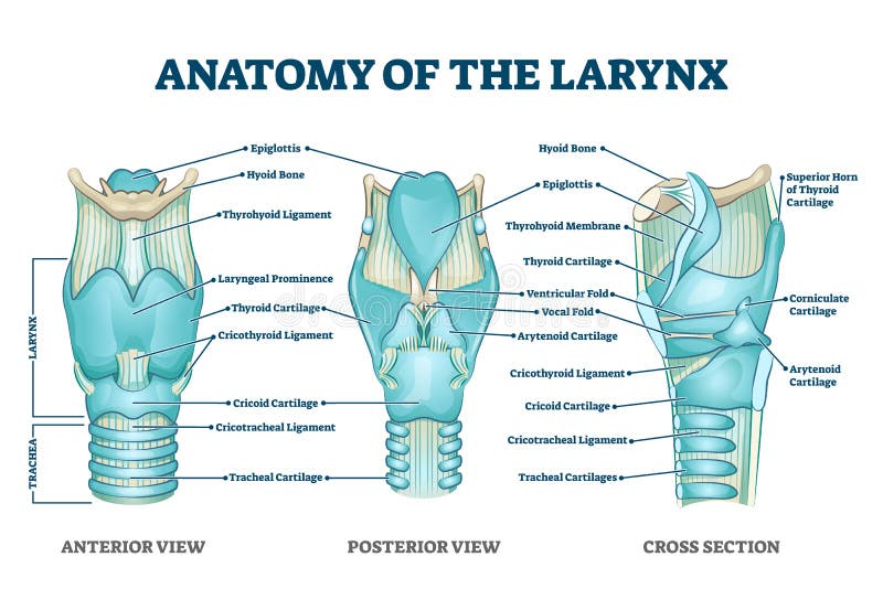
Free with trial Larynx anatomy with labeled structure scheme and educational medical views. Anterior, posterior and cross section examination with trachea parts vector illustration. Vocal cords housing description. Ventricular vectors Larynx anatomy with labeled structure scheme and educational medical views. Anterior, posterior and cross section examination with trachea parts vector
Free with trial Liver, hepatitis and yellowing of eyes and hands. Illustration of the liver, hepatitis and yellowing of eyes and hands. Ventricular vectors Liver, hepatitis and yellowing of eyes and hands. Illustration of the liver, hepatitis and yellowing of eyes and hands
Free with trial Heart structure. The organ of the circulatory system. Types of cardiac arrhythmia: sinus tachycardia, sinus arrhythmia, sinus bradycardia, normal rhythm. Cardiogram. Ventricular vectors Heart structure. Types of cardiac arrhythmia. Heart structure. The organ of the circulatory system. Types of cardiac arrhythmia: sinus tachycardia, sinus. Heart structure. The organ of the circulatory system. Types of cardiac arrhythmia: sinus tachycardia, sinus arrhythmia, sinus bradycardia, normal rhythm. Cardiogram.
Free with trial Heart with mitral valve prolapse. On the blue background. Ventricular vectors Heart with mitral valve prolapse. On the blue background
Free with trial Cyanosis of the hand of a patient with low oxygen saturations. Peripheral cyanosis. Ventricular vectors Cyanosis of the hand of a patient with low oxygen saturations. Peripheral cyanosis. Cyanosis of the hand of a patient with low oxygen saturations
Free with trial Abdominal aneurysm 3d medical vector illustration isolated on white background. Eps 10 education vector. Ventricular vectors Abdominal aneurysm 3d medical vector illustration isolated on white background. Eps 10 education vector
Free with trial Look on atrial fibrillation through magnifying glass that human hand holds in round vector picture on white. Long piece of paper with ECG scheme of improper heart working, emergency cardiology. Ventricular vectors Look at atrial fibrillation through magnifying glass held by hand. Look on atrial fibrillation through magnifying glass that human hand holds in round vector. Look on atrial fibrillation through magnifying glass that human hand holds in round vector picture on white. Long piece of paper with ECG scheme of improper heart working, emergency cardiology.
Free with trial Dilated cardiomyopathy, Heart disease, Vector image. Dilated cardiomyopathy, Heart disease, - Vector image file for use. Ventricular vectors Dilated cardiomyopathy, Heart disease, Vector image. Dilated cardiomyopathy, Heart disease, - Vector image file for use
Free with trial AED - automated external defibrillator illustration - isolated vector medical healthcare device equipment. Ventricular vectors AED - automated external defibrillator device. AED - automated external defibrillator illustration - isolated vector medical healthcare device equipment. AED - automated external defibrillator illustration - isolated vector medical healthcare device equipment
Free with trial Automated external defibrillator AED icon - isolated vector medical equipment. Ventricular vectors Automated external defibrillator AED icon. Isolated vector medical equipment
Free with trial Heart checkup, magnifying glass on an ecg diagram. Ventricular vectors Heart checkup. Magnifying glass on an ecg diagram
Free with trial A Ventricular Assist Device (VAD) is a mechanical pump that helps the heart circulate blood throughout the body in patients with severe heart failure. Medical health vector illustration. Ventricular vectors A Ventricular Assist Device (VAD) is a mechanical pump. That helps the heart circulate blood throughout the body in patients with severe heart failure. Medical. A Ventricular Assist Device (VAD) is a mechanical pump that helps the heart circulate blood throughout the body in patients with severe heart failure. Medical health vector illustration
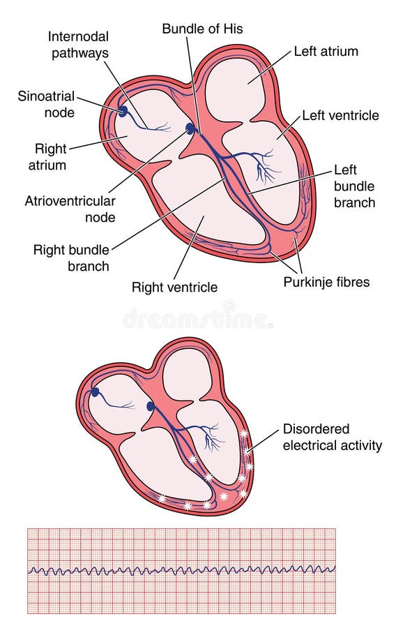
Free with trial Drawing of heart electrical conducting system, the site of ventricular fibrillation disordered electrical activity and the resulting ecg trace. Created in Adobe Illustrator. EPS 10. Ventricular vectors Ventricular fibrillation of heart. Drawing of heart electrical conducting system, the site of ventricular fibrillation disordered electrical activity and the. Drawing of heart electrical conducting system, the site of ventricular fibrillation disordered electrical activity and the resulting ecg trace. Created in Adobe Illustrator. EPS 10.
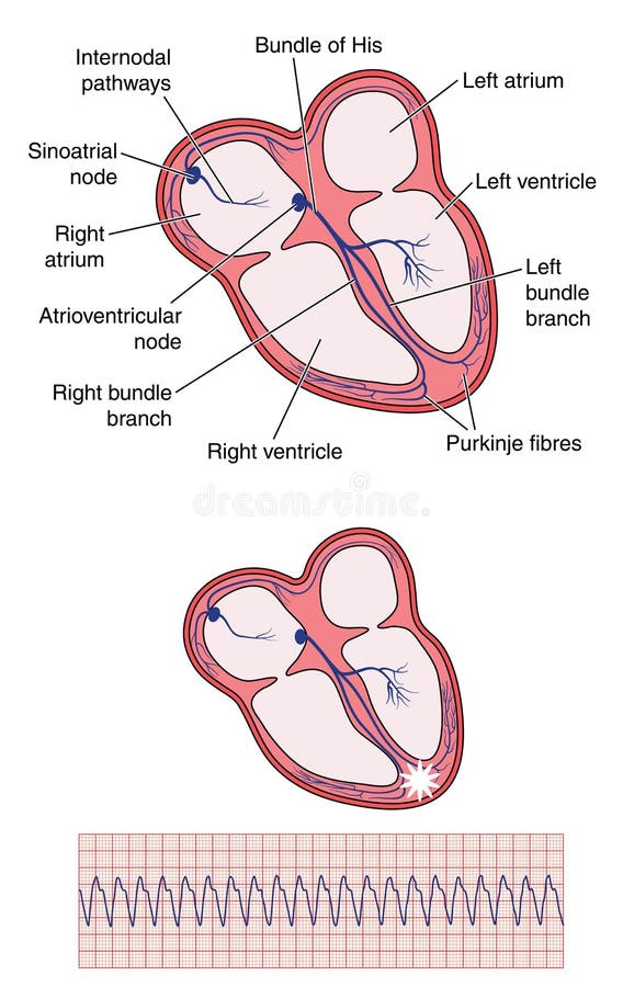
Free with trial Drawing of heart electrical conducting system, the site of ventricular tachycardia and the resulting ecg trace. Created in Adobe Illustrator. EPS 10. Ventricular vectors Ventricular tachycardia of heart. Drawing of heart electrical conducting system, the site of ventricular tachycardia and the resulting ecg trace. Created in. Drawing of heart electrical conducting system, the site of ventricular tachycardia and the resulting ecg trace. Created in Adobe Illustrator. EPS 10.
Free with trial A ventricular assist device (VAD) is a device that helps pump blood from the lower chambers of the heart to the rest of the body. It's a treatment for a weakened heart or heart failure. Medical health. Ventricular vectors A ventricular assist device (VAD) is a device that helps pump blood. From the lower chambers of the heart to the rest of the body. It's a treatment for a. A ventricular assist device (VAD) is a device that helps pump blood from the lower chambers of the heart to the rest of the body. It's a treatment for a weakened heart or heart failure.Medical health
Free with trial Electrocardiogram show ventricular fibrillation VF pattern. Cardiac fibrillation. Heart beat. CPR. ECG. EKG. Vital sign. Life support. Defib. Emergency. Medical healthcare symbol. Ventricular vectors Electrocardiogram show ventricular fibrillation VF pattern. Cardiac fibrillation. Heart beat. CPR. ECG. EKG. Vital sign. Life support. Defib. Emergency
Free with trial Cerebrospinal fluid. Cross section of a human brain with Ventricular system. Close-up of the meninges. CSF and Membranes that envelop the brain. Vector illustration. Ventricular vectors Cerebrospinal fluid. Cross section of a human brain with Ventricular system. Close-up of the meninges. CSF and Membranes that envelop the brain. Vector
Free with trial Electrocardiogram show monomorphic ventricular tachycardia VT. Cardiac fibrillation. Heart beat. CPR. ECG. EKG. Vital sign. Life support. Medical healthcare symbol. Ventricular vectors Electrocardiogram show monomorphic ventricular tachycardia VT. Cardiac fibrillation. Heart beat. CPR. ECG. EKG. Vital sign. Life support. Medical healthcare
Free with trial Electrocardiogram show Premature Ventricular Contraction PVC pattern ,Heart beat ,ECG ,EKG interpretation ,Vital sign ,Life line ,Medical healthcare symbol. Ventricular vectors Electrocardiogram show Premature Ventricular Contraction PVC pattern. Electrocardiogram show Premature Ventricular Contraction PVC pattern ,Heart beat ,ECG ,EKG. Electrocardiogram show Premature Ventricular Contraction PVC pattern ,Heart beat ,ECG ,EKG interpretation ,Vital sign ,Life line ,Medical healthcare symbol.
Free with trial Ventricular septal defect with normal heart anatomy. Illustration of the congenital defect of the ventricular wall. Ventricular vectors Ventricular septal defect with normal heart anatomy. Illustration of the congenital heart defect. Ventricular septal defect with normal heart anatomy. Ventricular septal defect with normal heart anatomy. Illustration of the congenital defect of the ventricular wall
Free with trial Abnormal cardiogram ventricular tachycardia. , vector illustration. Ventricular vectors Abnormal cardiogram ventricular tachycardia. Vector illustration
Free with trial Ventricular system anatomy, set of communicating cavities within the brain. Cerebral ventricles concept. Production, transport and removal of cerebrospinal fluid. Flat vector anatomical illustration. Ventricular vectors Ventricular system concept. Ventricular system anatomy, set of communicating cavities within the brain. Cerebral ventricles concept. Production, transport and. Ventricular system anatomy, set of communicating cavities within the brain. Cerebral ventricles concept. Production, transport and removal of cerebrospinal fluid. Flat vector anatomical illustration.
Free with trial Hydrocephalus shunt in the child head poster. Pressure on the brain. Ventricular system anatomy. Cerebral ventricles, cerebrospinal fluid in kid silhouette. Medical flat vector illustration. Ventricular vectors Ventricular system concept. Hydrocephalus shunt in the child head poster. Pressure on the brain. Ventricular system anatomy. Cerebral ventricles, cerebrospinal. Hydrocephalus shunt in the child head poster. Pressure on the brain. Ventricular system anatomy. Cerebral ventricles, cerebrospinal fluid in kid silhouette. Medical flat vector illustration.
Free with trial Ventricular septal defect VSD. Human heart muscle diseases cross-section on white background. Cardiology concept. Ventricular vectors Ventricular septal defect VSD. Human heart muscle diseases cross-section. Cardiology concept. Ventricular septal defect VSD. Human heart muscle diseases cross. Ventricular septal defect VSD. Human heart muscle diseases cross-section on white background. Cardiology concept.
Free with trial Ventricular system anatomy, set of communicating cavities within the brain. Cerebral ventricles concept. Production, transport and removal of cerebrospinal fluid. Flat vector anatomical illustration. Ventricular vectors Ventricular system anatomy. Set of communicating cavities within the brain. Cerebral ventricles concept. Production, transport and removal of cerebrospinal. Ventricular system anatomy, set of communicating cavities within the brain. Cerebral ventricles concept. Production, transport and removal of cerebrospinal fluid. Flat vector anatomical illustration.
Free with trial Ventricular septal defect VSD on white background. Human heart muscle diseases cross-section. Cardiology concept. Ventricular vectors Ventricular septal defect VSD. Human heart muscle diseases cross-section. Cardiology concept. Ventricular septal defect VSD on white background. Human heart. Ventricular septal defect VSD on white background. Human heart muscle diseases cross-section. Cardiology concept.
Free with trial Pathophysiology of CTEPH depicts embolus becoming organized thrombus, narrowing pulmonary arteries and causing right ventricular strain, vessels, lungs, heart. Outline diagram. Ventricular vectors Pathophysiology of CTEPH depicts embolus becoming organized thrombus, narrowing pulmonary ... Pathophysiology of CTEPH depicts embolus becoming organized. Pathophysiology of CTEPH depicts embolus becoming organized thrombus, narrowing pulmonary arteries and causing right ventricular strain, vessels, lungs, heart. Outline diagram
Free with trial Medical illustration that compares a normal heart with hearts afflicted by ventricular septal defects, an abnormal opening hole in the heart, with annotations. Ventricular vectors Heart defects. Medical illustration that compares a normal heart with hearts afflicted by ventricular septal defects, an abnormal opening hole in the heart, with. Medical illustration that compares a normal heart with hearts afflicted by ventricular septal defects, an abnormal opening hole in the heart, with annotations.
Free with trial Atrial Septal Defect ASD and Ventricular Septal Defect VSD. Abnormal of the heart atrial and heart ventricle. From baby birth. Ventricular vectors Atrial Septal Defect ASD and Ventricular Septal Defect VSD. Abnormal of the heart atrial and heart ventricle. From baby birth
Free with trial Automated External Defibrillator. It is electronic device for life support that recognises ventricular fibrillation and other dysrhythmias and delivers an electric shock at the right time. Ventricular vectors Automated External Defibrillator. It is electronic device for life support that recognises ventricular fibrillation and other dysrhythmias and delivers an
Free with trial Tetralogy of Fallot (ToF) anatomical diagram highlights heart defects: ventricular septal defect, pulmonary stenosis, right ventricular hypertrophy, and overriding aorta. The heart's structure is color-coded to differentiate oxygen-rich (red) and oxygen-poor (blue) blood flow. Labels mark major components: aorta (AO), pulmonary artery (PA), left atrium (LA), right atrium (RA), left ventricle (LV), and right ventricle (RV). Arrows indicate the blood flow direction. The diagram aids in understanding ToF's impact on heart function. Ventricular vectors Tetralogy of Fallot (ToF) Heart Flashcard. Tetralogy of Fallot (ToF) anatomical diagram highlights heart defects: ventricular septal defect, pulmonary stenosis. Tetralogy of Fallot (ToF) anatomical diagram highlights heart defects: ventricular septal defect, pulmonary stenosis, right ventricular hypertrophy, and overriding aorta. The heart's structure is color-coded to differentiate oxygen-rich (red) and oxygen-poor (blue) blood flow. Labels mark major components: aorta (AO), pulmonary artery (PA), left atrium (LA), right atrium (RA), left ventricle (LV), and right ventricle (RV). Arrows indicate the blood flow direction. The diagram aids in understanding ToF's impact on heart function.
Free with trial Diagram illustrates normal and congenital heart defects, highlighting anatomical differences. Center left shows a healthy heart with labels for atria, ventricles, and major vessels. Four conditions depicted: Atrial Septal Defect (ASD) with an opening between atria Ventricular Septal Defect (VSD) showing a gap between ventricles Aortic Coarctation indicating narrowed aorta Fallot's Tetralogy featuring four defects including a ventricular septal defect and thickened right ventricle. Color-coded: red for oxygen-rich blood, blue for oxygen-poor, purple for mixed blood. Labels enhance visual comprehension. Ventricular vectors Congenital Heart Disease Illustration. Diagram illustrates normal and congenital heart defects, highlighting anatomical differences. Center left shows a healthy. Diagram illustrates normal and congenital heart defects, highlighting anatomical differences. Center left shows a healthy heart with labels for atria, ventricles, and major vessels. Four conditions depicted: Atrial Septal Defect (ASD) with an opening between atria Ventricular Septal Defect (VSD) showing a gap between ventricles Aortic Coarctation indicating narrowed aorta Fallot's Tetralogy featuring four defects including a ventricular septal defect and thickened right ventricle. Color-coded: red for oxygen-rich blood, blue for oxygen-poor, purple for mixed blood. Labels enhance visual comprehension.
Free with trial Heart failure. Cross section of human heart. Diastolic dysfunction, Systolic dysfunction. Filling problem and Pumping problem of circulatory system. Vector illustration. Medical diagram. Ventricular vectors Heart failure. Cross section of heart. Diastolic dysfunction, Systolic dysfunction. Heart failure. Cross section of human heart. Diastolic dysfunction, Systolic. Heart failure. Cross section of human heart. Diastolic dysfunction, Systolic dysfunction. Filling problem and Pumping problem of circulatory system. Vector illustration. Medical diagram
Free with trial Cerebrospinal fluid circulation visualizes CSF production, flow, and absorption through ventricles, choroid plexus, and arachnoid granulations in a sagittal brain cutaway. Outline diagram. Ventricular vectors Cerebrospinal fluid circulation visualizes CSF production, flow, and absorption through ... Cerebrospinal fluid circulation visualizes CSF production, flow, and. Cerebrospinal fluid circulation visualizes CSF production, flow, and absorption through ventricles, choroid plexus, and arachnoid granulations in a sagittal brain cutaway. Outline diagram
Free with trial Aortic valve function diagram explaining systole vs diastole, showing blood flow with aorta, left ventricle, and mitral valve emphasized. Outline diagram. Ventricular vectors Aortic valve function diagram explaining systole vs diastole, showing blood ... Aortic valve function diagram explaining systole vs diastole, showing blood flow. Aortic valve function diagram explaining systole vs diastole, showing blood flow with aorta, left ventricle, and mitral valve emphasized. Outline diagram
Free with trial A medical illustration of a normal heart and a heart affected by cardiac defect called tricuspid atresia, with annotations. Ventricular vectors Heart with tricuspid atresia. A medical illustration of a normal heart and a heart affected by cardiac defect called tricuspid atresia, with annotations. A medical illustration of a normal heart and a heart affected by cardiac defect called tricuspid atresia, with annotations.
Free with trial A coronary angiogram CAG is a test that uses X-rays and contrast dye to show how well blood is moving in coronary (heart) arteries by Fluoroscopy Machine (C-arm or Biplane Cath Lab. Ventricular vectors A coronary angiogram CAG is a test that uses X rays and contrast dye. A coronary angiogram CAG is a test that uses X-rays and contrast dye to show how well blood. A coronary angiogram CAG is a test that uses X-rays and contrast dye to show how well blood is moving in coronary (heart) arteries by Fluoroscopy Machine (C-arm or Biplane Cath Lab
Free with trial A coronary angiogram CAG is a test that uses X rays to look at the heart's blood vessels by Fluoroscopy Machine (C-arm or Biplane Cath Lab). Medical healthcare, vector illustration. Ventricular vectors A coronary angiogram CAG is a test that uses X rays to look at the heart. 's blood vessels by Fluoroscopy Machine (C-arm or Biplane Cath Lab).Medical healthcare
Free with trial Bradycardia. Sinus bradycardia and Sick sinus. Syndrome. Ventricular vectors Bradycardia. Sinus bradycardia and Sick sinus. Syndrome
Free with trial Congenital heart defects resulting in abnormal blood flow through the heart and to the lungs, resulting in Eisenmengers syndrome. Created in Adobe Illustrator. Contains transparent objects. EPS 10. Ventricular vectors Heart defects causing Eisenmenger syndrome. Congenital heart defects resulting in abnormal blood flow through the heart and to the lungs, resulting in. Congenital heart defects resulting in abnormal blood flow through the heart and to the lungs, resulting in Eisenmengers syndrome. Created in Adobe Illustrator. Contains transparent objects. EPS 10.
Free with trial Heart structure. The organ of the circulatory system. Ventricular vectors Heart structure. The organ of the circulatory system.
Free with trial Inflammation diseases of the throat. Illustration of the inflammatory diseases of the throat. Ventricular vectors Inflammation diseases of the throat. Illustration of the inflammatory diseases of the throat
Free with trial Use of medical instruments to measure blood pressure And the heart pulse. Ventricular vectors Measurement of arterial blood pressure. Use of medical instruments to measure blood pressure And the heart pulse. Use of medical instruments to measure blood pressure And the heart pulse.
Free with trial Congenital heart defects resulting in abnormal blood flow through the heart and to the lungs, resulting in Eisenmengers syndrome. Ventricular vectors Heart defects causing Eisenmenger syndrome. Congenital heart defects resulting in abnormal blood flow through the heart and to the lungs, resulting in. Congenital heart defects resulting in abnormal blood flow through the heart and to the lungs, resulting in Eisenmengers syndrome.
Free with trial Cartoon style illustration of a human heart anatomy, an organ that pumps blood throughout the body via the circulatory system on isolated white background. Ventricular vectors Human Heart Cartoon. Cartoon style illustration of a human heart anatomy, an organ that pumps blood throughout the body via the circulatory system on isolated. Cartoon style illustration of a human heart anatomy, an organ that pumps blood throughout the body via the circulatory system on isolated white background
 Sign in