Free with trial Mechanism of allergy (drug, hay fever, asthma,. ). Diagram of the process of allergy response, eps8, gradient and mesh printing compatible. Vesicle vectors Mechanism of allergy (drug, hay fever, asthma, . ). Diagram of the process of allergy response, eps8, gradient and mesh printing compatible
Free with trial Effect of Insulin on glucose uptake. Signal transduction pathway by insulin receptor, usually defective in type 2 diabetes, eps8. Vesicle vectors Effect of Insulin on glucose uptake. Signal transduction pathway by insulin receptor, usually defective in type 2 diabetes, eps8
Free with trial Glucose induces insulin release in beta cells. Glucose induces insulin exocytosis in beta cells of the pancreas, eps10. Vesicle vectors Glucose induces insulin release in beta cells. Glucose induces insulin exocytosis in beta cells of the pancreas, eps10
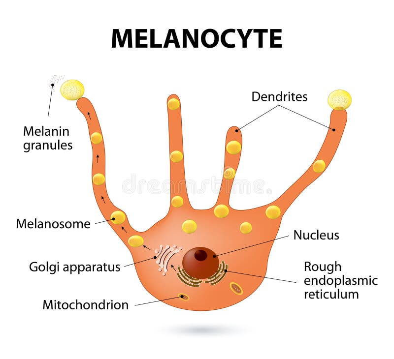
Free with trial Melanocyte, melanin and melanogenesis. Melanocyte - melanin producing cells. Melanin is the pigment responsible for skin color. Vesicle vectors Melanocyte. Melanin and melanogenesis. - melanin producing cells. Melanin is the pigment responsible for skin color
Free with trial Synapse detailed anatomy, beautiful colorful illustration. Neuron passes signal to another neuron. At the right side closer look at Ligand gated ion channel. Vesicle vectors Synapse. Detailed anatomy, beautiful colorful illustration. Neuron passes signal to another neuron. At the right side closer look at Ligand gated ion channel
Free with trial Endoplasmic reticulum and Golgi Apparatus. Protein synthesis and distribution detailed drawing. Vesicle vectors Endoplasmic reticulum and Golgi Apparatus. Protein synthesis and distribution detailed drawing
Free with trial Plant cell. Structure of a vegetal cell. Vesicle vectors Plant cell. Structure of a vegetal cell
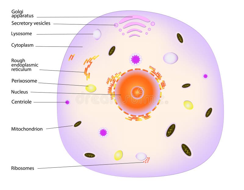
Free with trial Structure of a typical animal cell. Vector. Vesicle vectors Animal cell. Structure of a typical animal cell. Vector. Structure of a typical animal cell. Vector.
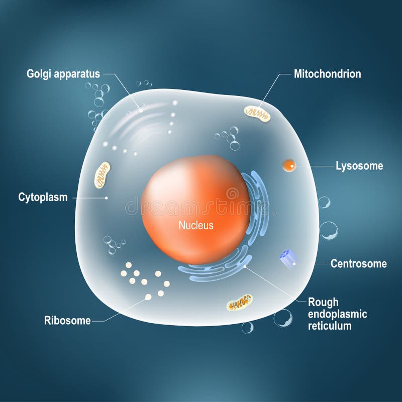
Free with trial Anatomy of cell. All organelles: Nucleus, Ribosome, Rough endoplasmic reticulum, Golgi apparatus, mitochondrion, cytoplasm, lysosome, Centrosome. Animal cell on the dark background. Illustration easy editable for Your color. Vesicle vectors Anatomy of animal cell. Anatomy of cell. All organelles: Nucleus, Ribosome, Rough endoplasmic reticulum, Golgi apparatus, mitochondrion, cytoplasm, lysosome. Anatomy of cell. All organelles: Nucleus, Ribosome, Rough endoplasmic reticulum, Golgi apparatus, mitochondrion, cytoplasm, lysosome, Centrosome. Animal cell on the dark background. Illustration easy editable for Your color
Free with trial Steps of phagocytosis. antigen presenting cell in human immune response. Antigen-presenting cell engulf antigens and process them so that they can be recognized by lymphocytes. Vesicle vectors Antigen-presenting cell. Steps of phagocytosis. antigen presenting cell in human immune response. Antigen-presenting cell engulf antigens and process them so. Steps of phagocytosis. antigen presenting cell in human immune response. Antigen-presenting cell engulf antigens and process them so that they can be recognized by lymphocytes.
Free with trial Vector human cell structure. Cell structure, cross section of the cell detailed anatomy with description. Vesicle vectors Vector human cell structure. Cell structure, cross section of the cell detailed anatomy with description
Free with trial Structure of a synapse, textbook accuracy. Scientifically accurate structure of a synapse, eps8, gradient and mesh printing compatible. Vesicle vectors Structure of a synapse, textbook accuracy. Scientifically accurate structure of a synapse, eps8, gradient and mesh printing compatible
Free with trial Dopamine vector illustration. Labeled diagram with its action and pathways. Scheme with closeup presynaptic axon, terminal, synaptic cleft, dendrite and receiving cells. Vesicle vectors Dopamine vector illustration. Labeled diagram with its action and pathways. Scheme with closeup presynaptic axon, terminal, synaptic cleft, dendrite and
Free with trial Mouth sores. Medical illustration of symptoms of mouth sores. Vesicle vectors Mouth sores. Medical illustration of symptoms of mouth sores
Free with trial Set the cell organelles. Nucleus, endoplasmic reticulum, Golgi apparatus, mitochondria, centrosome, lysosome, the ribosome. Infographics. Vector illustration on isolated background. Vesicle vectors Set the cell organelles. Nucleus, endoplasmic reticulum, Golgi a. Pparatus, mitochondria, centrosome, lysosome, the ribosome. Infographics. Vector illustration. Set the cell organelles. Nucleus, endoplasmic reticulum, Golgi apparatus, mitochondria, centrosome, lysosome, the ribosome. Infographics. Vector illustration on isolated background.
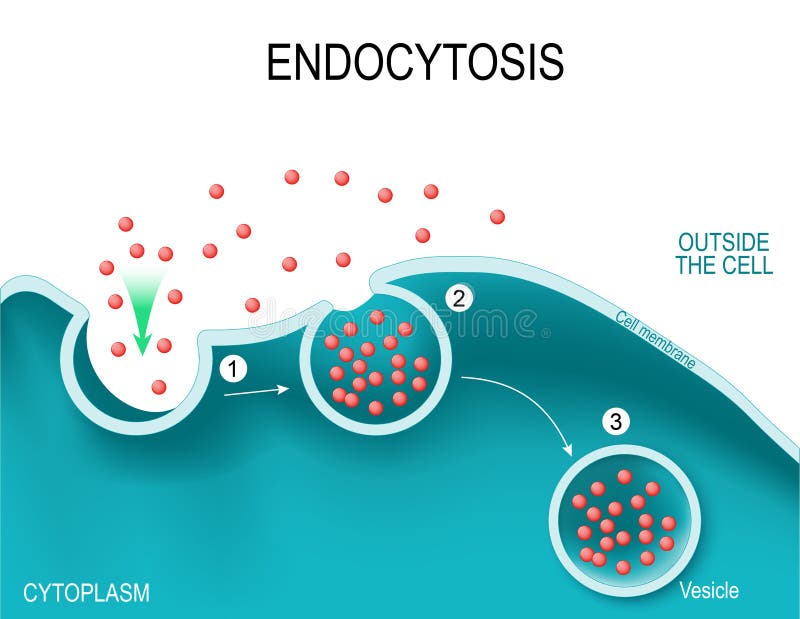
Free with trial Endocytosis Vesicle Transport Cell Membrane. Endocytosis. Type of vesicle transport that carry very large molecules across the cell membrane. Vector illustration. Vesicle vectors Endocytosis Vesicle Transport Cell Membrane. Endocytosis. Type of vesicle transport that carry very large molecules across the cell membrane. Vector illustration
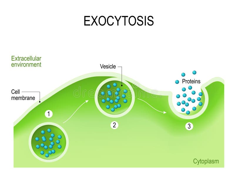
Free with trial Exocytosis Vesicle Transport Cell Membrane. Exocytosis - vesicle transport that carry very large molecules across the cell membrane. Vector illustration. Vesicle vectors Exocytosis Vesicle Transport Cell Membrane. Exocytosis - vesicle transport that carry very large molecules across the cell membrane. Vector illustration
Free with trial Golgi apparatus vector illustration. Labeled microscopic scheme with cisternae, lumen, cis or trans face, cell, secretory and newly forming vesicle. Closeup diagram with receiving and shipping side. Vesicle vectors Golgi apparatus vector illustration. Labeled microscopic scheme and diagram with cisternae, lumen, secretory forming vesicle. Golgi apparatus vector. Golgi apparatus vector illustration. Labeled microscopic scheme with cisternae, lumen, cis or trans face, cell, secretory and newly forming vesicle. Closeup diagram with receiving and shipping side.
Free with trial The different types of endocytosis: receptor-mediated endocytosis, pinocytosis cell drinking and phagocytosis cell eating. vesicle, coated vesicle, and phagosome. vector illustration for medical, educational and science use. Vesicle vectors The different types of endocytosis. Receptor-mediated endocytosis, pinocytosis cell drinking and phagocytosis cell eating. vesicle, coated vesicle, and. The different types of endocytosis: receptor-mediated endocytosis, pinocytosis cell drinking and phagocytosis cell eating. vesicle, coated vesicle, and phagosome. vector illustration for medical, educational and science use
Free with trial Acetylcholine vector illustration. Labeled scheme with structure of neurotransmitter, neuromuscular junction, synaptic vesicle, axon and cleft. Anatomical closeup diagram. Vesicle vectors Acetylcholine vector illustration. Labeled scheme with neurotransmitter. Acetylcholine vector illustration. Labeled scheme with structure of neurotransmitter. Acetylcholine vector illustration. Labeled scheme with structure of neurotransmitter, neuromuscular junction, synaptic vesicle, axon and cleft. Anatomical closeup diagram
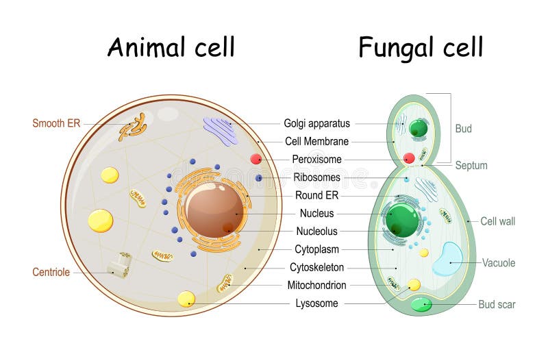
Free with trial Animal cell and fungal yeast cell structure. cross section and anatomy of cell. Biology Chart. Vector illustration on a white background. detailed diagram for use in education. Vesicle vectors Animal cell and fungal yeast cell structure. Cross section and anatomy of cell. Biology Chart. Vector illustration on a white background. detailed diagram for
Free with trial Plant cell anatomy. Cross section of a plant cel. Vesicle vectors Plant cell anatomy. Cross section of a plant cel
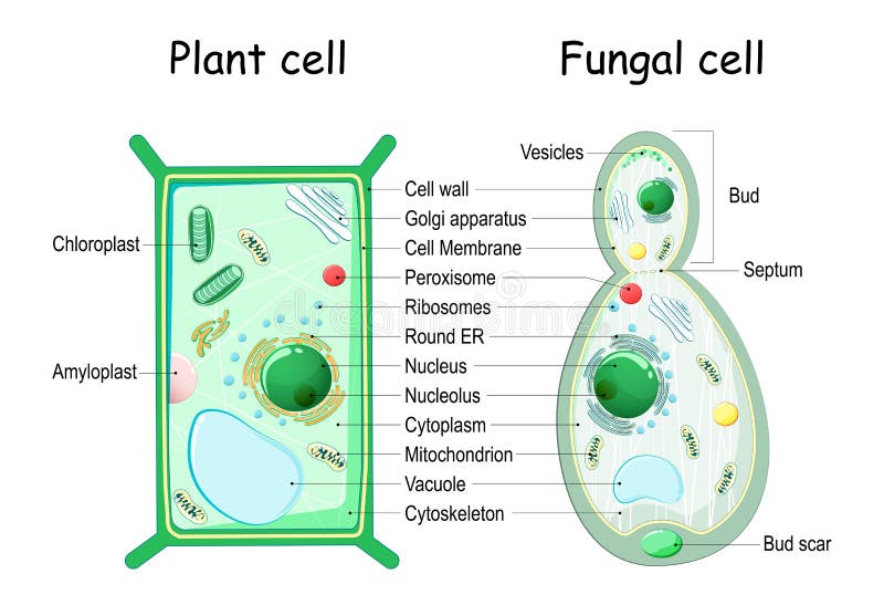
Free with trial Plant cell and fungal yeast cell structure. cross section and anatomy of cell. Biology Chart. Vector illustration on a white background. detailed diagram for use in education. Vesicle vectors Plant cell and fungal yeast cell structure. Cross section and anatomy of cell. Biology Chart. Vector illustration on a white background. detailed diagram for
Free with trial Chlamydia life cycle. bacteria. Sexually transmitted disease and Chlamydia infection. Vesicle vectors The chlamydial life cycle. Chlamydia life cycle. bacteria. Sexually transmitted disease and Chlamydia infection. Chlamydia life cycle. bacteria. Sexually transmitted disease and Chlamydia infection.
Free with trial Male Reproductive System. Easy to edit vector illustration of male reproductive system. Vesicle vectors Male Reproductive System. Easy to edit vector illustration of male reproductive system
Free with trial Exocytosis. Cell transports molecules out of the cell. vesicles are carried to the cell membrane, fuses with membrane, contents are secreted into the extracellular environment. Vesicle vectors Exocytosis. Cell transports molecules out of the cell. vesicles are carried to the cell membrane, fuses with membrane, contents are secreted into the
Free with trial Golgi complex. Part of the eukaryotic cell. Detailed illustration. Vesicle vectors Golgi complex. Part of the eukaryotic cell. Detailed illustration
Free with trial Synapse detailed anatomy, abstract technology background. Neuron passes signal to another neuron. Vesicle vectors Neuron synapse. Synapse detailed anatomy, abstract technology background. Neuron passes signal to another neuron. Synapse detailed anatomy, abstract technology background. Neuron passes signal to another neuron.
Free with trial Golgi apparatus. A part of the eukaryotic cell. Detailed labeled illustration. Vesicle vectors Golgi apparatus. A part of the eukaryotic cell. Detailed labeled illustration
Free with trial Cross section of a Fungal hyphae cells septum; bud scar, mitochondrion; vacuole; nucleus; endoplasmic reticulum; lipid granule; membrane; golgi apparatus, and growth tip and vesicles. Vector diagram for educational, biological, and science use. Vesicle vectors Fungal hyphae cells. Cross section of a Fungal hyphae cells septum; bud scar, mitochondrion; vacuole; nucleus; endoplasmic reticulum; lipid granule; membrane. Cross section of a Fungal hyphae cells septum; bud scar, mitochondrion; vacuole; nucleus; endoplasmic reticulum; lipid granule; membrane; golgi apparatus, and growth tip and vesicles. Vector diagram for educational, biological, and science use
Free with trial Herpesvirus infection. Eps8, gradient and mesh printing compatible. Vesicle vectors Herpesvirus infection. Eps8, gradient and mesh printing compatible
Free with trial Gold fish with bubbles. Illustration of Gold fish with bubbles. Vesicle vectors Gold fish with bubbles. Illustration of Gold fish with bubbles
Free with trial Herpes virus structure. Structure of herpes virus. A portion of the virus has been removed to reveal its inner structure. Vector scheme. Vesicle vectors Herpes virus structure. Structure of herpes virus. A portion of the virus has been removed to reveal its inner structure. Vector scheme
Free with trial Golgi apparatus or Golgi body. Golgi apparatus. Golgi Complex plays an important role in the modification and transport of proteins within the cell. Vesicle vectors Golgi apparatus or Golgi body. Golgi apparatus. Golgi Complex plays an important role in the modification and transport of proteins within the cell
Free with trial Insulin secretion vector illustration. Biological pancreas function labeled scheme. Full cycle diagram with glucose transporter, metabolism, ATP and potassium channel. Vesicle vectors Insulin secretion vector illustration. Biological pancreas function. Insulin secretion vector illustration. Biological pancreas function labeled scheme. Full. Insulin secretion vector illustration. Biological pancreas function labeled scheme. Full cycle diagram with glucose transporter, metabolism, ATP and potassium channel.
Free with trial Synapse vector illustration. Labeled diagram with neuromuscular junction, glandular and other neirons example. Closeup with isolated axon, cleft and dendrite structure. Vesicle vectors Synapse vector illustration. Labeled diagram with neuromuscular example. Synapse vector illustration. Labeled diagram with neuromuscular junction, glandular and. Synapse vector illustration. Labeled diagram with neuromuscular junction, glandular and other neirons example. Closeup with isolated axon, cleft and dendrite structure.
Free with trial Endocytosis and exocytosis in the cell. endocytosis: transports proteins into the cell. exocytosis: transports molecules out of the cytoplasm. vesicles fuses with membrane, contents are secreted into the extracellular environment. Vesicle vectors Endocytosis and exocytosis in the cell. Endocytosis: transports proteins into the cell. exocytosis: transports molecules out of the cytoplasm. vesicles fuses
Free with trial Budding. asexual reproduction of yeast cell. Cross section of a Fungal hyphae cells septum; bud scar, vacuole. Vector diagram for educational, biological, and science use. Vesicle vectors Budding. asexual reproduction of yeast cell. Cross section of a Fungal hyphae cells septum; bud scar, vacuole. Vector diagram for educational, biological, and
Free with trial Synaptic transmission. Structure of a typical chemical synapse. Neurotransmitter release mechanisms. Neurotransmitters are packaged into synaptic vesicles transmit signals from a neuron to a target cell. Vesicle vectors Synaptic transmission. Structure of chemical synapse. Synaptic transmission. Structure of a typical chemical synapse. Neurotransmitter release mechanisms. Synaptic transmission. Structure of a typical chemical synapse. Neurotransmitter release mechanisms. Neurotransmitters are packaged into synaptic vesicles transmit signals from a neuron to a target cell.
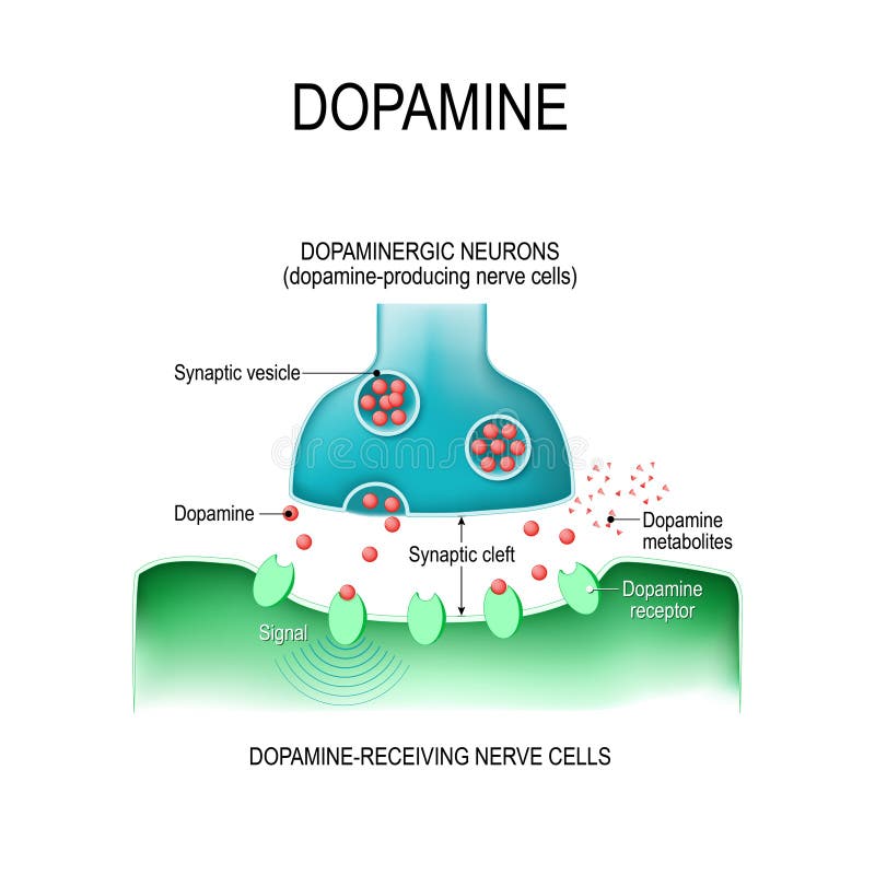
Free with trial Dopamine. two neurons dopamine-producing and dopamine-receiving nerve cells, receptors, and synaptic cleft with dopamine. Vesicle vectors Dopamine. two neurons with receptors, and synaptic cleft with d. Dopamine. two neurons dopamine-producing and dopamine-receiving nerve cells, receptors, and. Dopamine. two neurons dopamine-producing and dopamine-receiving nerve cells, receptors, and synaptic cleft with dopamine.
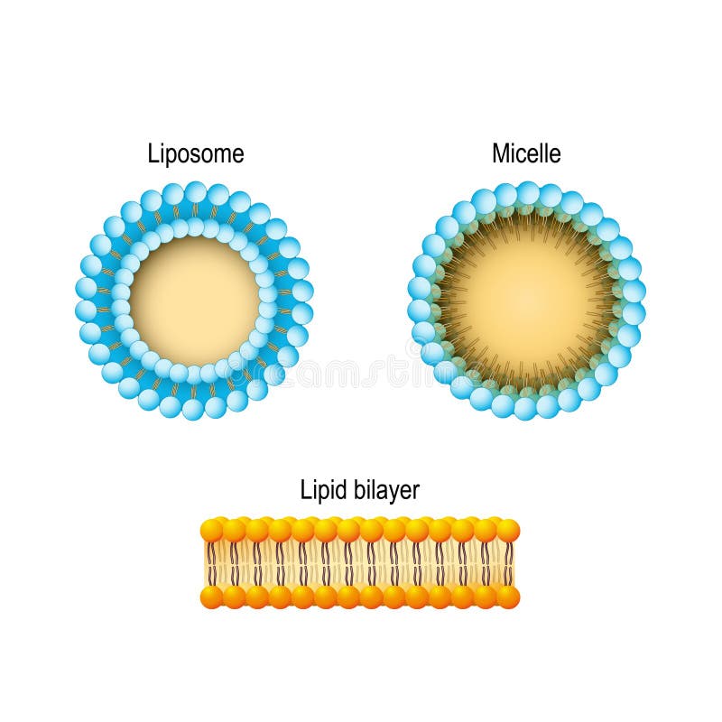
Free with trial Cell membrane Lipid bilayer, Micelle, Liposome. Phospholipids. Cell membrane. Vesicle vectors Cell membrane Lipid bilayer, Micelle, Liposome. Phospholipids. Cell membrane
Free with trial Healthy prostate and BPH. Vector illustration of a healthy prostate and BPH. Vesicle vectors Healthy prostate and BPH. Vector illustration of a healthy prostate and BPH
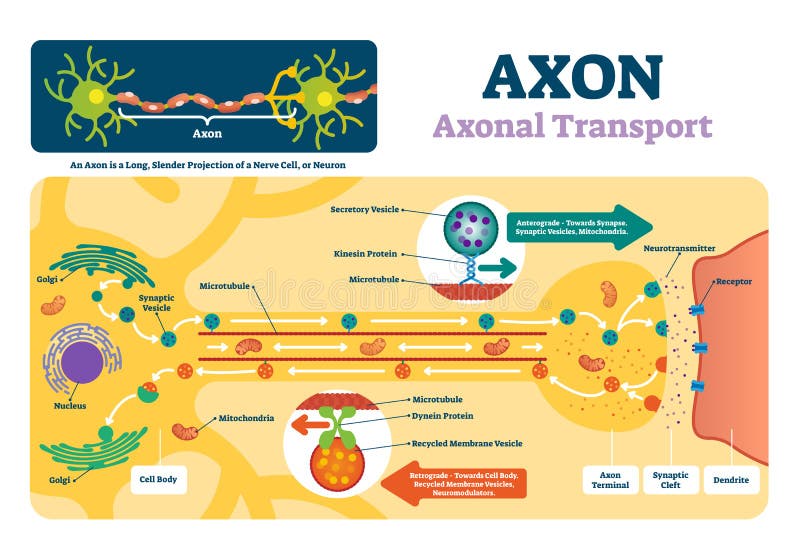
Free with trial Axon vector illustration. Labeled diagram with explanation and structure. Closeup with cell body, terminal, synaptic cleft and dendrite. Nerve cell projection or neuron. Vesicle vectors Axon vector illustration. Labeled diagram with explanation and structure. Closeup with cell body, terminal, synaptic cleft and dendrite. Nerve cell projection
Free with trial Human cell vector illustration. Flat tiny stylized microbiology persons concept. Scientists examine and research life blocks structure. Laboratory for basic structural, functional and biological unit. Vesicle vectors Human cell vector illustration. Tiny stylized microbiology persons concept. Human cell vector illustration. Flat tiny stylized microbiology persons concept. Human cell vector illustration. Flat tiny stylized microbiology persons concept. Scientists examine and research life blocks structure. Laboratory for basic structural, functional and biological unit.
Free with trial Tongue sores. Medical illustration of symptoms of tongue sores. Vesicle vectors Tongue sores. Medical illustration of symptoms of tongue sores
Free with trial Anatomy of the Lysosome: Hydrolytic enzymes, Membrane and transport proteins. This organelle use the enzymes to break down and digest food particles, engulfed viruses or bacteria in the cell. Vector diagram for medical use. Vesicle vectors Anatomy of the Lysosome. Vector diagram for medical use. Anatomy of the Lysosome: Hydrolytic enzymes, Membrane and transport proteins. This organelle use the. Anatomy of the Lysosome: Hydrolytic enzymes, Membrane and transport proteins. This organelle use the enzymes to break down and digest food particles, engulfed viruses or bacteria in the cell. Vector diagram for medical use
Free with trial Anatomy of the Lysosome: Hydrolytic enzymes, Membrane and transport proteins. This organelle use the enzymes to break down and digest food particles, engulfed viruses or bacteria in the cell. Vector diagram for medical use. Vesicle vectors Anatomy of the Lysosome: Hydrolytic enzymes, Membrane and transport proteins. This organelle use the enzymes to break down and digest food particles, engulfed
Free with trial Vesicle and Bulla. Skin lesion. Bulla is a large vesicle containing fluid. Vesicle vectors Vesicle and Bulla. Skin lesion. Bulla is a large vesicle containing fluid
Free with trial Secretory vesicle vector illustration. Labeled closeup infographic scheme with constitutive and regulated secretion. Diagram with golgi apparatus and secreted proteins. Vesicle vectors Secretory vesicle vector illustration. Labeled closeup infographic scheme. Secretory vesicle vector illustration. Labeled closeup infographic scheme with. Secretory vesicle vector illustration. Labeled closeup infographic scheme with constitutive and regulated secretion. Diagram with golgi apparatus and secreted proteins.
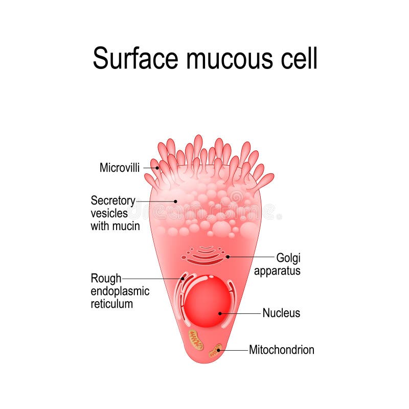
Free with trial Surface mucous cell is a foveolar mucus-producing cell that covering the inside of the stomach. Structure cell: golgi apparatus, secretory vesicle, mucin, nucleus, mitochondrion, microvilli. Vesicle vectors Surface mucous cell. Is a foveolar mucus-producing cell that covering the inside of the stomach. Structure cell: golgi apparatus, secretory vesicle, mucin. Surface mucous cell is a foveolar mucus-producing cell that covering the inside of the stomach. Structure cell: golgi apparatus, secretory vesicle, mucin, nucleus, mitochondrion, microvilli
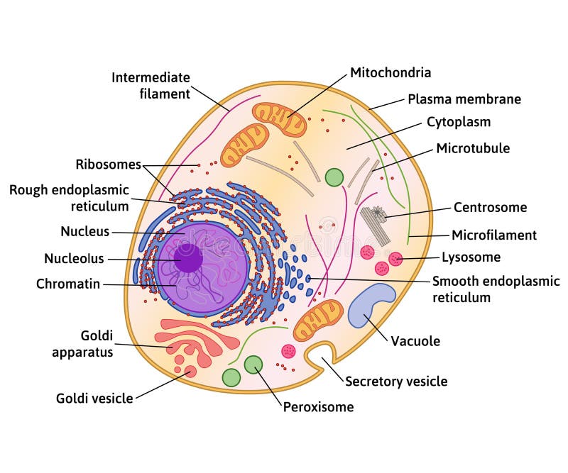
Free with trial Components of a typical cell. structure of human or animal cell. cross section with all organelles: Nucleolus and Nucleus, Ribosome, Vesicle, Rough and Smooth endoplasmic reticulum, Golgi apparatus, Cytoskeleton, mitochondrion, cytoplasm, lysosome, Centrosome, Cell membrane. Vesicle vectors Structure of a typical cell. Components of a typical cell. structure of human or animal cell. cross section with all organelles: Nucleolus and Nucleus, Ribosome. Components of a typical cell. structure of human or animal cell. cross section with all organelles: Nucleolus and Nucleus, Ribosome, Vesicle, Rough and Smooth endoplasmic reticulum, Golgi apparatus, Cytoskeleton, mitochondrion, cytoplasm, lysosome, Centrosome, Cell membrane
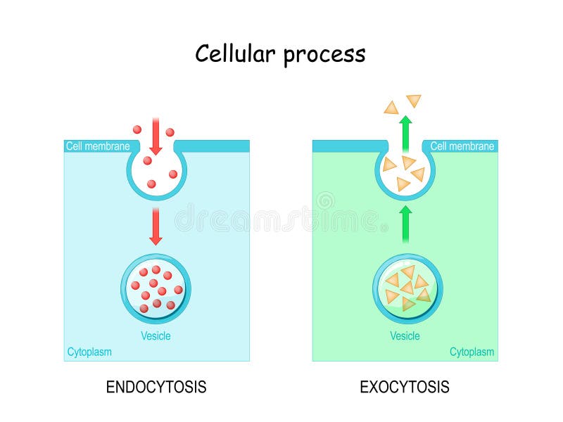
Free with trial Endocytosis and exocytosis are types of vesicle transport that carry very large molecules across the cell membrane. Vector illustration. Vesicle vectors Endocytosis and Exocytosis Diagram. Endocytosis and exocytosis are types of vesicle transport that carry very large molecules across the cell membrane. Vector. Endocytosis and exocytosis are types of vesicle transport that carry very large molecules across the cell membrane. Vector illustration
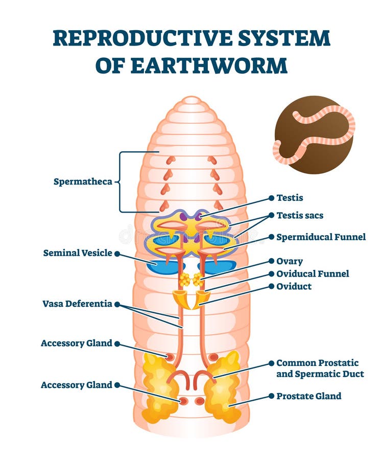
Free with trial Reproductive system of earthworm vector illustration. Labeled educational scheme with anatomical worm structure. Ground soil organism diagram with spermatheca, seminal vesicle, testis sacs and ovary. Vesicle vectors Reproductive system of anatomical earthworm labeled scheme vector illustration. Reproductive system of earthworm vector illustration. Labeled educational scheme. Reproductive system of earthworm vector illustration. Labeled educational scheme with anatomical worm structure. Ground soil organism diagram with spermatheca, seminal vesicle, testis sacs and ovary.
Free with trial Illustration explain the origin of exosomes (also called microvesicles, epididimosomes, argosomes, exosome-like vesicles, microparticles, promininosomes, prostasomes, dexosomes, texosomes, dex, tex, archeosomes and oncosomes). Vesicle vectors Exosomes. Illustration explain the origin of exosomes (also called microvesicles, epididimosomes, argosomes, exosome-like vesicles, microparticles. Illustration explain the origin of exosomes (also called microvesicles, epididimosomes, argosomes, exosome-like vesicles, microparticles, promininosomes, prostasomes, dexosomes, texosomes, dex, tex, archeosomes and oncosomes)
Free with trial Vitiligo. Is a skin condition characterized by portions of the skin losing their pigment. It occurs when skin pigment cells (melanocytes) die or are unable to function. Vesicle vectors Vitiligo. Is a skin condition characterized by portions of the skin losing their pigment. It occurs when skin pigment cells (melanocytes) die or are unable to
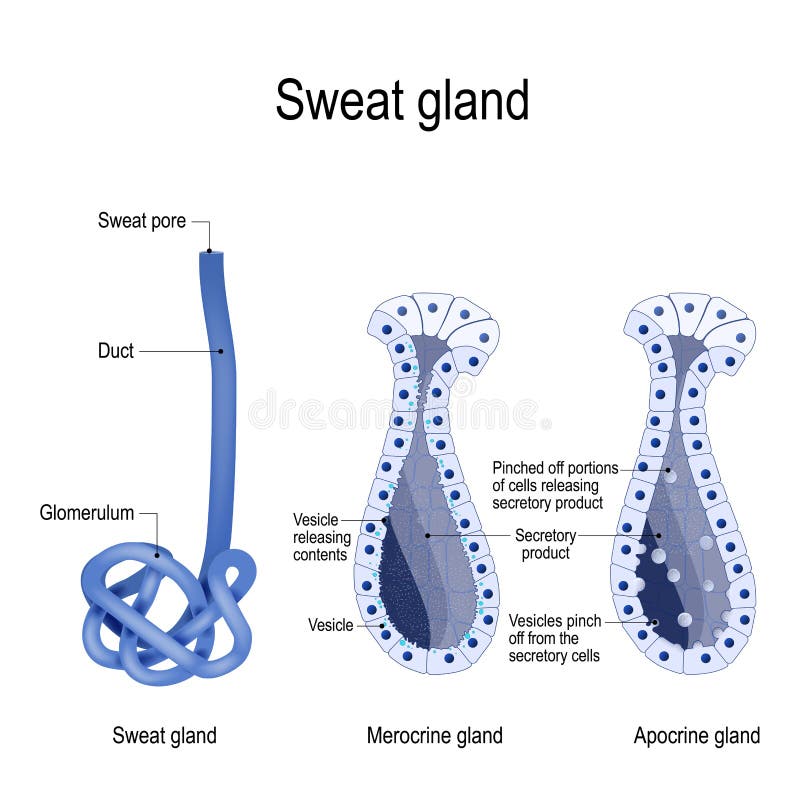
Free with trial Sweat gland. Merocrine and apocrine. different of manner of secretion. cross-section of the human skin, with the sweat gland. Close-up of dark and clear cells, lumen, sweat duct, glomerulum and pore. labeled Vector diagram for educational, medical, biological and science use. Vesicle vectors Sweat gland. cross-section. Sweat gland. Merocrine and apocrine. different of manner of secretion. cross-section of the human skin, with the sweat gland. Close. Sweat gland. Merocrine and apocrine. different of manner of secretion. cross-section of the human skin, with the sweat gland. Close-up of dark and clear cells, lumen, sweat duct, glomerulum and pore. labeled Vector diagram for educational, medical, biological and science use
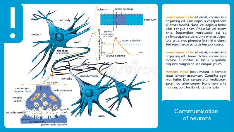
Free with trial Communication of neurons. Infographic diagram. Vesicle vectors Communication of neurons. Infographic diagram
Free with trial Endocytosis. The cell transports proteins into the cell. Vesicle vectors Endocytosis. The cell transports proteins into the cell
Free with trial Autophagy steps. Schematic diagram. Natural mechanism in the cell that removes unnecessary components. Vector illustration. Vesicle vectors Autophagy steps. Schematic diagram. Natural mechanism in the cell that removes unnecessary components. Vector illustration
Free with trial Animal cell anatomy. Cartoon vector illustration of structure of animal cell. Illustration showing the animal cell anatomy. Vesicle vectors Animal cell anatomy. Cartoon vector illustration of structure of animal cell. Illustration showing the animal cell anatomy
Free with trial Melanocyte, melanin and melanogenesis. Structure of Melanocyte melanin producing cells. Melanin is the pigment responsible for skin color. Vesicle vectors Melanocyte, melanin and melanogenesis. Structure of Melanocyte melanin producing cells. Melanin is the pigment responsible for skin color
Free with trial Liver. And gallbladder detailed anatomy illustration on white background. Vesicle vectors Liver. And gallbladder detailed anatomy illustration on white background
Free with trial Dopamine and Antipsychotics. Neuroleptics or tranquilizers are medications that use to manage psychosis hallucinations, paranoia, schizophrenia, bipolar disorder. Neuroleptics blocks the ability of dopamine to activate receptor. two neurons, receptors, and synaptic cleft with dopamine and Neuroleptic. Vesicle vectors Dopamine and Antipsychotic. S. Neuroleptics or tranquilizers are medications that use to manage psychosis hallucinations, paranoia, schizophrenia, bipolar. Dopamine and Antipsychotics. Neuroleptics or tranquilizers are medications that use to manage psychosis hallucinations, paranoia, schizophrenia, bipolar disorder. Neuroleptics blocks the ability of dopamine to activate receptor. two neurons, receptors, and synaptic cleft with dopamine and Neuroleptic.
Free with trial Communication of neurons. 1 diagram with neurotransmitters. Vesicle vectors Communication of neurons. Neurotransmitters. Communication of neurons. 1 diagram with neurotransmitters. Communication of neurons. 1 diagram with neurotransmitters.
Free with trial Golgi apparatus and exocytosis. Cell transports molecules out of the cell. vesicles are carried to the cell membrane, fuses with membrane, contents are secreted into the extracellular environment. Vesicle vectors Golgi apparatus and exocytosis. Cell transports molecules out of the cell. vesicles are carried to the cell membrane, fuses with membrane, contents are
Free with trial Structure of kinesin. Motor proteins. kinesin walking on a microtubule. Vesicle vectors Structure of kinesin. Motor proteins. kinesin walking on a microtubule
Free with trial Cell anatomy vector illustration. Labeled educational structure diagram. Isolated microscopic biological scheme with cytoplasm, mitochondria, ribosome and endomlasmic reticulum location explanation. Vesicle vectors Cell anatomy vector illustration. Labeled educational structure diagram. Isolated microscopic biological scheme with cytoplasm, mitochondria, ribosome and
Free with trial Cut and glue a house. Paper 3d model. Children art game for activity page. Vector illustration. Vesicle vectors Cut and glue a house. Children art game for activity page. Paper 3d model. Vector illustration. Cut and glue a house. Paper 3d model. Children art game for. Cut and glue a house. Paper 3d model. Children art game for activity page. Vector illustration.
Free with trial Endocytosis process with closeup cell side view in anatomical outline diagram. Phagocytosis, pinocytosis or receptor mediated stages explanation vector illustration. Labeled intracellular invagination. Vesicle vectors Endocytosis process with closeup cell side view in anatomical outline diagram. Phagocytosis, pinocytosis or receptor mediated stages explanation vector
Free with trial Fibroblast is a dermis cell vital to the skin`s strength and elasticity. Structure of Fibroblast cell. Vesicle vectors Fibroblast is vital to the skin`s strength and elasticity. Struc. Fibroblast is a dermis cell vital to the skin`s strength and elasticity. Structure of. Fibroblast is a dermis cell vital to the skin`s strength and elasticity. Structure of Fibroblast cell.
Free with trial Difference between exocytosis and endocytosis. The cell transports molecules into and from the cell. Vector illustration for science and educational use. Vesicle vectors Difference between exocytosis and endocytosis. The cell transports molecules into and from the cell. Vector illustration for science and educational use
Free with trial Lysosome Hydrolytic enzymes and Membrane cell / anatomy concept. / white background. Vesicle vectors Lysosome Hydrolytic enzymes and Membrane cell / anatomy concept. / white background
Free with trial Myasthenia gravis as autoimmune, neuromuscular disease outline diagram. Labeled educational medical scheme with skeletal muscles weakness symptoms vector illustration. Anatomical illness explanation. Vesicle vectors Myasthenia gravis as autoimmune, neuromuscular disease outline diagram. Labeled educational medical scheme with skeletal muscles weakness symptoms vector
Free with trial Lysosome Hydrolytic enzymes, Membrane and transport proteins,. Science and medical use, Vector Illustration. Vesicle vectors Lysosome Hydrolytic enzymes, Membrane and transport proteins,. Science and medical use, Vector Illustration
Free with trial Infographics of prostate cancer illustration. Prostate cancer symptoms, risk factors. Prostate cancer symptoms. Vesicle vectors Infographics of prostate cancer illustration. Prostate cancer symptoms, risk factors. Infographics of prostate cancer illustration. Prostate cancer symptoms
Free with trial Anatomy neyron and synapse. Signng in the brain. Cells neyron and synapse with descriptions. Structure neyron for educational, medical, biological use. Transmission of impulses in a living organism. Synapse electronic pulses. Vector illustration. Vesicle vectors Anatomy neyron and synapse. Signaling in the brain. Cells neyron and synapse with descriptions. Structure neyron for. Anatomy neyron and synapse. Signng in the. Anatomy neyron and synapse. Signng in the brain. Cells neyron and synapse with descriptions. Structure neyron for educational, medical, biological use.Transmission of impulses in a living organism. Synapse electronic pulses. Vector illustration.
Free with trial Anatomy neyron cells. Transmission signal of impulse in a living organism. Signaling in the brain. Neural connections in the brain form thoughts, concept learning. Structure neyron for educational, medical, biological use. Synapse electronic pulses. Vector illustration. Vesicle vectors Anatomy neyron cells. Transmission signal of impulse in a living organism. Signaling in the brain. Neural connections in. The brain form thoughts, concept. Anatomy neyron cells. Transmission signal of impulse in a living organism. Signaling in the brain. Neural connections in the brain form thoughts, concept learning. Structure neyron for educational, medical, biological use. Synapse electronic pulses. Vector illustration.
Free with trial Anatomy neyron cells. Transmission signal of impulse in a living organism. Signaling in the brain. Neural connections in the brain form thoughts, concept learning. Structure neyron for educational, medical, biological use. Synapse electronic pulses. Vector illustration. Vesicle vectors Anatomy neyron cells. Transmission signal of impulse in a living organism. Signaling in the brain. Neural connections in. The brain form thoughts, concept. Anatomy neyron cells. Transmission signal of impulse in a living organism. Signaling in the brain. Neural connections in the brain form thoughts, concept learning. Structure neyron for educational, medical, biological use. Synapse electronic pulses. Vector illustration.
Free with trial Neurons communication. Transmission of the nerve signal between two neurons. Close-up of a Synaptic cleft, chemical synapse, synaptic vesicle, Neurotransmitter. Vector illustration. Vesicle vectors Close-up of a Synaptic cleft, chemical synapse, synaptic vesicle, Neurotransmitter. Neurons communication. Transmission of the nerve signal between two neurons. Neurons communication. Transmission of the nerve signal between two neurons. Close-up of a Synaptic cleft, chemical synapse, synaptic vesicle, Neurotransmitter. Vector illustration
Free with trial Neurotransmitter process with synapse, vesicle and receptors outline diagram. Labeled educational scheme with neurology chemical messengers for serotonin or dopamine production vector illustration. Vesicle vectors Neurotransmitter process with synapse, vesicle and receptors outline diagram. Labeled educational scheme with neurology chemical messengers for serotonin or
 Sign in