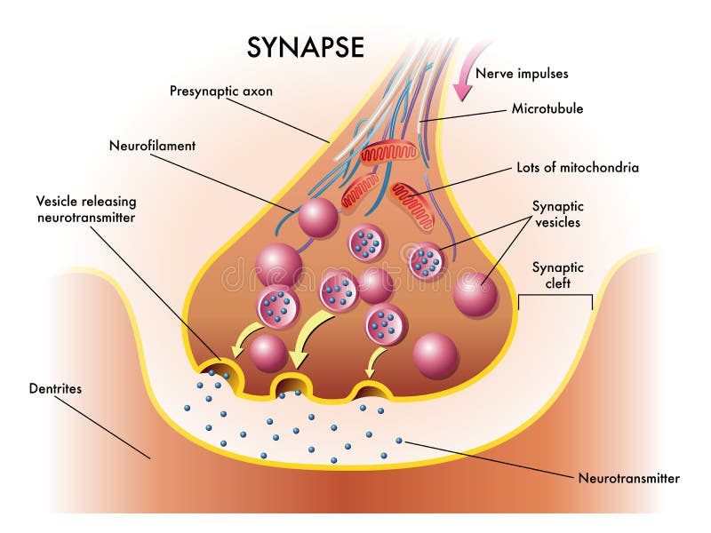
Free with trial Synapse. Medical illustration of elements of synapse. Vesicles vectors Synapse. Medical illustration of elements of synapse
Free with trial Structure of a typical chemical synapse. neurotransmitter release mechanisms. Neurotransmitters are packaged into synaptic vesicles transmit signals from a neuron to a target cell across a synapse. Vector diagram. Vesicles vectors Structure of a typical chemical synapse. Neurotransmitter release mechanisms. Neurotransmitters are packaged into synaptic vesicles transmit signals from a
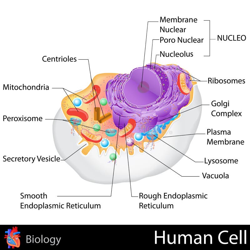
Free with trial Human Cell. Easy to edit vector illustration of human cell structure. Vesicles vectors Human Cell. Easy to edit vector illustration of human cell structure
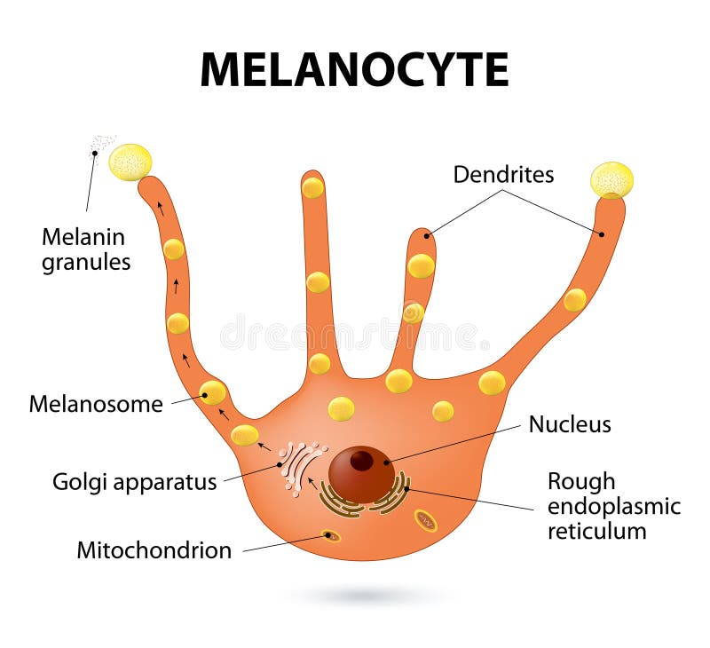
Free with trial Melanocyte, melanin and melanogenesis. Melanocyte - melanin producing cells. Melanin is the pigment responsible for skin color. Vesicles vectors Melanocyte. Melanin and melanogenesis. - melanin producing cells. Melanin is the pigment responsible for skin color
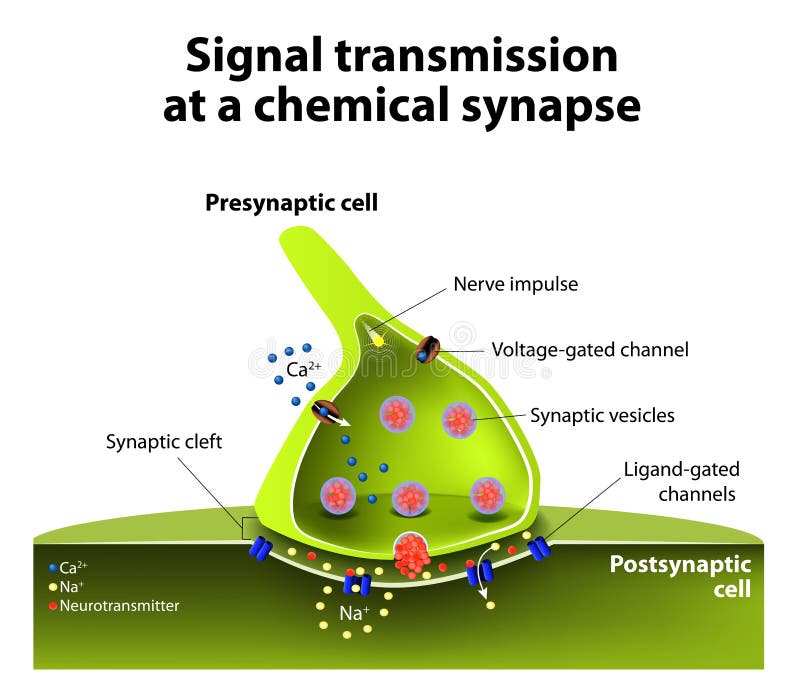
Free with trial Signal transmission at a chemical synapse. one neuron releases neurotransmitter molecules into a synaptic cleft that is adjacent to another neuron. Vesicles vectors Chemical synapse. Signal transmission at a chemical synapse. one neuron releases neurotransmitter molecules into a synaptic cleft that is adjacent to another. Signal transmission at a chemical synapse. one neuron releases neurotransmitter molecules into a synaptic cleft that is adjacent to another neuron.
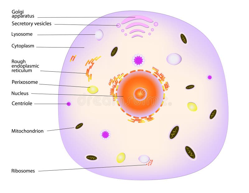
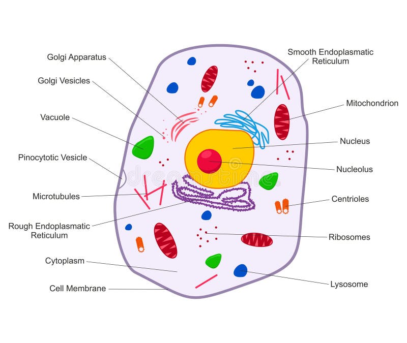
Free with trial A typical cell, labeled. Major components of an animal cell, eps10. Vesicles vectors A typical cell, labeled. Major components of an animal cell, eps10
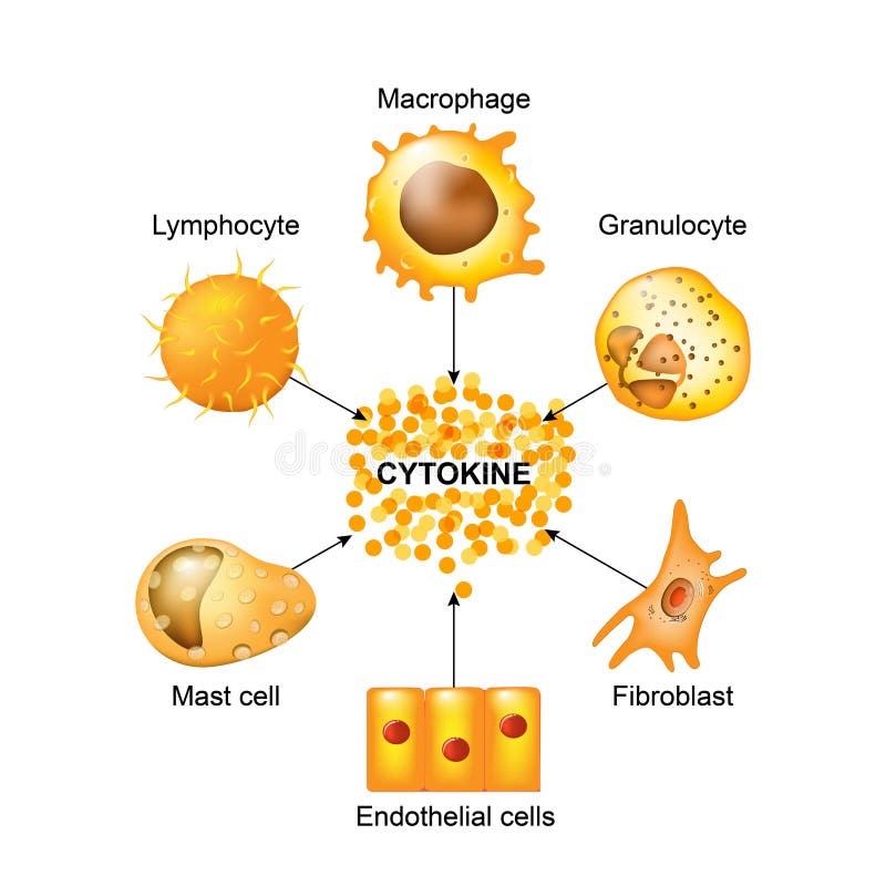
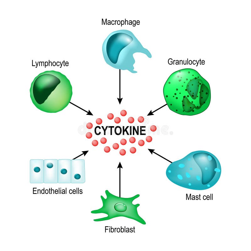
Free with trial Cytokines are produced by macrophages, lymphocytes, mast cells, endothelial cells and fibroblasts. Cytokines include chemokines, interferons, interleukins, lymphokines, and tumour necrosis factors, but not hormones or growth factors. Vesicles vectors Cells that produce cytokines. Cytokines are produced by macrophages, lymphocytes, mast cells, endothelial cells and fibroblasts. Cytokines include chemokines. Cytokines are produced by macrophages, lymphocytes, mast cells, endothelial cells and fibroblasts. Cytokines include chemokines, interferons, interleukins, lymphokines, and tumour necrosis factors, but not hormones or growth factors
Free with trial Structure of a typical animal cell. Vector. Vesicles vectors Animal cell. Structure of a typical animal cell. Vector. Structure of a typical animal cell. Vector.
Free with trial Labial herpes. Medical illustration of the effects of the labial herpes infection. Vesicles vectors Labial herpes. Medical illustration of the effects of the labial herpes infection
Free with trial Varicella zoster virus. Medical illustration of the varicella zoster virus. Vesicles vectors Varicella zoster virus. Medical illustration of the varicella zoster virus
Free with trial Neuromuscular junction vector illustration scheme. Labeled medical infographic. Motor neuron and muscle cell structure closeup. Diagram with myofibril and muscle fibers. Vesicles vectors Neuromuscular junction vector illustration scheme. Labeled cell infographic. Neuromuscular junction vector illustration scheme. Labeled medical infographic. Neuromuscular junction vector illustration scheme. Labeled medical infographic. Motor neuron and muscle cell structure closeup. Diagram with myofibril and muscle fibers.
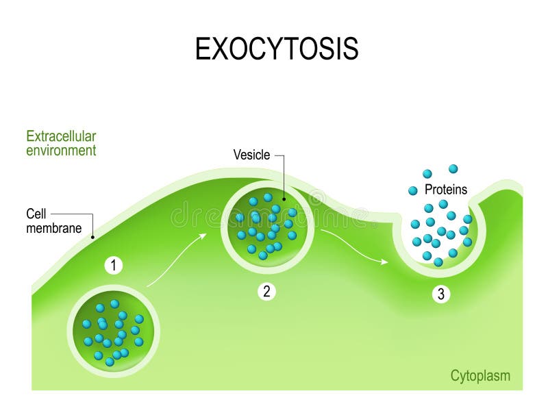
Free with trial Exocytosis. Cell transports molecules out of the cell. vesicles are carried to the cell membrane, fuses with membrane, contents are secreted into the extracellular environment. Vesicles vectors Exocytosis. Cell transports molecules out of the cell. vesicles are carried to the cell membrane, fuses with membrane, contents are secreted into the
Free with trial Endocytosis and exocytosis in the cell. endocytosis: transports proteins into the cell. exocytosis: transports molecules out of the cytoplasm. vesicles fuses with membrane, contents are secreted into the extracellular environment. Vesicles vectors Endocytosis and exocytosis in the cell. Endocytosis: transports proteins into the cell. exocytosis: transports molecules out of the cytoplasm. vesicles fuses
Free with trial Neurotransmitter release mechanisms. Neurotransmitters are packaged into synaptic vesicles transmit signals from a neuron to a target cell across a synapse. Vesicles vectors Structure of a typical chemical synapse. Neurotransmitter release mechanisms. Neurotransmitters are packaged into synaptic vesicles transmit signals from a. Neurotransmitter release mechanisms. Neurotransmitters are packaged into synaptic vesicles transmit signals from a neuron to a target cell across a synapse.
Free with trial Synaptic transmission. Structure of a typical chemical synapse. Neurotransmitter release mechanisms. Neurotransmitters are packaged into synaptic vesicles transmit signals from a neuron to a target cell. Vesicles vectors Synaptic transmission. Structure of chemical synapse. Synaptic transmission. Structure of a typical chemical synapse. Neurotransmitter release mechanisms. Synaptic transmission. Structure of a typical chemical synapse. Neurotransmitter release mechanisms. Neurotransmitters are packaged into synaptic vesicles transmit signals from a neuron to a target cell.
Free with trial Structure of a typical chemical synapse. neurotransmitter release mechanisms. Neurotransmitters are packaged into synaptic vesicles transmit signals from a neuron to a target cell across a synapse. Vesicles vectors Structure of a typical chemical synapse. neurotransmitter release mechanisms. Neurotransmitters are packaged into synaptic vesicles transmit signals from a
Free with trial Eczema. Medical illustration of the symptoms of eczema. Vesicles vectors Eczema. Medical illustration of the symptoms of eczema
Free with trial Cytokines are produced by macrophages, lymphocytes, mast cells, endothelial cells and fibroblasts. Cytokines include chemokines, interferons, interleukins, lymphokines, and tumour necrosis factors, but not hormones or growth factors. Vesicles vectors Cytokine. Vector concept. Cytokines are produced by macrophages, lymphocytes, mast cells, endothelial cells and fibroblasts. Cytokines include chemokines. Cytokines are produced by macrophages, lymphocytes, mast cells, endothelial cells and fibroblasts. Cytokines include chemokines, interferons, interleukins, lymphokines, and tumour necrosis factors, but not hormones or growth factors
Free with trial Eosinophil granulocytes. type of White Blood Cell that responsible for combating multicellular parasites and certain infections in vertebrates, they also control mechanisms associated with allergy and asthma. Characteristics and structure of lymphocytes. Vesicles vectors Eosinophil. Granulocytes. type of White Blood Cell that responsible for combating multicellular parasites and certain infections in vertebrates, they also. Eosinophil granulocytes. type of White Blood Cell that responsible for combating multicellular parasites and certain infections in vertebrates, they also control mechanisms associated with allergy and asthma. Characteristics and structure of lymphocytes
Free with trial Cross section of a Fungal hyphae cells septum; bud scar, mitochondrion; vacuole; nucleus; endoplasmic reticulum; lipid granule; membrane; golgi apparatus, and growth tip and vesicles. Vector diagram for educational, biological, and science use. Vesicles vectors Fungal hyphae cells. Cross section of a Fungal hyphae cells septum; bud scar, mitochondrion; vacuole; nucleus; endoplasmic reticulum; lipid granule; membrane. Cross section of a Fungal hyphae cells septum; bud scar, mitochondrion; vacuole; nucleus; endoplasmic reticulum; lipid granule; membrane; golgi apparatus, and growth tip and vesicles. Vector diagram for educational, biological, and science use
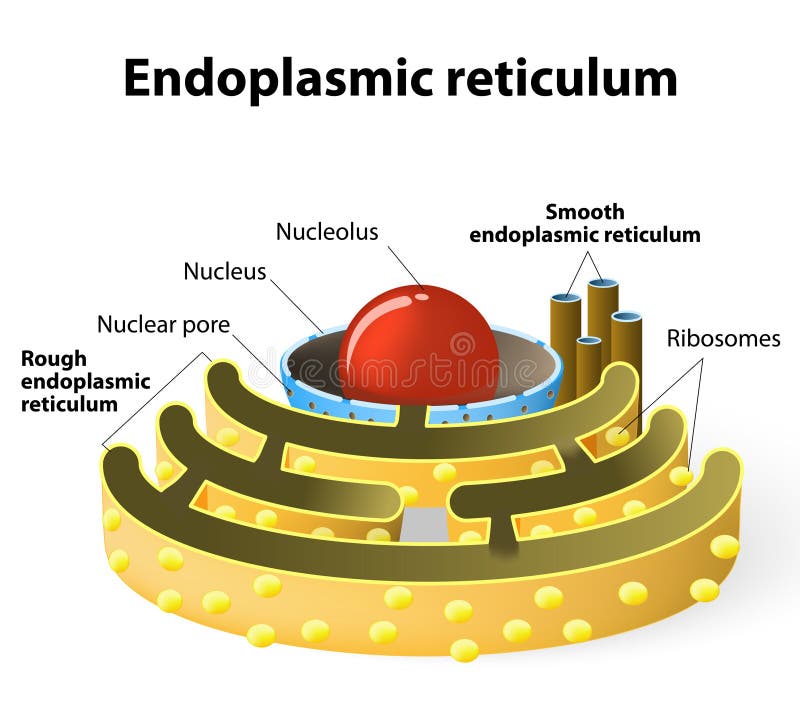
Free with trial Endoplasmic reticulum is a continuous membrane, which is present in both plant cells, animal cells and absent in prokaryotic cells. Vesicles vectors Endoplasmic reticulum. Is a continuous membrane, which is present in both plant cells, animal cells and absent in prokaryotic cells
Free with trial Human neuron on a white background. Human nerve cell on a white background. Vesicles vectors Human neuron on a white background. Human nerve cell on a white background
Free with trial Plant cell isolated on white vector. Plant cell isolated on white photo-realistic vector illustration. Vesicles vectors Plant cell isolated on white vector. Plant cell isolated on white photo-realistic vector illustration
Free with trial Chickenpox. Medical illustration of the symptoms of chickenpox. Vesicles vectors Chickenpox. Medical illustration of the symptoms of chickenpox
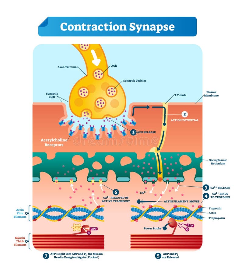
Free with trial Contraction synapse vector illustration. Labeled closeup medical structure scheme. Diagram with full cycle of ACH release, action potential, troponin bonding and filament. Vesicles vectors Contraction synapse vector illustration. Labeled medical structure scheme. Contraction synapse vector illustration. Labeled closeup medical structure scheme. Contraction synapse vector illustration. Labeled closeup medical structure scheme. Diagram with full cycle of ACH release, action potential, troponin bonding and filament
Free with trial Cell signaling. Signalling mechanism in cells: intracrine, autocrine and endocrine signals. Vesicles vectors Cell signaling. intracrine, autocrine and endocrine signals. Cell signaling. Signalling mechanism in cells: intracrine, autocrine and endocrine signals. Cell signaling. Signalling mechanism in cells: intracrine, autocrine and endocrine signals.
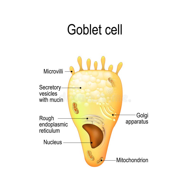
Free with trial Goblet cell. simple columnar epithelial cell for secrete mucus. They are found inside the trachea, bronchi, small and large intestine, and conjunctiva in the eyes. Structure cell nucleus and other organelles. Vesicles vectors Goblet cell. Structure cell nucleus and other organelles. Goblet cell. simple columnar epithelial cell for secrete mucus. They are found inside the trachea. Goblet cell. simple columnar epithelial cell for secrete mucus. They are found inside the trachea, bronchi, small and large intestine, and conjunctiva in the eyes. Structure cell nucleus and other organelles.
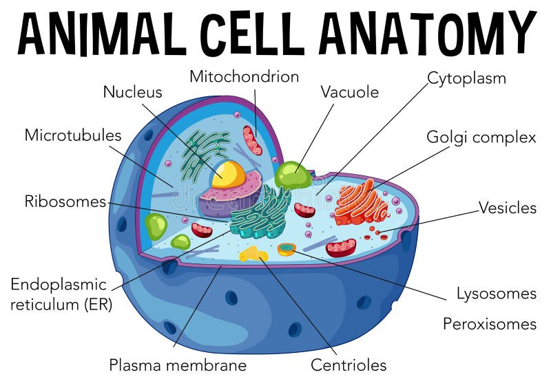
Free with trial Diagram of animal cell anatomy. Illustration. Vesicles vectors Diagram of animal cell anatomy. Illustration
Free with trial Biosynthesis of protein on ribosome in vector. Vesicles vectors Biosynthesis of protein on ribosome in vector.
Free with trial Plant Cell Anatomy diagram. Illustration. Vesicles vectors Plant Cell Anatomy diagram. Illustration
Free with trial Illustration explain the origin of exosomes (also called microvesicles, epididimosomes, argosomes, exosome-like vesicles, microparticles, promininosomes, prostasomes, dexosomes, texosomes, dex, tex, archeosomes and oncosomes). Vesicles vectors Exosomes. Illustration explain the origin of exosomes (also called microvesicles, epididimosomes, argosomes, exosome-like vesicles, microparticles. Illustration explain the origin of exosomes (also called microvesicles, epididimosomes, argosomes, exosome-like vesicles, microparticles, promininosomes, prostasomes, dexosomes, texosomes, dex, tex, archeosomes and oncosomes)
Free with trial Golgi apparatus and exocytosis. Cell transports molecules out of the cell. vesicles are carried to the cell membrane, fuses with membrane, contents are secreted into the extracellular environment. Vesicles vectors Golgi apparatus and exocytosis. Cell transports molecules out of the cell. vesicles are carried to the cell membrane, fuses with membrane, contents are
Free with trial Vitiligo. Is a skin condition characterized by portions of the skin losing their pigment. It occurs when skin pigment cells (melanocytes) die or are unable to function. Vesicles vectors Vitiligo. Is a skin condition characterized by portions of the skin losing their pigment. It occurs when skin pigment cells (melanocytes) die or are unable to
Free with trial Cup of coffee. An illustration of cup of coffee or hot chocolate. Vesicles vectors Cup of coffee. An illustration of cup of coffee or hot chocolate
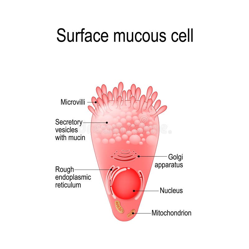
Free with trial Surface mucous cell is a foveolar mucus-producing cell that covering the inside of the stomach. Structure cell: golgi apparatus, secretory vesicle, mucin, nucleus, mitochondrion, microvilli. Vesicles vectors Surface mucous cell. Is a foveolar mucus-producing cell that covering the inside of the stomach. Structure cell: golgi apparatus, secretory vesicle, mucin. Surface mucous cell is a foveolar mucus-producing cell that covering the inside of the stomach. Structure cell: golgi apparatus, secretory vesicle, mucin, nucleus, mitochondrion, microvilli
Free with trial Factors involved in damage to and inflammation of perineal skin. Vesicles vectors Perineal skin damage. Factors involved in damage to and inflammation of perineal skin. Factors involved in damage to and inflammation of perineal skin.
Free with trial Signal transmission at a chemical synapse. one neuron releases neurotransmitter molecules into a synaptic cleft that is adjacent to another neuron. Vesicles vectors Signal transmission at a chemical synapse. One neuron releases neurotransmitter molecules into a synaptic cleft that is adjacent to another neuron
Free with trial Glucose transport through cell membrane via. Transporters activated by insulin affecting its receptors. Vesicles vectors Glucose transport through cell membrane via. Transporters activated by insulin affecting its receptors
Free with trial Melanocyte, melanin and melanogenesis. Structure of Melanocyte melanin producing cells. Melanin is the pigment responsible for skin color. Vesicles vectors Melanocyte, melanin and melanogenesis. Structure of Melanocyte melanin producing cells. Melanin is the pigment responsible for skin color
Free with trial Vector beer background. Vector beer foam background horizontally seamless pattern. Vesicles vectors Vector beer background. Vector beer foam background horizontally seamless pattern
Free with trial Glucose transport through cell membrane via. Transporters activated by insulin. Vesicles vectors Glucose transport through cell membrane via. Transporters activated by insulin
Free with trial Anatomy of animal cell (Biology Diagram. ) illustration. Vesicles vectors Anatomy of animal cell (Biology Diagram. ) illustration
Free with trial Animal Cell structure. Cross section of the cell detailed colorful anatomy with description. Animal cell in flat style on white background. Vesicles vectors Animal Cell structure. Cross section of the cell detailed colorful anatomy with description. Animal cell in flat style. On white background
Free with trial Anatomy neyron and synapse. Signng in the brain. Cells neyron and synapse with descriptions. Structure neyron for educational, medical, biological use. Transmission of impulses in a living organism. Synapse electronic pulses. Vector illustration. Vesicles vectors Anatomy neyron and synapse. Signaling in the brain. Cells neyron and synapse with descriptions. Structure neyron for. Anatomy neyron and synapse. Signng in the. Anatomy neyron and synapse. Signng in the brain. Cells neyron and synapse with descriptions. Structure neyron for educational, medical, biological use.Transmission of impulses in a living organism. Synapse electronic pulses. Vector illustration.
Free with trial Neurotransmission. the presynaptic neuron releases a neurotransmitter, which activates receptors on the postsynaptic neuron. Synapse Structure. Neurotransmitter, synaptic vesicles and synaptic cleft. Anatomy of neurons. Vector Illustration. Vesicles vectors Synapse Structure. Neurotransmitter, synaptic vesicles and synaptic cleft. Neurotransmission. the presynaptic neuron releases a neurotransmitter, which activates. Neurotransmission. the presynaptic neuron releases a neurotransmitter, which activates receptors on the postsynaptic neuron. Synapse Structure. Neurotransmitter, synaptic vesicles and synaptic cleft. Anatomy of neurons. Vector Illustration
Free with trial Posterior face of the bladder, seminal vesicles and prostate, vi. Ntage engraved illustration. Usual Medicine Dictionary by Dr Labarthe - 1885. Vesicles vectors Posterior face of the bladder, seminal vesicles and prostate, vi. Ntage engraved illustration. Usual Medicine Dictionary by Dr Labarthe - 1885
Free with trial Chemical synapse structure. Neuron communication. Close-up of axon terminal with Synaptic vesicles, Voltage-gated channel, and Neurotransmitter transporters. neurotransmitter binds to receptor that located in the membrane of another neuron on the opposite side of the synaptic cleft. Vesicles vectors Chemical synapse structure. Neuron communication. Close-up of axon terminal with Synaptic vesicles, Voltage-gated channel, and Neurotransmitter transporters
Free with trial Neuronal communication. The dendrites contain receptors for neurotransmitters released by nearby neurons. Soma, dendrites, axons, terminal buttons, and synaptic vesicles. Vesicles vectors Neuronal communication. The dendrites contain receptors for neurotransmitters released by nearby neurons. Soma, dendrites, axons, terminal buttons, and
Free with trial Location pulmonary alveoli clipart. Pink lungs with red vessels and healthy bronchial vesicles good breathing and resistance vector pneumonia. Vesicles vectors Location pulmonary alveoli clipart. Pink lungs with red vessels and healthy bronchial vesicles good breathing. Location pulmonary alveoli clipart. Pink lungs. Location pulmonary alveoli clipart. Pink lungs with red vessels and healthy bronchial vesicles good breathing and resistance vector pneumonia.
Free with trial Botulinum toxin mechanism illustration shows a nerve terminal blocking acetylcholine release, key objects, synaptic vesicles, SNARE proteins, ACh receptor. Outline diagram. Vesicles vectors Botulinum toxin mechanism illustration shows a nerve terminal blocking acetylcholine ... Botulinum toxin mechanism illustration shows a nerve terminal blocking. Botulinum toxin mechanism illustration shows a nerve terminal blocking acetylcholine release, key objects, synaptic vesicles, SNARE proteins, ACh receptor. Outline diagram
Free with trial Anatomy of a G-cell. This cells are located in the gastric glands into stomach. G cell is a type of cell in the stomach and duodenum that secretes hormone gastrin. Vesicles vectors Anatomy of a G-cell. gastrin. Anatomy of a G-cell. This cells are located in the gastric glands into stomach. G cell is a type of cell in the stomach and. Anatomy of a G-cell. This cells are located in the gastric glands into stomach. G cell is a type of cell in the stomach and duodenum that secretes hormone gastrin.
Free with trial Mucous neck cell is a foveolar mucus-producing cell that covering the inside of the stomach. Structure cell: golgi apparatus, secretory vesicle, nucleus, mitochondrion, and microvilli. Vesicles vectors Mucous neck cell. Is a foveolar mucus-producing cell that covering the inside of the stomach. Structure cell: golgi apparatus, secretory vesicle, nucleus. Mucous neck cell is a foveolar mucus-producing cell that covering the inside of the stomach. Structure cell: golgi apparatus, secretory vesicle, nucleus, mitochondrion, and microvilli
Free with trial Three bottles of alcoholic drinks, vodka, cognac and brandy. Vesicles vectors Three bottles of alcohol drinks. Three bottles of alcoholic drinks, vodka, cognac and brandy. Three bottles of alcoholic drinks, vodka, cognac and brandy.
Free with trial Paneth cell it is the epithelium cells of the small intestine. their granules consist of anti-microbial compounds for important in immunity. Structure of the Paneth cell nucleus, mitochondria and other organelles. Vesicles vectors Paneth cell. It is the epithelium cells of the small intestine. their granules consist of anti-microbial compounds for important in immunity. Structure of the. Paneth cell it is the epithelium cells of the small intestine. their granules consist of anti-microbial compounds for important in immunity. Structure of the Paneth cell nucleus, mitochondria and other organelles.
Free with trial Gastric chief peptic, gastric zymogenic cell. release the zymogen enzyme. this cells are located in the gastric glands into stomach. Structure of a chief cell: golgi apparatus, nucleus, mitochondrion, microvilli. Vesicles vectors Gastric chief peptic, gastric zymogenic cell. Release the zymogen enzyme. this cells are located in the gastric glands into stomach. Structure of a chief cell
Free with trial An image of a human male reproductive systems chart. Vesicles vectors Human Male Reproductive Systems Chart. An image of a human male reproductive systems chart. An image of a human male reproductive systems chart.
Free with trial Plant cell isolated on white photo-realistic vector illustration. Vesicles vectors Plant cell isolated on white photo-realistic vector illustration.
Free with trial Structure of an animal cell. Illustration of structure of an animal cell. Vesicles vectors Structure of an animal cell. Illustration of structure of an animal cell
Free with trial Herpangina Caused by Coxsackievirus. Medical Illustration of Coxsackievirus Infection in Throat and Mouth. Oral Herpangina Ulcers from Coxsackie Virus. Vesicles vectors Herpangina Caused by Coxsackievirus. Medical Illustration of Coxsackievirus Infection in Throat and Mouth. Oral Herpangina Ulcers from Coxsackie Virus
Free with trial Neuron anatomy. Close-up of a Chemical synapse, Synaptic vesicle with neurotransmitter, and Receptors. Structure of Synaptic cleft. Vector poster. Vesicles vectors Neuron anatomy. Close-up of a Chemical synapse. Synaptic vesicle with neurotransmitter, and Receptors. Structure of Synaptic cleft. Vector poster
Free with trial Sunburn. Symbolic medical illustration of the consequences of sunburn. Vesicles vectors Sunburn. Symbolic medical illustration of the consequences of sunburn
Free with trial Plant cell isolated on white photo-realistic vector illustration. Vesicles vectors Plant cell isolated on white photo-realistic vector illustration.
Free with trial Epithelial Tissue with Goblet Cells. Goblet Cell Structure Illustration. Mucus Secreting. Human Goblet Cell Anatomy. Educational Chart. Vesicles vectors Epithelial Tissue with Goblet Cells. Goblet Cell Structure Illustration. Mucus Secreting. Human Goblet Cell Anatomy. Educational Chart
Free with trial Severe sunburn. Medical illustration of the consequences of severe sunburn. Vesicles vectors Severe sunburn. Medical illustration of the consequences of severe sunburn
Free with trial In the nervous system, Synapse is a connection between nerve cells. Vesicles vectors Synapse is a structure that permits a neuron. In the nervous system, Synapse is a connection between nerve cells. In the nervous system, Synapse is a connection between nerve cells.
Free with trial Neurons communication. Transmission of the nerve signal between two neurons. Close-up of a Synaptic cleft, chemical synapse, synaptic vesicle, Neurotransmitter. Vector illustration. Vesicles vectors Close-up of a Synaptic cleft, chemical synapse, synaptic vesicle, Neurotransmitter. Neurons communication. Transmission of the nerve signal between two neurons. Neurons communication. Transmission of the nerve signal between two neurons. Close-up of a Synaptic cleft, chemical synapse, synaptic vesicle, Neurotransmitter. Vector illustration
Free with trial Centriole is a cylindrical organelle in the cell. Vesicles vectors Anatomy of centriole. Centriole is a cylindrical organelle in the cell. Centriole is a cylindrical organelle in the cell.
Free with trial Diagram of animal cell eukaryotic cell labeled. Animal cell eukaryotic cell diagram, A typical cell labeled vector illustration on white background. Vesicles vectors Diagram of animal cell eukaryotic cell labeled. Animal cell eukaryotic cell diagram, A typical cell labeled vector illustration on white background
Free with trial Melanocyte structure and anatomy. melanin producing cells. Melanin is the pigment responsible for skin color. vector poster. Vesicles vectors Melanocyte structure and anatomy. Melanin producing cells. Melanin is the pigment responsible for skin color. vector poster
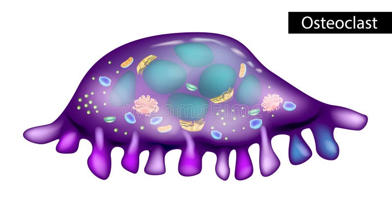
Free with trial Osteoclast is a type of bone cell that breaks down bone tissue. Structure of osteoclasts. Vesicles vectors Structure of osteoclasts. Osteoclast is a type of bone cell that breaks down bone tissue. Osteoclast is a type of bone cell that breaks down bone tissue. Osteoclast is a type of bone cell that breaks down bone tissue. Structure of osteoclasts
Free with trial Anatomy of trichome with biological model structure closeup outline diagram. Labeled educational scheme with microscopic side view of cuticle, striae, stipe, basal cells and bract vector illustration. Vesicles vectors Anatomy of trichome with biological model structure closeup outline diagram. Labeled educational scheme with microscopic side view of cuticle, striae, stipe
Free with trial Reproductive system of planaria flatworm. Isolated on white background. Vesicles vectors Reproductive system of planaria flatworm. Isolated on white background
Free with trial Autophagy of mitochondria. Diagram of the process of autophagy from Forming a membrane and autophagosome to fuse phagosome and lysosome when contents of the vesicles are degraded and recycled. Autophagy defects linked to various diseases and cancer development. Vector poster. Vesicles vectors Autophagy of mitochondria. Diagram of the process of autophagy from Forming a membrane and autophagosome to fuse phagosome and lysosome when contents of the
Free with trial An animal cell is a type of eukaryotic cell. Animal cells are typical of the eukaryotic cell. The basic unit of life in organisms. Vesicles vectors An animal cell is a type of eukaryotic cell. Animal cells are typical of the eukaryotic cell. The basic unit of life in organisms
Free with trial Water bubbles isolated on a transparent checkered background. Underwater effervescent sparkling oxygen bubbles in water. Iridescent soap bubbles with reflex and reflection, realistic vector effect. Vesicles vectors Water bubbles isolated. On a transparent checkered background. Underwater effervescent sparkling oxygen bubbles in water. Iridescent soap bubbles with reflex. Water bubbles isolated on a transparent checkered background. Underwater effervescent sparkling oxygen bubbles in water. Iridescent soap bubbles with reflex and reflection, realistic vector effect.
Free with trial Nervous, Reproductive, Digestive and Excretory system of planaria flatworm. Isolated on white background. Vesicles vectors Nervous, Reproductive, Digestive and Excretory system of planaria flatworm. Isolated on white background
Free with trial Reproductive system of planaria flatworm. Isolated on white background. Vesicles vectors Reproductive system of planaria flatworm. Isolated on white background
Free with trial Water bubbles isolated on a transparent checkered background. Iridescent soap bubbles with reflex and reflection, realistic vector effect. Vesicles vectors Water bubbles isolated on a transparent checkered background. Iridescent soap bubbles with reflex and reflection, realistic vector effect
Free with trial Fizzy sparkles in aquarium, champagne, soda drink. Soap bubbles with reflex and reflection, realistic vector effect. Vesicles vectors Water bubbles isolated on a transparent checkered background. Fizzy sparkles in aquarium, champagne, soda drink. Soap bubbles with reflex and reflection. Fizzy sparkles in aquarium, champagne, soda drink. Soap bubbles with reflex and reflection, realistic vector effect
Free with trial The structure of Paneth cells. Davidoff`s cell. fographics. Vector illustration on isolated background. Vesicles vectors The structure of Paneth cells. Davidoff`s cell. fographics. Vector illustration on isolated background. The structure of Paneth cells. Davidoff`s cell
 Sign in