Free with trial Intestinal bacterial flora. Medical illustration of the good and the bad bacterial flora. Villi vectors Intestinal bacterial flora. Medical illustration of the good and the bad bacterial flora
Free with trial The human gut. The human alimentary canal from mouth to anus showing detail of intestinal mucosal cells. Villi vectors The human gut. The human alimentary canal from mouth to anus showing detail of intestinal mucosal cells
Free with trial Epithelial types. The structure and formation of different types of epithelial tissue. Villi vectors Epithelial types. The structure and formation of different types of epithelial tissue
Free with trial Intestinal villi anatomy, small intestine lining, villi and epithelial cells with microvilli detailed illustration. Villi vectors Intestinal villi anatomy. Small intestine lining, villi and epithelial cells with microvilli detailed illustration
Free with trial Intestinal villi and microvilli. Detailed anatomy on a white background. Villi vectors Intestinal villi and microvilli. Detailed anatomy on a white background
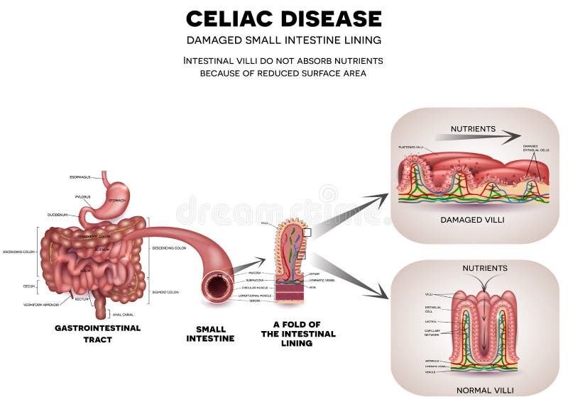
Free with trial Gastrointestinal tract anatomy and Celiac disease affected small intestine villi. Unhealthy villi with damaged cells and healthy villi. Intestinal villi do not absorb nutrients because of reduced surface area. Villi vectors Celiac disease. Gastrointestinal tract anatomy and Celiac disease affected small intestine villi. Unhealthy villi with damaged cells and healthy villi. Gastrointestinal tract anatomy and Celiac disease affected small intestine villi. Unhealthy villi with damaged cells and healthy villi. Intestinal villi do not absorb nutrients because of reduced surface area.
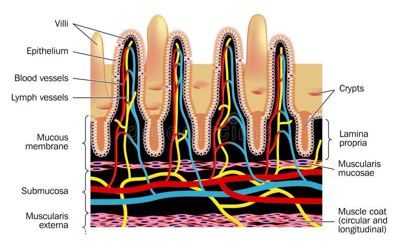
Free with trial Microvilli. Detail of small intestinal villim, showing the villi and the crypts, blood supply and lymph vessels. Villi vectors Microvilli. Detail of small intestinal villim, showing the villi and the crypts, blood supply and lymph vessels
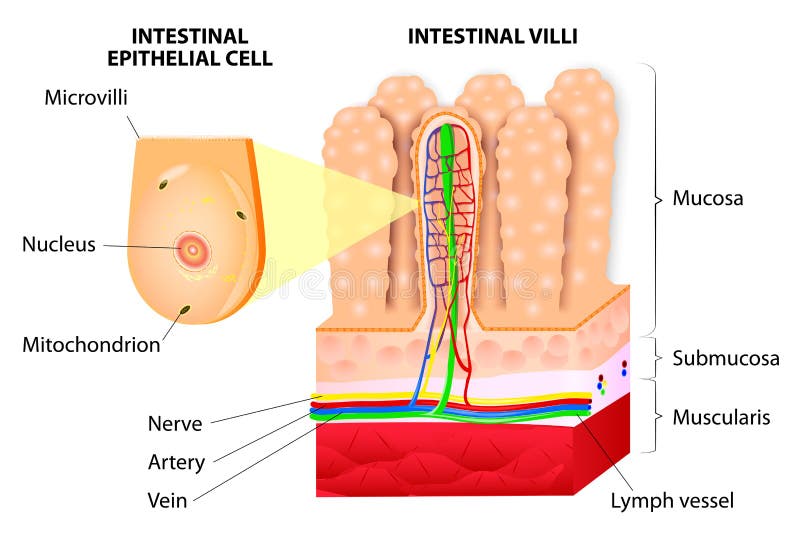
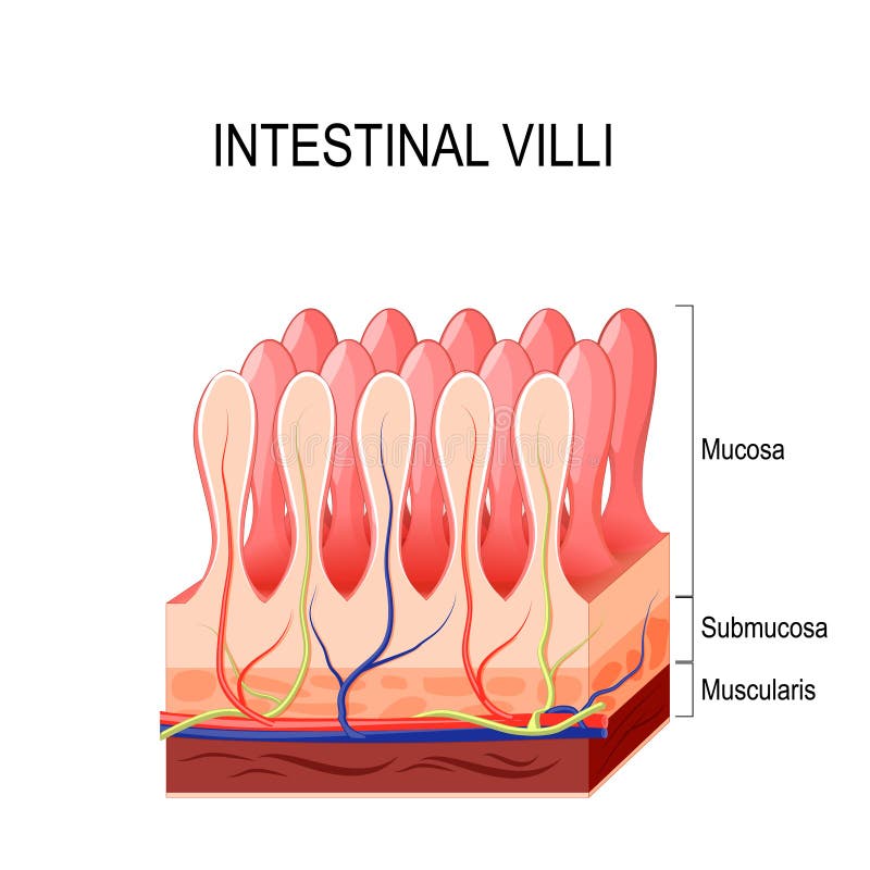
Free with trial Structure of Villi and microvilli showing arteries, veins, nerve and lymph vessel. Villi vectors Microvilli. Detail of the small intestine. Vector. Structure of Villi and microvilli showing arteries, veins, nerve and lymph vessel. Structure of Villi and microvilli showing arteries, veins, nerve and lymph vessel.
Free with trial Cerebrospinal fluid vector illustration. Anatomical labeled scheme with human head and inside of skull. Diagram with superior sigittal sinus, ventricles, arachnoid Villi and spinal cord central canal. Villi vectors Cerebrospinal fluid vector illustration. Anatomical labeled diagram - human superior sigittal sinus and spinal cord central canal. Cerebrospinal fluid vector. Cerebrospinal fluid vector illustration. Anatomical labeled scheme with human head and inside of skull. Diagram with superior sigittal sinus, ventricles, arachnoid Villi and spinal cord central canal.
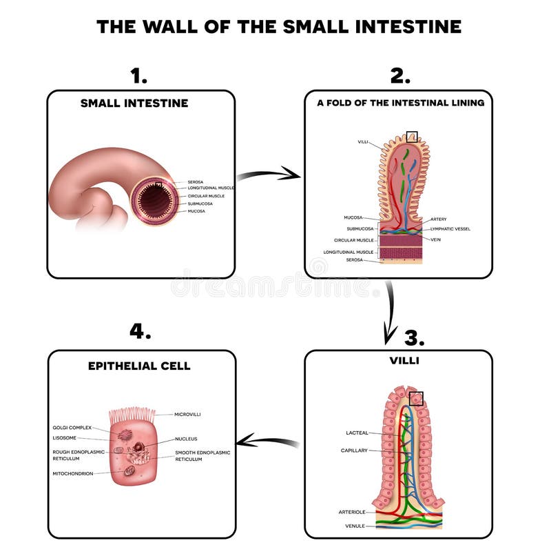
Free with trial Small intestine lining anatomy, a fold of the intestinal lining, villi and epithelial cell with microvilli detailed illustrations. Villi vectors Small intestine lining. Anatomy, a fold of the intestinal lining, villi and epithelial cell with microvilli detailed illustrations
Free with trial Leaky gut. cells on gut lining held tightly together. in intestine with celiac disease and gluten sensitivity these tight junctions come apart. autoimmune disorder. Vector diagram for educational, medical, biological and science use. Villi vectors Leaky gut. Cells on gut lining held tightly together. in intestine with celiac disease and gluten sensitivity these tight junctions come apart. autoimmune
Free with trial Anatomy of the placenta. Cross section through the placenta showing umbilical cord, placental wall and uterus. Villi vectors Anatomy of the placenta. Cross section through the placenta showing umbilical cord, placental wall and uterus
Free with trial Gastrointestinal system small intestine detailed wall anatomy. Small intestine villi and epithelial cell with microvilli detailed illustration. Villi vectors Gastrointestinal system small intestine anatomy. Gastrointestinal system small intestine detailed wall anatomy. Small intestine villi and epithelial cell with. Gastrointestinal system small intestine detailed wall anatomy. Small intestine villi and epithelial cell with microvilli detailed illustration.
Free with trial Leaky gut Syndrome. difference between Healthy cells, and inflamed intestinal cells. Comparison normal tissue of the gastrointestinal tract, and leaky gut. Villi vectors Leaky gut Syndrome. Difference between Healthy cells, and inflamed intestinal cells. Comparison normal tissue of the gastrointestinal tract, and leaky gut
Free with trial Intestinal bacterial flora. Symbolic medical illustration of the intestinal bacterial flora. Villi vectors Intestinal bacterial flora. Symbolic medical illustration of the intestinal bacterial flora
Free with trial Enterocytes. intestinal absorptive cells. Intestinal epithelial cell with microvilli. Villi vectors Enterocytes. intestinal absorptive cells. Intestinal epithelial cell with microvilli
Free with trial Soy bean. Realistic illustration. Villi vectors Soybean. Realistic illustration. Soy bean. Realistic illustration. Soy bean. Realistic illustration.
Free with trial Intestinal mucosa immunity. Cells medical vector illustration. Villi vectors Intestinal mucosa immunity. Cells medical vector illustration
Free with trial Anatomy of the wall of the small intestine villi into the lumen of the intestine increasing its surface area for secretion of enzymes, water, mucus, and for absorption of nutrients. Vector diagram. Villi vectors Intestinal villi. Vector diagram. Anatomy of the wall of the small intestine villi into the lumen of the intestine increasing its surface area for secretion of. Anatomy of the wall of the small intestine villi into the lumen of the intestine increasing its surface area for secretion of enzymes, water, mucus, and for absorption of nutrients. Vector diagram
Free with trial Wall of small intestine with villi and epithelial cells Enterocyte, Goblet and Paneth cell. Villi vectors Small intestine with villi and epithelial cells. Wall of small intestine with villi and epithelial cells Enterocyte, Goblet and Paneth cell. Wall of small intestine with villi and epithelial cells Enterocyte, Goblet and Paneth cell.
Free with trial Celiac disease Small intestine lining damage. good and damaged villi. leaky gut progression. On white. Villi vectors Celiac disease Small intestine lining damage. good and damaged villi . leaky gut progression. On white
Free with trial Celiac disease Small intestine lining damage. good and damaged villi. leaky gut progression. On white background. Villi vectors Celiac disease Small intestine lining damage. good and damaged villi . leaky gut progression. On white background
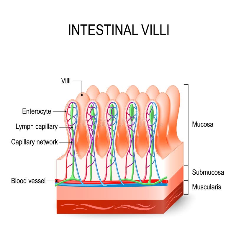
Free with trial Intestinal villi in the small intestine. showing arteries, veins and lymph vessel. Human anatomy. Villi vectors Intestinal villi in the small intestine. Human anatomy. Intestinal villi in the small intestine. showing arteries, veins and lymph vessel. Human anatomy. Intestinal villi in the small intestine. showing arteries, veins and lymph vessel. Human anatomy.
Free with trial Celiac disease Small intestine lining damage. good and damaged villi. leaky gut progression. On white. Villi vectors Celiac disease Small intestine lining damage. good and damaged villi . leaky gut progression. On white
Free with trial Celiac disease Small intestine lining damage. good and damaged villi. leaky gut progression. On white background. Villi vectors Celiac disease Small intestine lining damage. good and damaged villi . leaky gut progression. On white background
Free with trial Celiac disease. small intestinal with normal villi, and villous atrophy. Diagram showing changes in intestinal. coeliac disease manifested by blunting of villi. Villi vectors Celiac disease. small intestinal with normal villi, and villous. Atrophy. Diagram showing changes in intestinal. coeliac disease manifested by blunting of villi
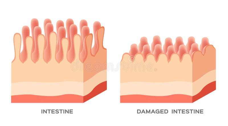
Free with trial Celiac disease detailed anatomy, healthy intestinal villi and damaged unhealthy villi. Intestinal villi do not absorb nutrients because of reduced surface area. Villi vectors Celiac disease. Detailed anatomy, healthy intestinal villi and damaged unhealthy villi. Intestinal villi do not absorb nutrients because of reduced surface area
Free with trial Celiac disease affected small intestine villi. Unhealthy villi with damaged cells and healthy villi. Intestinal villi do not absorb nutrients because of reduced surface area. Villi vectors Celiac disease. Affected small intestine villi. Unhealthy villi with damaged cells and healthy villi. Intestinal villi do not absorb nutrients because of
Free with trial Celiac disease. Affected small intestine villi on a white background. Healthy villi and unhealthy villi with damaged cells. Villi vectors Celiac disease. Affected small intestine villi on a white background. Healthy villi and unhealthy villi with damaged cells
Free with trial Celiac disease Small intestine lining damage. Healthy villi and damaged villi. Small intestine, a fold of the intestinal lining and villi. Villi vectors Celiac disease Small intestine lining damage. Healthy villi and damaged villi. Small intestine, a fold of the intestinal lining and villi
Free with trial Coeliac celiac disease. normal villi and villous atrophy. small bowel showing coeliac disease manifested by blunting of vill. Vector diagram for medical use. Villi vectors Coeliac disease. celiac disease. Coeliac celiac disease. normal villi and villous atrophy. small bowel showing coeliac disease manifested by blunting of vill. Coeliac celiac disease. normal villi and villous atrophy. small bowel showing coeliac disease manifested by blunting of vill. Vector diagram for medical use
Free with trial Celiac disease vector illustration. Labeled diagram with its structure. Autoimmune illness in stomach and intestine. Scheme with nutrients, healthy and destroyed villi. Villi vectors Celiac disease vector illustration. Labeled diagram with its structure. Autoimmune illness in stomach and intestine. Scheme with nutrients, healthy and
Free with trial Gastrointestinal tract. Stomach, small intestine and colon. Beautiful bright illustration on a white background. Villi vectors Gastrointestinal system. Gastrointestinal tract. Stomach, small intestine and colon. Beautiful bright illustration on a white background. Gastrointestinal tract. Stomach, small intestine and colon. Beautiful bright illustration on a white background.
Free with trial Small intestine wall anatomy, a fold of the intestinal lining, villi and epithelial cell with microvilli detailed illustrations. Villi vectors Small intestine wall anatomy. A fold of the intestinal lining, villi and epithelial cell with microvilli detailed illustrations
Free with trial Leaky gut. Intestinal permeability. cells on gut lining held tightly together. in intestine with celiac disease and gluten sensitivity these tight junctions are open. substances, microbes and toxines uncontrolled passage into the bloodstream. development of immune and inflammatory reactions. autoimmune disorder. Villi vectors Leaky gut. Intestinal permeability. Cells on gut lining held tightly together. in intestine with celiac disease and gluten sensitivity these tight junctions
Free with trial Gastrointestinal tract with description. Stomach, small intestine and colon. Beautiful bright illustration. Villi vectors Gastrointestinal tract with description. Stomach, small intestine and colon. Beautiful bright illustration
Free with trial Small intestine and stomach anatomy. Healthy small intestine and stomach, bright anatomy illustration, white background. Villi vectors Small intestine and stomach anatomy. Healthy small intestine and stomach, bright anatomy illustration, white background
Free with trial Gastrointestinal tract. Stomach, small intestine and colon, abstract blue technology background with lights and human silhouette. Villi vectors Gastrointestinal system. Gastrointestinal tract. Stomach, small intestine and colon, abstract blue technology background with lights and human silhouette. Gastrointestinal tract. Stomach, small intestine and colon, abstract blue technology background with lights and human silhouette.
Free with trial Leaky gut. The opening of intercellular tight junctions increased intestinal permeability. cells on gut lining held tightly together. in intestine with celiac disease and gluten sensitivity these tight junctions come apart. autoimmune disorder. Vector diagram. Villi vectors Leaky gut. The opening of intercellular tight junctions increased intestinal permeability. Cells on gut lining held tightly together. in intestine with celiac
Free with trial Peristalsis, or wave-like contractions of the muscles in the outer walls of the digestive tract, carries the bolus by the esophagus. Peristalsis does not only occur in the esophagus. It continues through the stomach, small intestine, and large intestine. Vector diagram. Villi vectors Peristalsis. involuntary wavelike muscular contrac. Peristalsis, or wave-like contractions of the muscles in the outer walls of the digestive tract, carries the. Peristalsis, or wave-like contractions of the muscles in the outer walls of the digestive tract, carries the bolus by the esophagus. Peristalsis does not only occur in the esophagus. It continues through the stomach, small intestine, and large intestine. Vector diagram
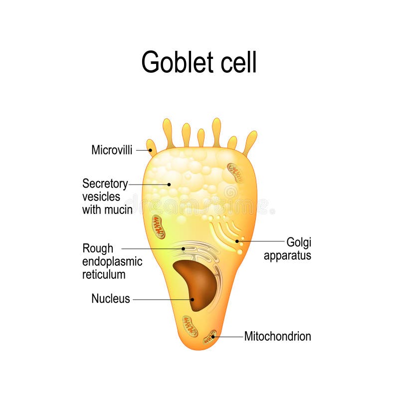
Free with trial Goblet cell. simple columnar epithelial cell for secrete mucus. They are found inside the trachea, bronchi, small and large intestine, and conjunctiva in the eyes. Structure cell nucleus and other organelles. Villi vectors Goblet cell. Structure cell nucleus and other organelles. Goblet cell. simple columnar epithelial cell for secrete mucus. They are found inside the trachea. Goblet cell. simple columnar epithelial cell for secrete mucus. They are found inside the trachea, bronchi, small and large intestine, and conjunctiva in the eyes. Structure cell nucleus and other organelles.
Free with trial Placental structure and circulation. Prenatal development. Villi vectors Placental structure and circulation. Prenatal development
Free with trial Gut brain connection, dysbiosis and microbiome. Normal and abnormal microbiota. Enteric nervous system, intestine anatomy. Signals from brain to digestive tract. Colon and cerebrum vector illustration. Villi vectors Human gut microbiota. Gut brain connection, dysbiosis and microbiome. Normal and abnormal microbiota. Enteric nervous system, intestine anatomy. Signals from. Gut brain connection, dysbiosis and microbiome. Normal and abnormal microbiota. Enteric nervous system, intestine anatomy. Signals from brain to digestive tract. Colon and cerebrum vector illustration
Free with trial Gut brain connection, dysbiosis and microbiome. Normal and abnormal microbiota. Enteric nervous system, intestine anatomy. Signals from brain to digestive tract. Colon and cerebrum vector illustration. Villi vectors Gut brain connection. Dysbiosis and microbiome. Normal and abnormal microbiota. Enteric nervous system, intestine anatomy. Signals from brain to digestive. Gut brain connection, dysbiosis and microbiome. Normal and abnormal microbiota. Enteric nervous system, intestine anatomy. Signals from brain to digestive tract. Colon and cerebrum vector illustration
Free with trial Leaky gut Syndrome concept. Comparison of healthy organ and inflamed tissue cells. Diseases of gastrointestinal tract. Toxins and viruses. Cartoon flat vector illustration isolated on white background. Villi vectors Leaky gut Syndrome concept. Comparison of healthy organ and inflamed tissue cells. Diseases of gastrointestinal tract. Toxins and viruses. Cartoon flat vector
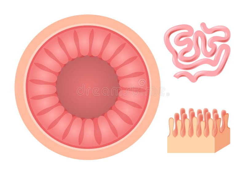
Free with trial Intestinal villi. Small intestine lining. Villi vectors Intestinal villi. Small intestine lining
Free with trial Intestinal villi in the surface area of intestinal walls. Folds, villus, microvilli, epithelial and goblet cells. Small intestine anatomical poster. Digestive system medical flat vector illustration. Villi vectors Intestinal villi anatomy. Intestinal villi in the surface area of intestinal walls. Folds, villus, microvilli, epithelial and goblet cells. Small intestine. Intestinal villi in the surface area of intestinal walls. Folds, villus, microvilli, epithelial and goblet cells. Small intestine anatomical poster. Digestive system medical flat vector illustration.
Free with trial Coeliac disease or celiac disease. small bowel showing coeliac disease manifested by blunting of villi. Villi vectors Coeliac disease or celiac disease. Small bowel showing coeliac disease manifested by blunting of villi
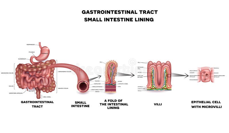
Free with trial Gastrointestinal tract anatomy. Intestinal villi, small intestine lining, epithelial cells with microvilli detailed illustration. Villi vectors Gastrointestinal tract detailed anatomy. Gastrointestinal tract anatomy. Intestinal villi, small intestine lining, epithelial cells with microvilli detailed. Gastrointestinal tract anatomy. Intestinal villi, small intestine lining, epithelial cells with microvilli detailed illustration.
Free with trial Celiac disease. Affected small intestine villi. Healthy villi and unhealthy villi with damaged cells on a blue technology background. Villi vectors Celiac disease. Affected small intestine villi. Healthy villi and unhealthy villi with damaged cells on a blue technology background
Free with trial Celiac disease treatment. Gluten free diet leads to healthy intestinal villi. Illustration shows unhealthy villi with damaged cells and healthy villi. Unhealthy intestinal villi do not absorb nutrients because of reduced surface area. Villi vectors Celiac disease treatment gluten free diet. Celiac disease treatment. Gluten free diet leads to healthy intestinal villi. Illustration shows unhealthy villi with. Celiac disease treatment. Gluten free diet leads to healthy intestinal villi. Illustration shows unhealthy villi with damaged cells and healthy villi. Unhealthy intestinal villi do not absorb nutrients because of reduced surface area.
Free with trial Intestinal wall anatomy. Intestinal wall medical science vector illustration. Internal human organ: mucosa, muscularis externa, serosa and villi. Human intestinal wall anatomy education illustration. Villi vectors Intestinal wall anatomy. Intestinal wall medical science vector illustration. Internal human organ: mucosa, muscularis externa, serosa and villi. Human
Free with trial Celiac disease affected small intestine villi. Damaged cells by body's reaction to gluten. Intestinal villi do not absorb nutrients because of reduced surface area. Villi vectors Celiac disease damaged intestine. Celiac disease affected small intestine villi. Damaged cells by body's reaction to gluten. Intestinal villi do not absorb. Celiac disease affected small intestine villi. Damaged cells by body's reaction to gluten. Intestinal villi do not absorb nutrients because of reduced surface area.
Free with trial Absorption of nutrients in the small intestine. Healthy and damaged villi. Medical vector illustration. Villi vectors Absorption of nutrients in the small intestine. Healthy and damaged villi. Medical vector illustration
Free with trial Leaky gut. Progression from Healthy gut to Dysbiosis and Intestinal permeability. Close-up of cells of a intestinal epithelium. Detailed Vector poster. Villi vectors Leaky gut. gut microbiota. Leaky gut. Progression from Healthy gut to Dysbiosis and Intestinal permeability. Close-up of cells of a intestinal epithelium. Leaky gut. Progression from Healthy gut to Dysbiosis and Intestinal permeability. Close-up of cells of a intestinal epithelium. Detailed Vector poster
Free with trial Coeliac disease or celiac disease gluten-sensitive enteropathy, sometimes called sprue or coeliac. Villi vectors Coeliac disease or celiac disease. Gluten-sensitive enteropathy, sometimes called sprue or coeliac
Free with trial Leaky gut. cells on gut lining held tightly together. in intestine with celiac disease and gluten sensitivity these tight junctions come apart. autoimmune disorder. Vector illustration. Villi vectors Leaky gut. Cells on gut lining held tightly together. in intestine with celiac disease and gluten sensitivity these tight junctions come apart. autoimmune
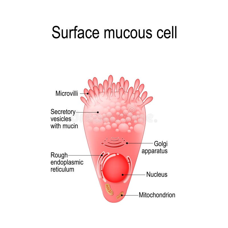
Free with trial Surface mucous cell is a foveolar mucus-producing cell that covering the inside of the stomach. Structure cell: golgi apparatus, secretory vesicle, mucin, nucleus, mitochondrion, microvilli. Villi vectors Surface mucous cell. Is a foveolar mucus-producing cell that covering the inside of the stomach. Structure cell: golgi apparatus, secretory vesicle, mucin. Surface mucous cell is a foveolar mucus-producing cell that covering the inside of the stomach. Structure cell: golgi apparatus, secretory vesicle, mucin, nucleus, mitochondrion, microvilli
Free with trial Gastrointestinal tract abstract health care and treatment concept. Stomach, small intestine and colon. Beautiful bright illustration. Villi vectors Gastrointestinal tract health care. Gastrointestinal tract abstract health care and treatment concept. Stomach, small intestine and colon. Beautiful bright. Gastrointestinal tract abstract health care and treatment concept. Stomach, small intestine and colon. Beautiful bright illustration.
Free with trial Celiac disease signs and symptoms. Illustrations depict celiac disease problems in adult and children. Villi vectors Celiac disease signs and symptoms. Illustrations depict celiac disease problems in adult and children
Free with trial Small intestine lining. Detailed anatomy, a fold of the intestinal lining, villi and epithelial cell with microvilli. Villi vectors Small intestine lining. Detailed anatomy, a fold of the intestinal lining, villi and epithelial cell with microvilli
Free with trial Hormones of the gastric glands cholecystokinin, serotonin, endorphins, histamine, gastrin, somatostatin. Stomach. Human endocrine system. Vector illustration for medical, education and science use. Villi vectors Hormones of the gastric glands. Stomach. Hormones of the gastric glands cholecystokinin, serotonin, endorphins, histamine, gastrin, somatostatin. Stomach. Human. Hormones of the gastric glands cholecystokinin, serotonin, endorphins, histamine, gastrin, somatostatin. Stomach. Human endocrine system. Vector illustration for medical, education and science use
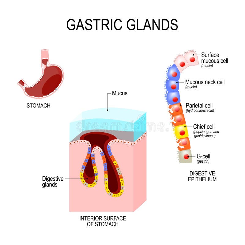
Free with trial Structure of the stomach: interior surface and cells of digestive epithelium. Human anatomy. Vector illustration for medical, educational and science use. Villi vectors Structure of the stomach: interior surface and cells of digestive epithelium. Human anatomy. Vector illustration for medical, educational and science use
Free with trial Placental structure Vector. Placental structure and circulation. Prenatal development. Villi vectors Placental structure Vector. Placental structure and circulation. Prenatal development
Free with trial Gastrointestinal tract. Stomach, small intestine and colon, abstract grey background with lights and human silhouette. Villi vectors Gastrointestinal tract abstract background. Gastrointestinal tract. Stomach, small intestine and colon, abstract grey background with lights and human silhouette. Gastrointestinal tract. Stomach, small intestine and colon, abstract grey background with lights and human silhouette.
Free with trial Hormones of the gastrointestinal tract and Enteroendocrine cell. Enterocyte. Human endocrine system. Vector illustration for medical, education and science use. Villi vectors Hormones of the gastrointestinal tract and Enteroendocrine cell. Enterocyte. Human endocrine system. Vector illustration for medical, education and science use
Free with trial Intestinal cell system, human medical anatomy closeup. Vector flat style cartoon illustration isolated on white background. Villi vectors Intestinal cell system, human medical anatomy closeup. Vector flat style cartoon illustration isolated on white background
Free with trial Vector illustration of epithelial tissue. types of epithelium. Villi vectors Epithelial tissue. types of epithelium. Vector illustration of epithelial tissue. types of epithelium. Vector illustration of epithelial tissue. types of epithelium.
Free with trial Types of muscle tissue. Vector illustration of types of muscle tissue. Villi vectors Types of muscle tissue. Vector illustration of types of muscle tissue
Free with trial Small intestine and stomach anatomy. Healthy small intestine and stomach, bright anatomy illustration, white background. Villi vectors Small intestine and stomach anatomy. Healthy small intestine and stomach, bright anatomy illustration, white background
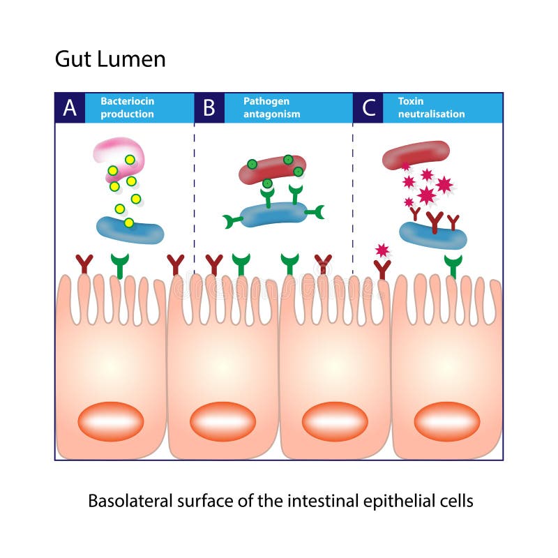
Free with trial Gut Lumen. Columnar epithelial cells. Gut lumen. Enterocytes, or intestinal absorptive cells. Small intestine. Villi vectors Gut Lumen. Columnar epithelial cells. Gut lumen. Enterocytes, or intestinal absorptive cells. Small intestine. Columnar epithelial cells
Free with trial Gut brain connection, dysbiosis and microbiome. Healthy CNS and depression anex. Enteric nervous system, intestine anatomy. Signal from brain to digestive tract. Colon and cerebrum vector illustration. Villi vectors Gut brain connection. Dysbiosis and microbiome. Healthy CNS and depression anex. Enteric nervous system, intestine anatomy. Signal from brain to digestive. Gut brain connection, dysbiosis and microbiome. Healthy CNS and depression anex. Enteric nervous system, intestine anatomy. Signal from brain to digestive tract. Colon and cerebrum vector illustration
Free with trial Gut brain connection, dysbiosis and microbiome. Normal and abnormal microbiota. Enteric nervous system, intestine anatomy. Signals from brain to digestive tract. Colon and cerebrum vector illustration. Villi vectors Gut brain connection. Dysbiosis and microbiome. Normal and abnormal microbiota. Enteric nervous system, intestine anatomy. Signals from brain to digestive. Gut brain connection, dysbiosis and microbiome. Normal and abnormal microbiota. Enteric nervous system, intestine anatomy. Signals from brain to digestive tract. Colon and cerebrum vector illustration
Free with trial Gut brain connection, dysbiosis and microbiome. Normal and abnormal microbiota. Enteric nervous system, intestine anatomy. Signals from brain to digestive tract. Colon and cerebrum vector illustration. Villi vectors Gut brain connection. Dysbiosis and microbiome. Normal and abnormal microbiota. Enteric nervous system, intestine anatomy. Signals from brain to digestive. Gut brain connection, dysbiosis and microbiome. Normal and abnormal microbiota. Enteric nervous system, intestine anatomy. Signals from brain to digestive tract. Colon and cerebrum vector illustration
Free with trial Intestinal villi blue technology background. Intestinal villi anatomy on a beautiful abstract technology background. Villi vectors Intestinal villi blue technology background. Intestinal villi anatomy on a beautiful abstract technology background
Free with trial Healthy intestine villi. Intestinal villi anatomy on a beautiful abstract technology background. Villi vectors Healthy intestine villi. Intestinal villi anatomy on a beautiful abstract technology background
Free with trial Intestinal villi anatomy, small intestine lining. organ. / white background. Villi vectors Intestinal villi anatomy, small intestine lining . organ. / white background
Free with trial Structure of colon villi. Illustration of the colon villi. Villi vectors Structure of colon villi. Illustration of the colon villi
Free with trial Intestinal Villi, Large and small intestine. Gastrointestinal tract. Human anatomy. Villi vectors Intestinal Villi, Large and small intestine. Gastrointestinal tract. Intestinal Villi, Large and small intestine. Human anatomy
Free with trial Small intestine Anatomy. Cross section of a ileum with Internal villi. Close-up of Epithelial cells with Microvilli. Vector illustration. Villi vectors Small intestine Anatomy. Cross section of a ileum with Internal villi. Close-up of Epithelial cells with Microvilli. Vector illustration
 Sign in