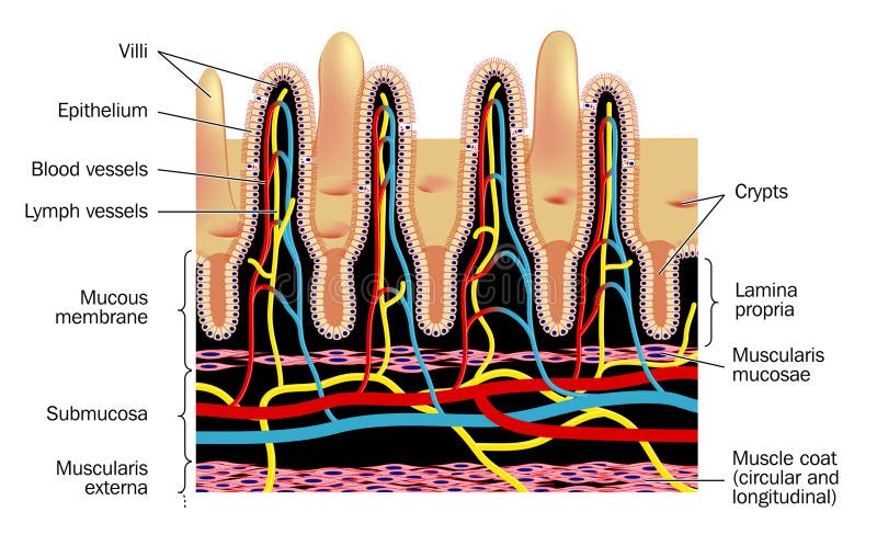
Free with trial Microvilli. Detail of small intestinal villim, showing the villi and the crypts, blood supply and lymph vessels. Villus vectors Microvilli. Detail of small intestinal villim, showing the villi and the crypts, blood supply and lymph vessels
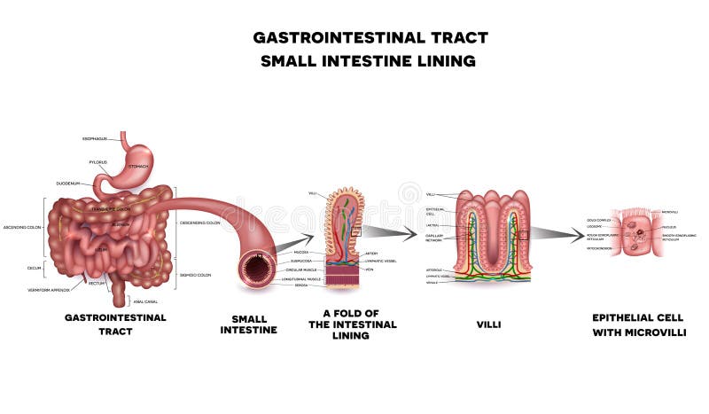
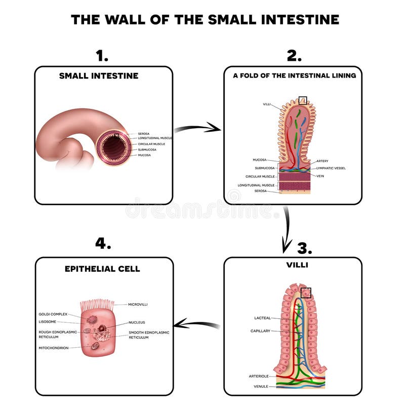
Free with trial Small intestine lining anatomy, a fold of the intestinal lining, villi and epithelial cell with microvilli detailed illustrations. Villus vectors Small intestine lining. Anatomy, a fold of the intestinal lining, villi and epithelial cell with microvilli detailed illustrations
Free with trial Meninges of the brain. Layers of the meninges of the brain, eps8. Villus vectors Meninges of the brain. Layers of the meninges of the brain, eps8
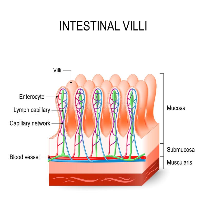
Free with trial Intestinal villus. Medical illustration of anatomy of the intestinal villus. Villus vectors Intestinal villus. Medical illustration of anatomy of the intestinal villus
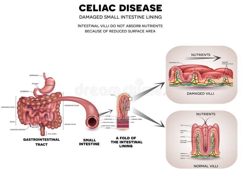
Free with trial Gastrointestinal tract anatomy and Celiac disease affected small intestine villi. Unhealthy villi with damaged cells and healthy villi. Intestinal villi do not absorb nutrients because of reduced surface area. Villus vectors Celiac disease. Gastrointestinal tract anatomy and Celiac disease affected small intestine villi. Unhealthy villi with damaged cells and healthy villi. Gastrointestinal tract anatomy and Celiac disease affected small intestine villi. Unhealthy villi with damaged cells and healthy villi. Intestinal villi do not absorb nutrients because of reduced surface area.
Free with trial Anatomy of the placenta. Cross section through the placenta showing umbilical cord, placental wall and uterus. Villus vectors Anatomy of the placenta. Cross section through the placenta showing umbilical cord, placental wall and uterus
Free with trial Gastrointestinal system small intestine detailed wall anatomy. Small intestine villi and epithelial cell with microvilli detailed illustration. Villus vectors Gastrointestinal system small intestine anatomy. Gastrointestinal system small intestine detailed wall anatomy. Small intestine villi and epithelial cell with. Gastrointestinal system small intestine detailed wall anatomy. Small intestine villi and epithelial cell with microvilli detailed illustration.
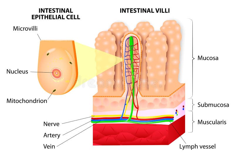
Free with trial Structure of Villi and microvilli showing arteries, veins, nerve and lymph vessel. Villus vectors Microvilli. Detail of the small intestine. Vector. Structure of Villi and microvilli showing arteries, veins, nerve and lymph vessel. Structure of Villi and microvilli showing arteries, veins, nerve and lymph vessel.
Free with trial Intestinal wall cells. Medical illustration of the Intestinal wall cells. Villus vectors Intestinal wall cells. Medical illustration of the Intestinal wall cells
Free with trial Intestinal villi anatomy, small intestine lining, villi and epithelial cells with microvilli detailed illustration. Villus vectors Intestinal villi anatomy. Small intestine lining, villi and epithelial cells with microvilli detailed illustration
Free with trial Intestinal villi and microvilli. Detailed anatomy on a white background. Villus vectors Intestinal villi and microvilli. Detailed anatomy on a white background
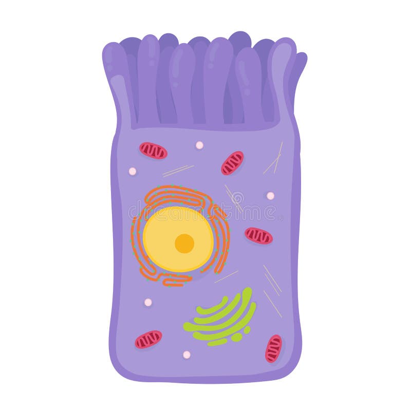
Free with trial Enterocytes. intestinal absorptive cells. Intestinal epithelial cell with microvilli. Villus vectors Enterocytes. intestinal absorptive cells. Intestinal epithelial cell with microvilli
Free with trial Celiac disease detailed anatomy, healthy intestinal villi and damaged unhealthy villi. Intestinal villi do not absorb nutrients because of reduced surface area. Villus vectors Celiac disease. Detailed anatomy, healthy intestinal villi and damaged unhealthy villi. Intestinal villi do not absorb nutrients because of reduced surface area
Free with trial Anatomy of the wall of the small intestine villi into the lumen of the intestine increasing its surface area for secretion of enzymes, water, mucus, and for absorption of nutrients. Vector diagram. Villus vectors Intestinal villi. Vector diagram. Anatomy of the wall of the small intestine villi into the lumen of the intestine increasing its surface area for secretion of. Anatomy of the wall of the small intestine villi into the lumen of the intestine increasing its surface area for secretion of enzymes, water, mucus, and for absorption of nutrients. Vector diagram
Free with trial Celiac disease affected small intestine villi. Unhealthy villi with damaged cells and healthy villi. Intestinal villi do not absorb nutrients because of reduced surface area. Villus vectors Celiac disease. Affected small intestine villi. Unhealthy villi with damaged cells and healthy villi. Intestinal villi do not absorb nutrients because of
Free with trial Celiac disease. Affected small intestine villi on a white background. Healthy villi and unhealthy villi with damaged cells. Villus vectors Celiac disease. Affected small intestine villi on a white background. Healthy villi and unhealthy villi with damaged cells
Free with trial Small intestine wall anatomy, a fold of the intestinal lining, villi and epithelial cell with microvilli detailed illustrations. Villus vectors Small intestine wall anatomy. A fold of the intestinal lining, villi and epithelial cell with microvilli detailed illustrations
Free with trial Gastroenteritis or infectious diarrhea. Gastroenteritis is an intestinal infection that causes vomiting cramping and diarrhea. Villus vectors Gastroenteritis or infectious diarrhea. Gastroenteritis is an intestinal infection that causes vomiting cramping and diarrhea
Free with trial Celiac disease Small intestine lining damage. Healthy villi and damaged villi. Small intestine, a fold of the intestinal lining and villi. Villus vectors Celiac disease Small intestine lining damage. Healthy villi and damaged villi. Small intestine, a fold of the intestinal lining and villi
Free with trial Wall of small intestine with villi and epithelial cells Enterocyte, Goblet and Paneth cell. Villus vectors Small intestine with villi and epithelial cells. Wall of small intestine with villi and epithelial cells Enterocyte, Goblet and Paneth cell. Wall of small intestine with villi and epithelial cells Enterocyte, Goblet and Paneth cell.
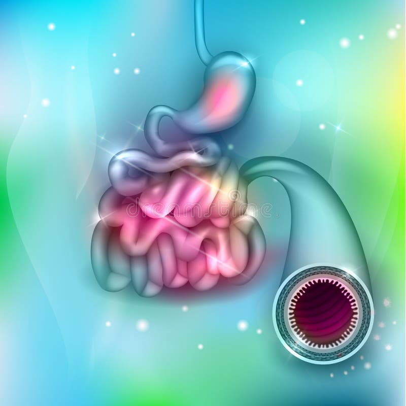
Free with trial Small intestine and stomach anatomy. Healthy small intestine and stomach, bright anatomy illustration, white background. Villus vectors Small intestine and stomach anatomy. Healthy small intestine and stomach, bright anatomy illustration, white background
Free with trial Coeliac celiac disease. normal villi and villous atrophy. small bowel showing coeliac disease manifested by blunting of vill. Vector diagram for medical use. Villus vectors Coeliac disease. celiac disease. Coeliac celiac disease. normal villi and villous atrophy. small bowel showing coeliac disease manifested by blunting of vill. Coeliac celiac disease. normal villi and villous atrophy. small bowel showing coeliac disease manifested by blunting of vill. Vector diagram for medical use
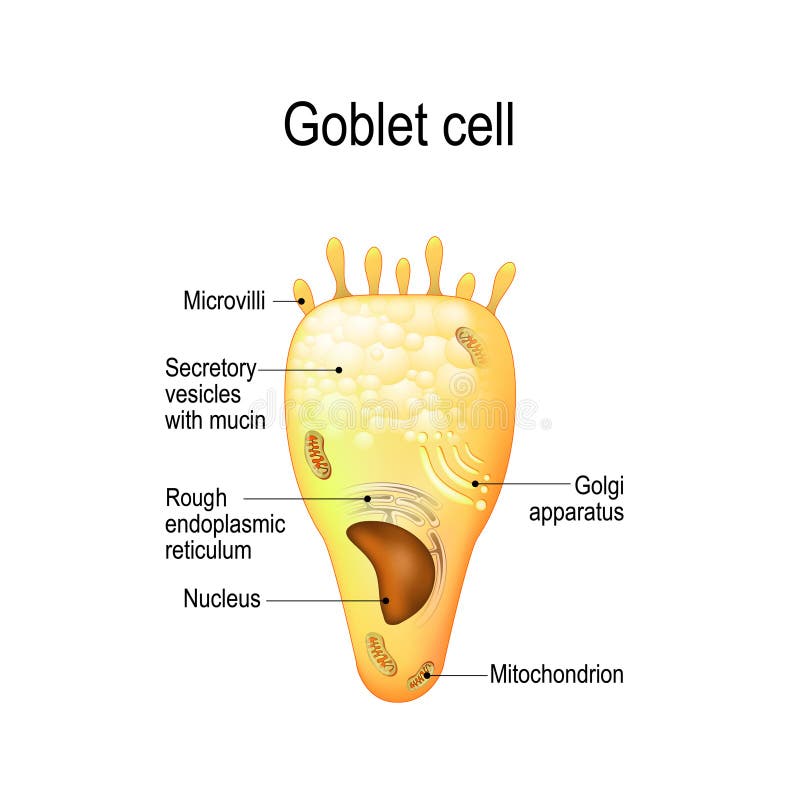
Free with trial Goblet cell. simple columnar epithelial cell for secrete mucus. They are found inside the trachea, bronchi, small and large intestine, and conjunctiva in the eyes. Structure cell nucleus and other organelles. Villus vectors Goblet cell. Structure cell nucleus and other organelles. Goblet cell. simple columnar epithelial cell for secrete mucus. They are found inside the trachea. Goblet cell. simple columnar epithelial cell for secrete mucus. They are found inside the trachea, bronchi, small and large intestine, and conjunctiva in the eyes. Structure cell nucleus and other organelles.
Free with trial Placental structure and circulation. Prenatal development. Villus vectors Placental structure and circulation. Prenatal development
Free with trial Intestinal villi in the small intestine. showing arteries, veins and lymph vessel. Human anatomy. Villus vectors Intestinal villi in the small intestine. Human anatomy. Intestinal villi in the small intestine. showing arteries, veins and lymph vessel. Human anatomy. Intestinal villi in the small intestine. showing arteries, veins and lymph vessel. Human anatomy.
Free with trial Celiac disease. small intestinal with normal villi, and villous atrophy. Diagram showing changes in intestinal. coeliac disease manifested by blunting of villi. Villus vectors Celiac disease. small intestinal with normal villi, and villous. Atrophy. Diagram showing changes in intestinal. coeliac disease manifested by blunting of villi
Free with trial Intestinal villi in the surface area of intestinal walls. Folds, villus, microvilli, epithelial and goblet cells. Small intestine anatomical poster. Digestive system medical flat vector illustration. Villus vectors Intestinal villi anatomy. Intestinal villi in the surface area of intestinal walls. Folds, villus, microvilli, epithelial and goblet cells. Small intestine. Intestinal villi in the surface area of intestinal walls. Folds, villus, microvilli, epithelial and goblet cells. Small intestine anatomical poster. Digestive system medical flat vector illustration.
Free with trial Coeliac disease or celiac disease gluten-sensitive enteropathy, sometimes called sprue or coeliac. Villus vectors Coeliac disease or celiac disease. Gluten-sensitive enteropathy, sometimes called sprue or coeliac
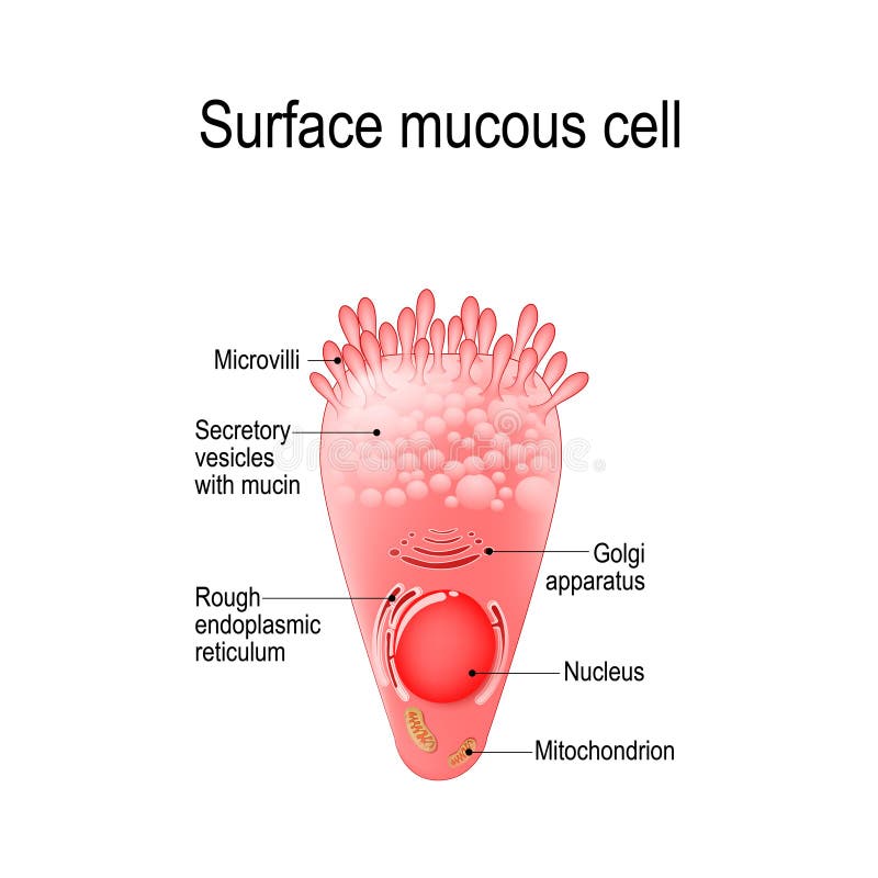
Free with trial Surface mucous cell is a foveolar mucus-producing cell that covering the inside of the stomach. Structure cell: golgi apparatus, secretory vesicle, mucin, nucleus, mitochondrion, microvilli. Villus vectors Surface mucous cell. Is a foveolar mucus-producing cell that covering the inside of the stomach. Structure cell: golgi apparatus, secretory vesicle, mucin. Surface mucous cell is a foveolar mucus-producing cell that covering the inside of the stomach. Structure cell: golgi apparatus, secretory vesicle, mucin, nucleus, mitochondrion, microvilli
Free with trial Normal Gastrointestinal tract. And small intestine detailed anatomy. Villus vectors Normal Gastrointestinal tract. And small intestine detailed anatomy
Free with trial Coeliac disease or celiac disease. small bowel showing coeliac disease manifested by blunting of villi. Villus vectors Coeliac disease or celiac disease. Small bowel showing coeliac disease manifested by blunting of villi
Free with trial Intestinal villi. Small intestine lining. Villus vectors Intestinal villi. Small intestine lining
Free with trial Gastrointestinal tract anatomy. Intestinal villi, small intestine lining, epithelial cells with microvilli detailed illustration. Villus vectors Gastrointestinal tract detailed anatomy. Gastrointestinal tract anatomy. Intestinal villi, small intestine lining, epithelial cells with microvilli detailed. Gastrointestinal tract anatomy. Intestinal villi, small intestine lining, epithelial cells with microvilli detailed illustration.
Free with trial Celiac disease. Affected small intestine villi. Healthy villi and unhealthy villi with damaged cells on a blue technology background. Villus vectors Celiac disease. Affected small intestine villi. Healthy villi and unhealthy villi with damaged cells on a blue technology background
Free with trial Hormones of the gastric glands cholecystokinin, serotonin, endorphins, histamine, gastrin, somatostatin. Stomach. Human endocrine system. Vector illustration for medical, education and science use. Villus vectors Hormones of the gastric glands. Stomach. Hormones of the gastric glands cholecystokinin, serotonin, endorphins, histamine, gastrin, somatostatin. Stomach. Human. Hormones of the gastric glands cholecystokinin, serotonin, endorphins, histamine, gastrin, somatostatin. Stomach. Human endocrine system. Vector illustration for medical, education and science use
Free with trial Placental structure Vector. Placental structure and circulation. Prenatal development. Villus vectors Placental structure Vector. Placental structure and circulation. Prenatal development
Free with trial Intestinal wall anatomy. Intestinal wall medical science vector illustration. Internal human organ: mucosa, muscularis externa, serosa and villi. Human intestinal wall anatomy education illustration. Villus vectors Intestinal wall anatomy. Intestinal wall medical science vector illustration. Internal human organ: mucosa, muscularis externa, serosa and villi. Human
Free with trial Celiac disease affected small intestine villi. Damaged cells by body's reaction to gluten. Intestinal villi do not absorb nutrients because of reduced surface area. Villus vectors Celiac disease damaged intestine. Celiac disease affected small intestine villi. Damaged cells by body's reaction to gluten. Intestinal villi do not absorb. Celiac disease affected small intestine villi. Damaged cells by body's reaction to gluten. Intestinal villi do not absorb nutrients because of reduced surface area.
Free with trial Hormones of the gastrointestinal tract and Enteroendocrine cell. Enterocyte. Human endocrine system. Vector illustration for medical, education and science use. Villus vectors Hormones of the gastrointestinal tract and Enteroendocrine cell. Enterocyte. Human endocrine system. Vector illustration for medical, education and science use
Free with trial Intestinal cell system, human medical anatomy closeup. Vector flat style cartoon illustration isolated on white background. Villus vectors Intestinal cell system, human medical anatomy closeup. Vector flat style cartoon illustration isolated on white background
Free with trial A fold of the intestinal lining. On a white background. Villus vectors A fold of the intestinal lining. On a white background
Free with trial Small intestine and stomach anatomy. Healthy small intestine and stomach, bright anatomy illustration, white background. Villus vectors Small intestine and stomach anatomy. Healthy small intestine and stomach, bright anatomy illustration, white background
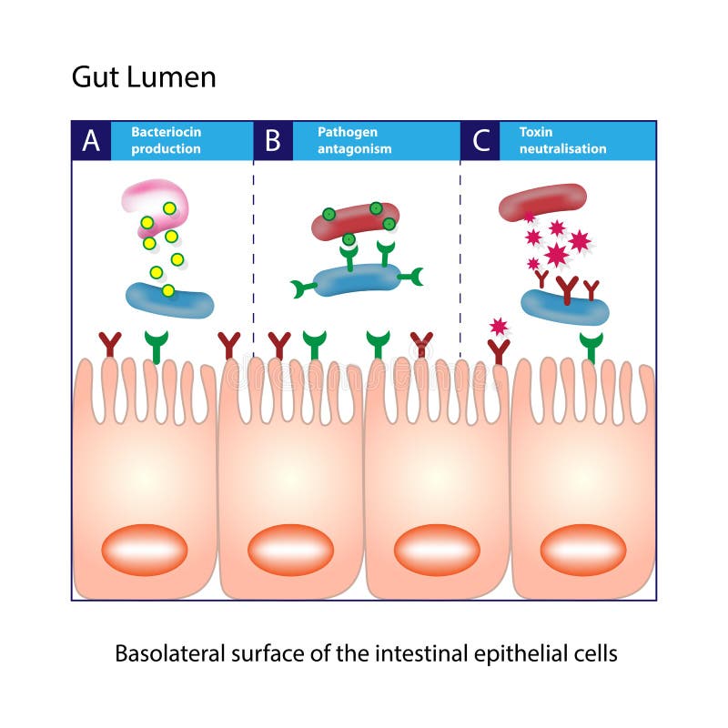
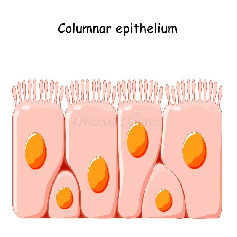
Free with trial Gut Lumen. Columnar epithelial cells. Gut lumen. Enterocytes, or intestinal absorptive cells. Small intestine. Villus vectors Gut Lumen. Columnar epithelial cells. Gut lumen. Enterocytes, or intestinal absorptive cells. Small intestine. Columnar epithelial cells
Free with trial Absorption of nutrients in the small intestine. Healthy and damaged villi. Medical vector illustration. Villus vectors Absorption of nutrients in the small intestine. Healthy and damaged villi. Medical vector illustration
Free with trial Human Intestines detailed anatomy. Vector Medical illustration. Villus vectors Human Intestines detailed anatomy. Vector Medical. Illustration
Free with trial Inflammation of the meninges - meningitis. Bacterial inflammation of the soft membrane of the human brain. Layout. Isolated on a white background. Vector. Villus vectors Meninges of the brain. Inflammation of the meninges - meningitis. Bacterial inflammation of the soft membrane of the human brain. Layout. Isolated on a white. Inflammation of the meninges - meningitis. Bacterial inflammation of the soft membrane of the human brain. Layout. Isolated on a white background. Vector.
Free with trial Noninvasive parental testing NIPT concept. Vector flat healthcare illustration. Genetic test. Baby in womb, blood vessel, dna spiral symbol. Design for healthcare, pharmacy, family planning. Villus vectors Noninvasive parental testing NIPT concept. Vector flat healthcare illustration. Genetic test. Baby in womb, blood vessel, dna. Spiral symbol. Design for. Noninvasive parental testing NIPT concept. Vector flat healthcare illustration. Genetic test. Baby in womb, blood vessel, dna spiral symbol. Design for healthcare, pharmacy, family planning
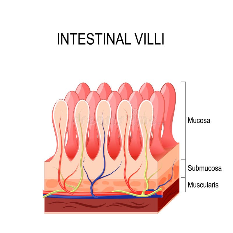
Free with trial Intestinal villus. Different between Villi and microvilli. Cross section of a duodenum with submucosa, mucosa and muscularis layers. Vertical section of a villus with Lymph capillary or Lacteal and Blood vessels. Close-up of a Columnar epithelium, and enterocytes. Vector illustration. Villus vectors Intestinal villus. Different between Villi and microvilli. Cross section of a duodenum with submucosa, mucosa and muscularis layers. Vertical section of a
Free with trial Chorionic villus sampling concept icon. CVS for prenatal testing. Genetic inheritance examination idea thin line illustration. Vector isolated outline RGB color drawing. Editable stroke. Villus vectors Chorionic villus sampling concept icon. CVS for prenatal testing. Genetic inheritance examination idea thin line illustration. Vector isolated outline RGB
Free with trial Structure of an Intestinal Villus, vintage engraved illustration. Usual Medicine Dictionary by Dr Labarthe - 1885. Villus vectors Structure of an Intestinal Villus, vintage engraving. Structure of an Intestinal Villus, vintage engraved illustration. Usual Medicine Dictionary by Dr Labarthe. Structure of an Intestinal Villus, vintage engraved illustration. Usual Medicine Dictionary by Dr Labarthe - 1885
Free with trial Coeliac disease. celiac disease. Gluten intolerance. Autoimmune disorder. Cross section of Small intestine with normal villus, and Villous atrophy. Leaky gut. Medical diagram. Vector illustration. Villus vectors Coeliac disease. celiac disease. Gluten intolerance. Autoimmune disorder. Cross section of Small intestine with normal villus, and Villous atrophy. Leaky gut
Free with trial Diagram showing fetal development and placenta structure. Left side illustrates a fetus inside the uterus, labeled: umbilical cord, placenta, fetus, uterus, cervix. Right side details placenta components: chorion, villus, umbilical cord, umbilical arteries, umbilical vein, placental septum. The depiction highlights the exchange mechanisms between mother and fetus. Villus vectors Human Fetus Placenta anatomy with the umbilical cord. Diagram showing fetal development and placenta structure. Left side illustrates a fetus inside the uterus. Diagram showing fetal development and placenta structure. Left side illustrates a fetus inside the uterus, labeled: umbilical cord, placenta, fetus, uterus, cervix. Right side details placenta components: chorion, villus, umbilical cord, umbilical arteries, umbilical vein, placental septum. The depiction highlights the exchange mechanisms between mother and fetus.
Free with trial Intestinal villi diagram. Surface area of intestinal walls. Small intestine cross section, fold, villus, microvilli and epithelial cells. Digestive system medical flat vector illustration for clinic. Villus vectors Intestinal villi concept. Intestinal villi diagram. Surface area of intestinal walls. Small intestine cross section, fold, villus, microvilli and epithelial. Intestinal villi diagram. Surface area of intestinal walls. Small intestine cross section, fold, villus, microvilli and epithelial cells. Digestive system medical flat vector illustration for clinic
Free with trial Small intestine anatomical poster. Surface area of intestinal walls. Intestinal villi, cross section, fold, villus and epithelial cells. Digestive system medical flat vector illustration in human body. Villus vectors Small intestine anatomy. Small intestine anatomical poster. Surface area of intestinal walls. Intestinal villi, cross section, fold, villus and epithelial cells. Small intestine anatomical poster. Surface area of intestinal walls. Intestinal villi, cross section, fold, villus and epithelial cells. Digestive system medical flat vector illustration in human body
Free with trial Intestinal villi in the surface area of intestinal walls. Folds, villus, microvilli, epithelial and goblet cells. Small intestine anatomical poster. Digestive system medical flat vector illustration. Villus vectors Intestinal villi anatomy. Intestinal villi in the surface area of intestinal walls. Folds, villus, microvilli, epithelial and goblet cells. Small intestine. Intestinal villi in the surface area of intestinal walls. Folds, villus, microvilli, epithelial and goblet cells. Small intestine anatomical poster. Digestive system medical flat vector illustration.
Free with trial Chromosomal abnormality diagnosis abstract concept vector illustration. Doctor deals with clinical genetics with pregnant woman, chorionic villus sampling, amniocentesis test abstract metaphor. Villus vectors Chromosomal abnormality diagnosis abstract concept vector illustration. Doctor deals with clinical genetics with pregnant woman, chorionic villus sampling
Free with trial Intestinal villi in the surface area of intestinal walls. Folds, villus, microvilli, epithelial and goblet cells. Small intestine anatomical poster. Digestive system medical flat vector illustration. Villus vectors Intestinal villi anatomy. Intestinal villi in the surface area of intestinal walls. Folds, villus, microvilli, epithelial and goblet cells. Small intestine. Intestinal villi in the surface area of intestinal walls. Folds, villus, microvilli, epithelial and goblet cells. Small intestine anatomical poster. Digestive system medical flat vector illustration.
Free with trial Intestinal villi in the surface area of intestinal walls. Folds, villus, microvilli, epithelial and goblet cells. Small intestine anatomical poster. Digestive system medical flat vector illustration. Villus vectors Intestinal villi anatomy. Intestinal villi in the surface area of intestinal walls. Folds, villus, microvilli, epithelial and goblet cells. Small intestine. Intestinal villi in the surface area of intestinal walls. Folds, villus, microvilli, epithelial and goblet cells. Small intestine anatomical poster. Digestive system medical flat vector illustration.
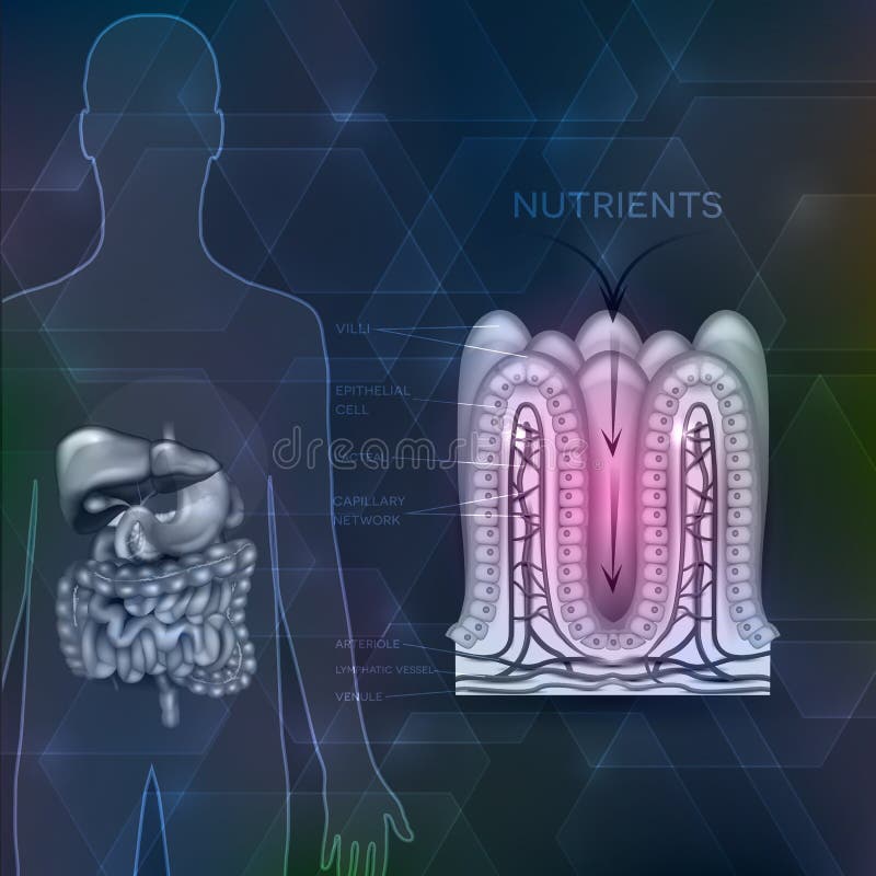
Free with trial Intestinal villi blue technology background. Intestinal villi anatomy on a beautiful abstract technology background. Villus vectors Intestinal villi blue technology background. Intestinal villi anatomy on a beautiful abstract technology background
Free with trial Celiac disease affected small intestine villi. Damaged cells by body's reaction to gluten. Intestinal villi do not absorb nutrients because of reduced surface area. Abstract grey background. Villus vectors Celiac disease. Affected small intestine villi. Damaged cells by body's reaction to gluten. Intestinal villi do not absorb nutrients because of reduced surface. Celiac disease affected small intestine villi. Damaged cells by body's reaction to gluten. Intestinal villi do not absorb nutrients because of reduced surface area. Abstract grey background.
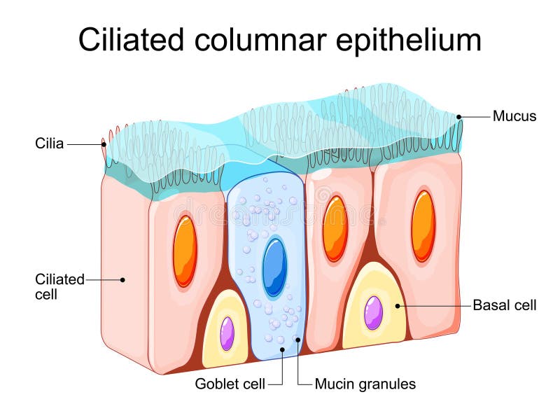
Free with trial Ciliated columnar epithelium. Epithelial cells forms the lining of the stomach and intestines, duodenum, fallopian tubes, uterus, central canal of the spinal cord, nose, ears and the taste buds. Nasal epithelium. Ciliated cells. Respiratory defense mechanisms. Vector illustration. Medical poster. Schematic diagram. Villus vectors Ciliated columnar epithelium. Nasal epithelium. Ciliated columnar epithelium. Epithelial cells forms the lining of the stomach and intestines, duodenum. Ciliated columnar epithelium. Epithelial cells forms the lining of the stomach and intestines, duodenum, fallopian tubes, uterus, central canal of the spinal cord, nose, ears and the taste buds. Nasal epithelium. Ciliated cells. Respiratory defense mechanisms. Vector illustration. Medical poster. Schematic diagram
Free with trial Paneth cell it is the epithelium cells of the small intestine. their granules consist of anti-microbial compounds for important in immunity. Structure of the Paneth cell nucleus, mitochondria and other organelles. Villus vectors Paneth cell. It is the epithelium cells of the small intestine. their granules consist of anti-microbial compounds for important in immunity. Structure of the. Paneth cell it is the epithelium cells of the small intestine. their granules consist of anti-microbial compounds for important in immunity. Structure of the Paneth cell nucleus, mitochondria and other organelles.
Free with trial The illustration is about cell in the human body. This is the intestinal cell. Villus vectors The intestinal cell. The illustration is about cell in the human body. This is the intestinal cell. The illustration is about cell in the human body. This is the intestinal cell.
Free with trial Celiac or Coeliac disease. leaky gut. Cross section of Healthy small intestine with normal villi, and Epithelial cells damaged. Villus vectors Celiac or Coeliac disease. leaky gut. Cross section of Healthy small intestine with normal villi, and Epithelial cells damaged
Free with trial Healthy intestine villi. Intestinal villi anatomy on a beautiful abstract technology background. Villus vectors Healthy intestine villi. Intestinal villi anatomy on a beautiful abstract technology background
Free with trial Ciliated columnar epithelium. epithelial cells forms the lining of the stomach and intestines, duodenum, fallopian tubes, uterus, central canal of the spinal cord, nose, ears and the taste buds. Villus vectors Ciliated columnar epithelium. Epithelial cells forms the lining of the stomach and intestines, duodenum, fallopian tubes, uterus, central canal of the spinal
Free with trial Coeliac disease or celiac disease. small bowel showing coeliac d. Isease manifested by blunting of villi. Villus vectors Coeliac disease or celiac disease. small bowel showing coeliac d. Isease manifested by blunting of villi
Free with trial Leaky gut syndrome and healthy gut. Cute medical cartoon. Vector. Villus vectors Leaky gut syndrome and healthy gut . Cute medical cartoon . Vector.
Free with trial Celiac disease, a very serious guy disease. Villus vectors Celiac Disease. A very serious guy disease
Free with trial Intestinal villi anatomy, small intestine lining. organ. / white background. Villus vectors Intestinal villi anatomy, small intestine lining . organ. / white background
Free with trial Structure of colon villi. Illustration of the colon villi. Villus vectors Structure of colon villi. Illustration of the colon villi
Free with trial Coeliac disease is a autoimmune disorder that affects the small intestine. Signs and symptoms of celiac disease. Cross section of small bowel with normal and shortening of the villi villous atrophy. Vector illustration. Villus vectors Coeliac disease. celiac autoimmune disorder. Coeliac disease is a autoimmune disorder that affects the small intestine. Signs and symptoms of celiac disease. Coeliac disease is a autoimmune disorder that affects the small intestine. Signs and symptoms of celiac disease. Cross section of small bowel with normal and shortening of the villi villous atrophy. Vector illustration
Free with trial Intestinal Villi, Large and small intestine. Gastrointestinal tract. Human anatomy. Villus vectors Intestinal Villi, Large and small intestine. Gastrointestinal tract. Intestinal Villi, Large and small intestine. Human anatomy
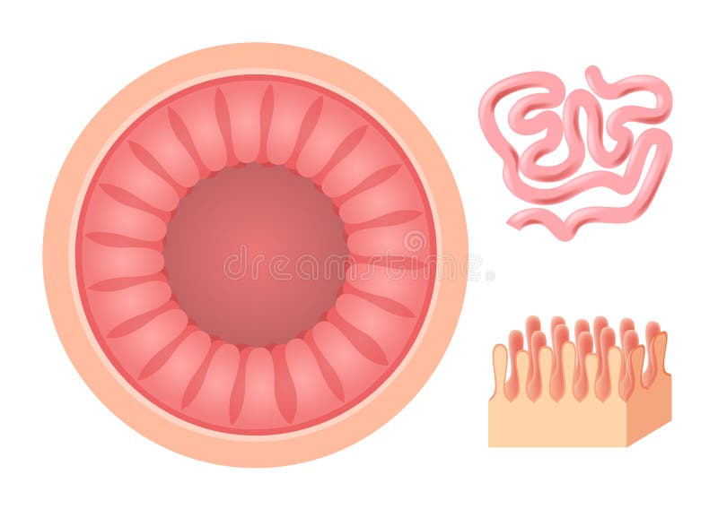
Free with trial Small intestine. general view and cross-section. Human anatomy. Villus vectors Small intestine. Human anatomy. Small intestine. general view and cross-section. Human anatomy. Small intestine. general view and cross-section. Human anatomy.
Free with trial Small Intestine Diagram. Cross-section of a typical segment of the intestinal wall showing the layers: mucosa, submucosa, muscularis, and serosa. Gut. Villus vectors Small Intestine Diagram. Cross-section of a typical segment of the intestinal wall showing the layers: mucosa, submucosa. Muscularis, and serosa. Gut
Free with trial Small intestine Anatomy. Cross section of a ileum with Internal villi. Close-up of Epithelial cells with Microvilli. Vector illustration. Villus vectors Small intestine Anatomy. Cross section of a ileum with Internal villi. Close-up of Epithelial cells with Microvilli. Vector illustration
Free with trial Small bowel and stomach. On an abstract light blue mesh background. Villus vectors Small bowel and stomach. On an abstract light blue mesh background
Free with trial Celiac disease affected small intestine villi on a beautiful grey hexagon background. Unhealthy intestinal villi do not absorb nutrients because of reduced surface area. Villus vectors Damaged intestinal villi. Celiac disease affected small intestine villi on a beautiful grey hexagon background. Unhealthy intestinal villi do not absorb. Celiac disease affected small intestine villi on a beautiful grey hexagon background. Unhealthy intestinal villi do not absorb nutrients because of reduced surface area.
Free with trial Intestinal lining anatomy, absorption of nutrients, human silhouette with digestive organs. Villus vectors Intestinal lining. Anatomy, absorption of nutrients, human silhouette with digestive organs
Free with trial Small intestine surface. Intestinal villi anatomy on a beautiful abstract background. Villus vectors Small intestine surface. Intestinal villi anatomy on a beautiful abstract background
 Sign in