Free with trial Osteoporosis stage 4 of 4 - upper limb bones. Limb bones 3d illustrations Osteoporosis stage 4 of 4 - upper limb bone - 3d rendering. Osteoporosis stage 4 of 4 - upper limb bones
Free with trial Osteoporosis stage 3 of 4 - upper limb bones - 3d rendering. Limb bones 3d illustrations Osteoporosis stage 3 of 4 - upper limb bones - 3d rendering
Free with trial Osteoporosis upper limb bones - human body. Limb bones 3d illustrations Osteoporosis - upper limb bones - 3d rendering. Osteoporosis upper limb bones - human body
Free with trial Osteoporosis stage 1 of 4 - upper limb bones - 3d rendering. Limb bones 3d illustrations Osteoporosis stage 1 of 4 - upper limb bones - 3d rendering
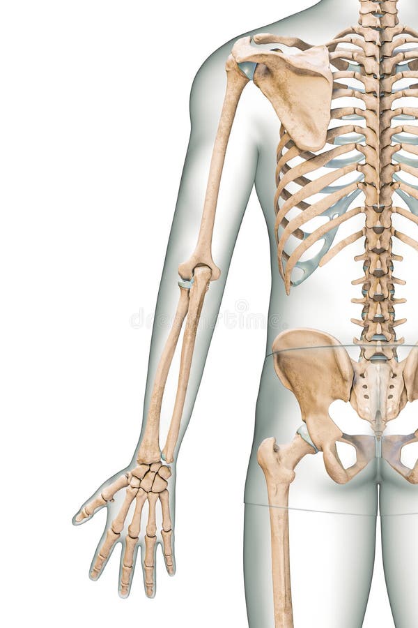
Free with trial Accurate posterior or rear view of the arm or upper limb bones of the human skeletal system with male body contours isolated on white background 3D rendering illustration. Anatomy, osteology concept. Limb bones 3d illustrations Accurate posterior or rear view of the arm or upper limb bones of the human skeletal system with male body contours isolated on
Free with trial Osteoporosis stage 2 of 4 - upper limb bones - 3d rendering. Limb bones 3d illustrations Osteoporosis stage 2 of 4 - upper limb bones - 3d rendering
Free with trial Osteoporosis upper limb bones - human body. Limb bones 3d illustrations Osteoporosis - upper limb bones - 3d rendering. Osteoporosis upper limb bones - human body
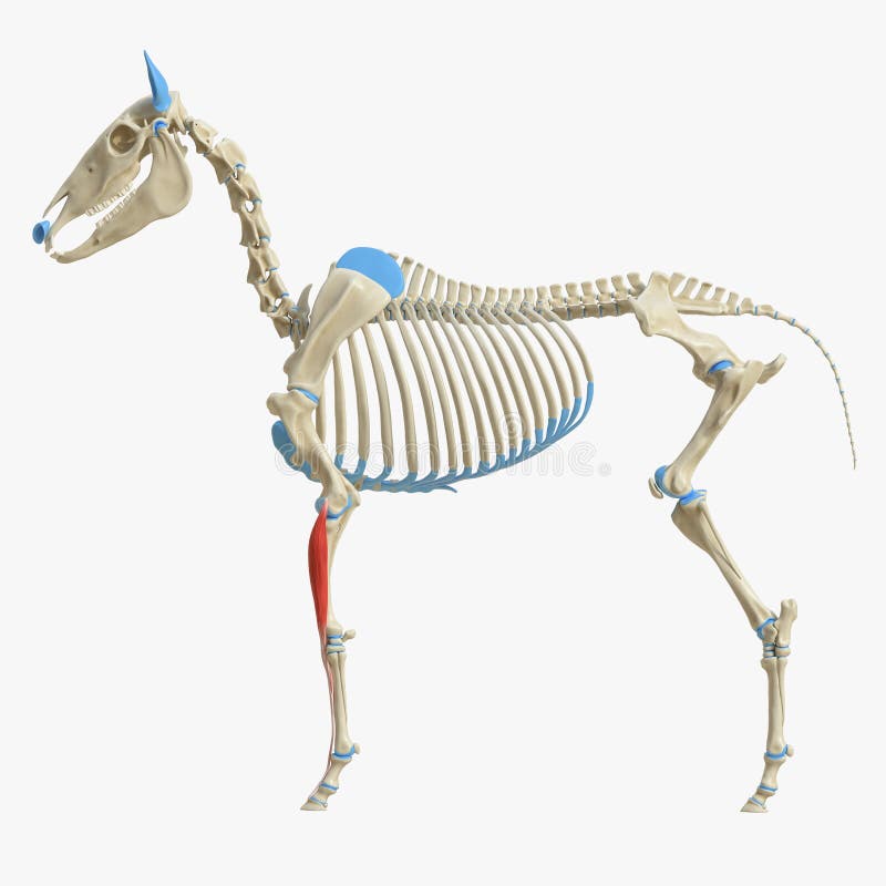
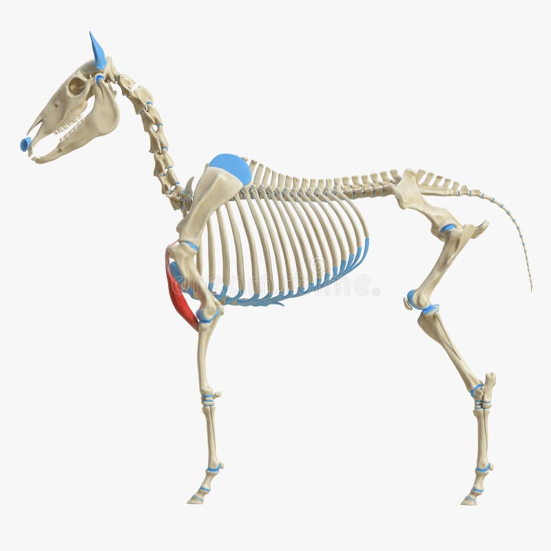
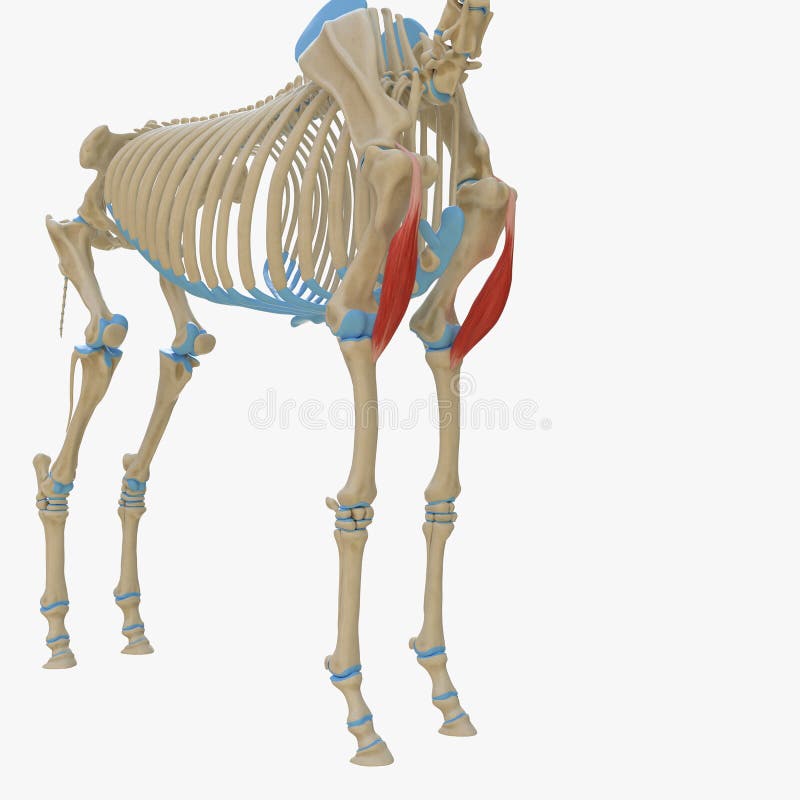
Free with trial 3d rendered medically accurate illustration of the equine muscle anatomy - Biceps Brachii. Limb bones 3d illustrations Biceps Brachii
Free with trial 3d rendered medically accurate illustration of the equine muscle anatomy - Digitorum Profundus. Limb bones 3d illustrations Digitorum Profundus
Free with trial 3d rendered medically accurate illustration of the equine muscle anatomy - Flexor Carpi Radialis. Limb bones 3d illustrations Flexor Carpi Radialis
Free with trial 3d rendered medically accurate illustration of the equine muscle anatomy - Extensor Carpi Radialis. Limb bones 3d illustrations Extensor Carpi Radialis
Free with trial 3d rendered medically accurate illustration of the equine muscle anatomy - Brachialis. Limb bones 3d illustrations Brachialis
Free with trial 3d rendered medically accurate illustration of the equine muscle anatomy - Superficial Digital Flexor. Limb bones 3d illustrations Superficial Digital Flexor
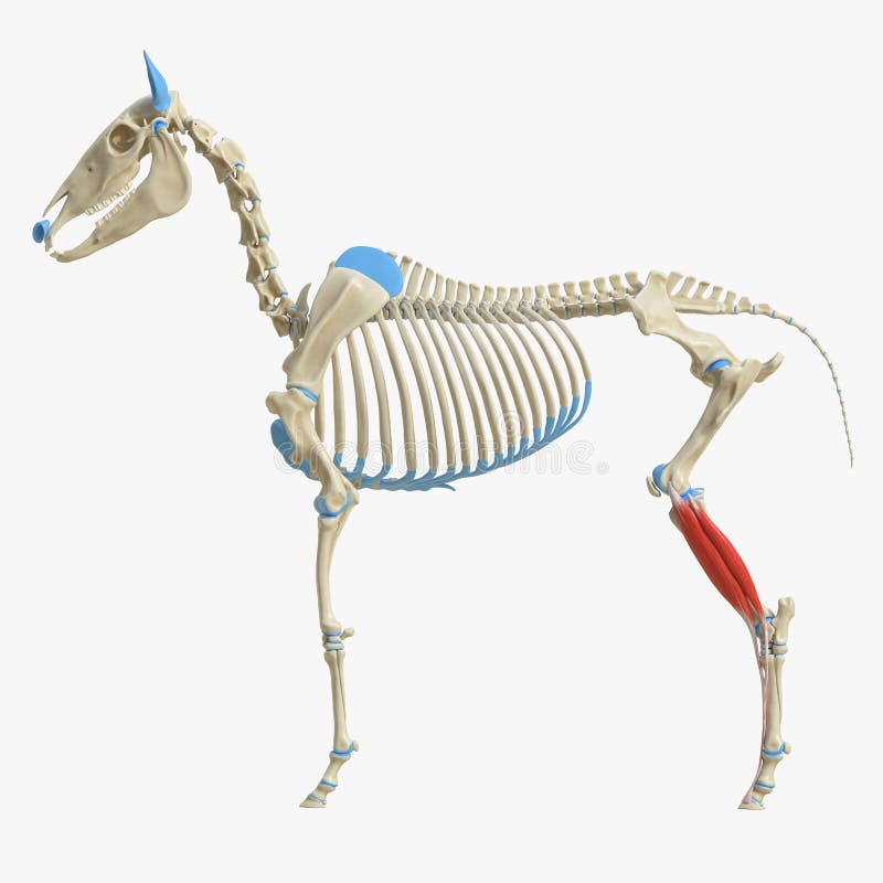
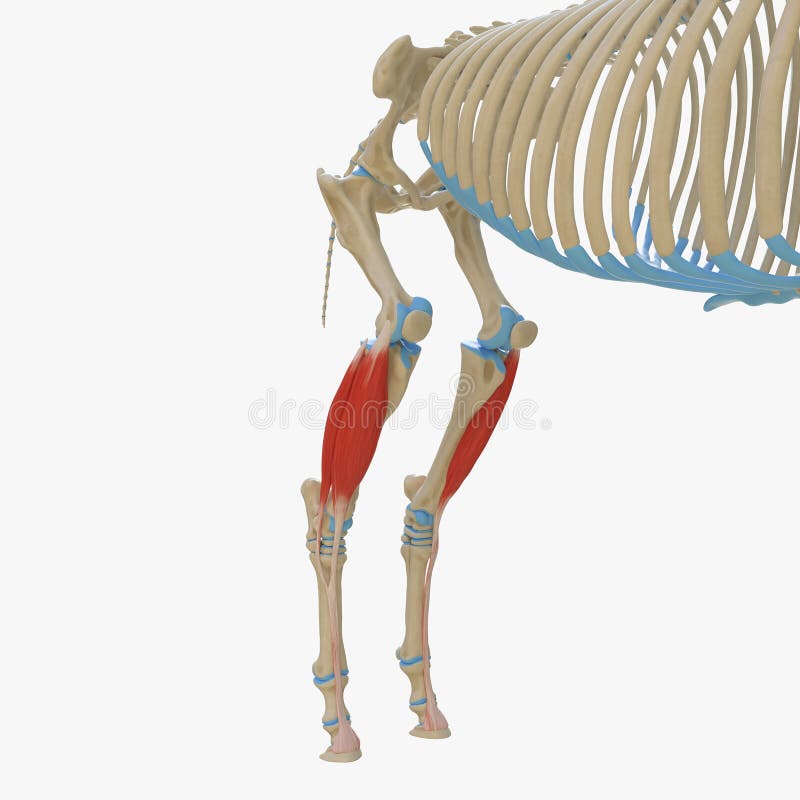
Free with trial 3d rendered medically accurate illustration of the equine muscle anatomy - Gastrocnemius. Limb bones 3d illustrations Gastrocnemius
Free with trial 3d rendered medically accurate illustration of the equine muscle anatomy - Carpi Radialis. Limb bones 3d illustrations Carpi Radialis
Free with trial 3d rendered medically accurate illustration of the equine muscle anatomy - Deep Digital Flexor. Limb bones 3d illustrations Deep Digital Flexor
Free with trial 3d rendered medically accurate illustration of the equine muscle anatomy - Flexor Carpi Ulnaris. Limb bones 3d illustrations Flexor Carpi Ulnaris
Free with trial 3d rendered medically accurate illustration of the equine muscle anatomy - Common Digital Extensor. Limb bones 3d illustrations Common Digital Extensor
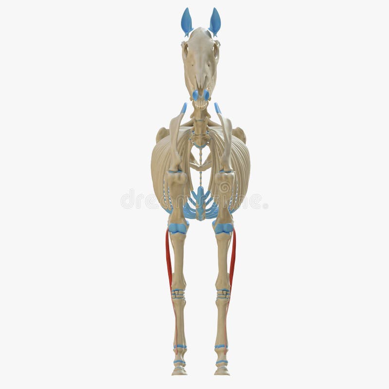
Free with trial 3d rendered medically accurate illustration of the equine muscle anatomy - Gracilis. Limb bones 3d illustrations Gracilis
Free with trial 3d rendered medically accurate illustration of the equine muscle anatomy - Extensor Digitorum Longus. Limb bones 3d illustrations Extensor Digitorum Longus
Free with trial 3d rendered medically accurate illustration of the equine muscle anatomy - Gastrocnemius. Limb bones 3d illustrations Gastrocnemius
Free with trial 3d rendered medically accurate illustration of the equine muscle anatomy - Gastrocnemius. Limb bones 3d illustrations Gastrocnemius
Free with trial 3d rendered medically accurate illustration of the equine muscle anatomy - Lateral Ulnar Muscle. Limb bones 3d illustrations Lateral Ulnar Muscle
Free with trial 3d rendered medically accurate illustration of the equine muscle anatomy - Tensor Fascia Lata. Limb bones 3d illustrations Tensor Fascia Lata
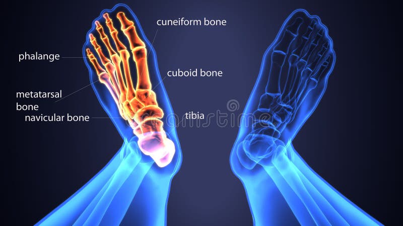
Free with trial The foot is an anatomical structure found in many vertebrates. It is the terminal portion of a limb which bears weight and allows locomotion. In many animals with feet, the foot is a separate organ at the terminal part of the leg made up of one or more segments or bones, generally including claws or nails. Limb bones 3d illustrations Human Skeleton Foot bones Anatomy. 3d illustration. The foot is an anatomical structure found in many vertebrates. It is the terminal portion of a limb which bears weight and allows locomotion. In many animals with feet, the foot is a separate organ at the terminal part of the leg made up of one or more segments or bones, generally including claws or nails.
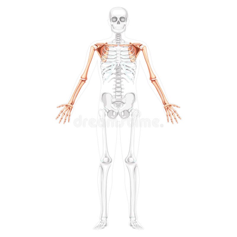
Free with trial Skeleton upper limb Arms with Shoulder girdle Human front view with two arm poses with transparent bones position. Forearms realistic flat Vector illustration of anatomy isolated on white background. Limb bones 3d vectors Skeleton upper limb Arms with Shoulder girdle Human front view with two arm poses with transparent bones position
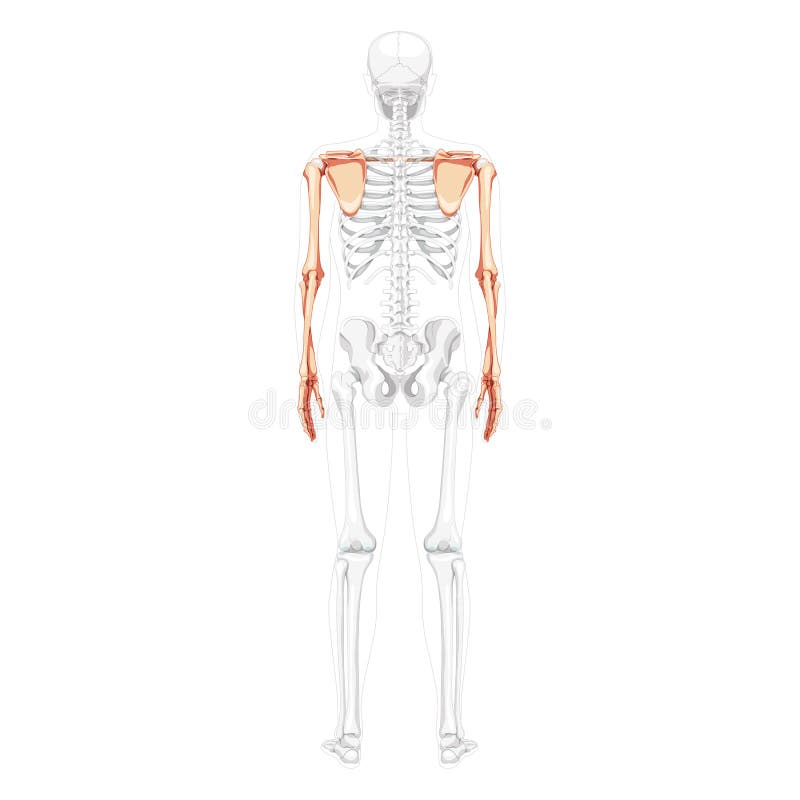
Free with trial Skeleton upper limb Arms with Shoulder girdle Human back view with partly transparent bones position. Hands realistic flat natural color Vector illustration of anatomy isolated on white background. Limb bones 3d vectors Skeleton upper limb Arms with Shoulder girdle Human back view with partly transparent bones position. Hands realistic
Free with trial 3d rendered medically accurate illustration of the bones of the foot. Limb bones 3d illustrations The bones of the foot
Free with trial Osteoporosis upper limb bones - human body. Limb bones 3d illustrations Osteoporosis - upper limb bones - 3d rendering. Osteoporosis upper limb bones - human body
Free with trial Osteoporosis upper limb bones - human body. Limb bones 3d illustrations Osteoporosis - upper limb bones - 3d rendering. Osteoporosis upper limb bones - human body
Free with trial Osteoporosis upper limb bones - human body. Limb bones 3d illustrations Osteoporosis - upper limb bones - 3d rendering. Osteoporosis upper limb bones - human body
Free with trial Detailed 3D illustration of human knee joint anatomy with labels. Educational medical diagram showing ligaments and bones on white background. Useful for healthcare presentations. Limb bones 3d illustrations Detailed 3D illustration of human knee joint anatomy with labels. Educational medical diagram showing ligaments and bones on white
Free with trial The foot plural feet is an anatomical structure found in many vertebrates. It is the terminal portion of a limb which bears weight and allows locomotion. In many animals with feet, the foot is a separate organ at the terminal part of the leg made up of one or more segments or bones, generally including claws or nails. Limb bones 3d illustrations 3d illustration of human body feet bone. The foot plural feet is an anatomical structure found in many vertebrates. It is the terminal portion of a limb which bears weight and allows locomotion. In many animals with feet, the foot is a separate organ at the terminal part of the leg made up of one or more segments or bones, generally including claws or nails.
Free with trial The foot plural feet is an anatomical structure found in many vertebrates. It is the terminal portion of a limb which bears weight and allows locomotion. In many animals with feet, the foot is a separate organ at the terminal part of the leg made up of one or more segments or bones, generally including claws or nails. Limb bones 3d illustrations 3d illustration of skeleton foot bone anatomy. The foot plural feet is an anatomical structure found in many vertebrates. It is the terminal portion of a limb which bears weight and allows locomotion. In many animals with feet, the foot is a separate organ at the terminal part of the leg made up of one or more segments or bones, generally including claws or nails.
Free with trial The foot is an anatomical structure found in many vertebrates. It is the terminal portion of a limb which bears weight and allows locomotion. In many animals with feet, the foot is a separate organ at the terminal part of the leg made up of one or more segments or bones, generally including claws or nails. Limb bones 3d illustrations 3d illustration of skeleton foot bone anatomy. The foot is an anatomical structure found in many vertebrates. It is the terminal portion of a limb which bears weight and allows locomotion. In many animals with feet, the foot is a separate organ at the terminal part of the leg made up of one or more segments or bones, generally including claws or nails.
Free with trial The two hip bones join at the pubic symphysis and together with the sacrum and coccyx the pelvic part of the spine comprise the skeletal component of the pelvis – the pelvic girdle which surrounds the pelvic cavity. They are connected to the sacrum, which is part of the axial skeleton, at the sacroiliac joint. Each hip bone is connected to the corresponding femur thigh bone forming the primary connection between the bones of the lower limb and the axial skeleton through the large ball and socket joint of the hip. [2]. Limb bones 3d illustrations 3d illustration of skeleton hip bone anatomy. The two hip bones join at the pubic symphysis and together with the sacrum and coccyx the pelvic part of the spine comprise the skeletal component of the pelvis – the pelvic girdle which surrounds the pelvic cavity. They are connected to the sacrum, which is part of the axial skeleton, at the sacroiliac joint. Each hip bone is connected to the corresponding femur thigh bone forming the primary connection between the bones of the lower limb and the axial skeleton through the large ball and socket joint of the hip.[2]
Free with trial The foot plural feet is an anatomical structure found in many vertebrates. It is the terminal portion of a limb which bears weight and allows locomotion. In many animals with feet, the foot is a separate organ at the terminal part of the leg made up of one or more segments or bones, generally including claws or nails. Limb bones 3d illustrations 3d illustration of skeleton feet bone anatomy. The foot plural feet is an anatomical structure found in many vertebrates. It is the terminal portion of a limb which bears weight and allows locomotion. In many animals with feet, the foot is a separate organ at the terminal part of the leg made up of one or more segments or bones, generally including claws or nails.
Free with trial The foot plural feet is an anatomical structure found in many vertebrates. It is the terminal portion of a limb which bears weight and allows locomotion. In many animals with feet, the foot is a separate organ at the terminal part of the leg made up of one or more segments or bones, generally including claws or nails. Limb bones 3d illustrations 3d illustration of skeleton feet bone anatomy. The foot plural feet is an anatomical structure found in many vertebrates. It is the terminal portion of a limb which bears weight and allows locomotion. In many animals with feet, the foot is a separate organ at the terminal part of the leg made up of one or more segments or bones, generally including claws or nails.
Free with trial This 3d illustration shows the knee pain condition in front view. Limb bones 3d illustrations 3d illustration of knee pain front view. This 3d illustration shows the knee pain condition in front view
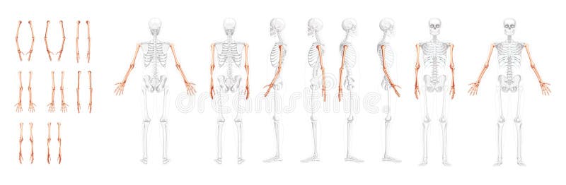
Free with trial Set of Skeleton Arms Human front back side view with partly transparent bones position. Hands, forearms realistic flat natural color concept Vector illustration of anatomy isolated on white background. Limb bones 3d vectors Set of Skeleton Arms Human front back side view with partly transparent bones position. Hands, forearms realistic flat
Free with trial 3d rendered medically accurate illustration of the equine muscle anatomy - Common Digital Extensor. Limb bones 3d illustrations Common Digital Extensor
Free with trial 3d rendered medically accurate illustration of the equine muscle anatomy - Gracilis. Limb bones 3d illustrations Gracilis
Free with trial 3d rendered medically accurate illustration of the equine muscle anatomy - Adductor. Limb bones 3d illustrations Adductor
Free with trial 3d rendered medically accurate illustration of the equine muscle anatomy - Popliteus. Limb bones 3d illustrations Popliteus
Free with trial 3d rendered medically accurate illustration of the equine muscle anatomy - Brachialis. Limb bones 3d illustrations Brachialis
Free with trial 3d rendered medically accurate illustration of the equine muscle anatomy - Flexor Digitorum Profundus. Limb bones 3d illustrations Flexor Digitorum Profundus
Free with trial 3d rendered medically accurate illustration of the equine muscle anatomy - Biceps Brachii. Limb bones 3d illustrations Biceps Brachii
Free with trial 3d rendered medically accurate illustration of the equine muscle anatomy - Policis Longus. Limb bones 3d illustrations Policis Longus
Free with trial 3d rendered medically accurate illustration of the equine muscle anatomy - Biceps Brachii. Limb bones 3d illustrations Biceps Brachii
Free with trial 3d rendered medically accurate illustration of the equine muscle anatomy - Flexor Digitorum Superficialis. Limb bones 3d illustrations Flexor Digitorum Superficialis
Free with trial 3d rendered medically accurate illustration of the equine muscle anatomy - Tibialis Cranialis. Limb bones 3d illustrations Tibialis Cranialis
Free with trial 3d rendered medically accurate illustration of the equine muscle anatomy - Tibialis Cranialis. Limb bones 3d illustrations Tibialis Cranialis
Free with trial 3d rendered medically accurate illustration of the equine muscle anatomy - Digitorum Profundus. Limb bones 3d illustrations Digitorum Profundus
Free with trial 3d rendered medically accurate illustration of the equine muscle anatomy - Deep Digital Flexor. Limb bones 3d illustrations Deep Digital Flexor
Free with trial 3d skeletal arm, over white. Limb bones 3d illustrations Skeleton Arm, Grey Scroll Sign. 3d skeletal arm, over white
Free with trial 3d rendered medically accurate illustration of the equine muscle anatomy - Lateral Ulnar Muscle. Limb bones 3d illustrations Lateral Ulnar Muscle
Free with trial 3d rendered medically accurate illustration of the equine muscle anatomy - Flexor Carpi Radialis. Limb bones 3d illustrations Flexor Carpi Radialis
Free with trial 3d rendered medically accurate illustration of the equine muscle anatomy - Extensor Carpi Radialis. Limb bones 3d illustrations Extensor Carpi Radialis
Free with trial 3d rendered medically accurate illustration of the equine muscle anatomy - Extensor Digitorum Longus. Limb bones 3d illustrations Extensor Digitorum Longus
Free with trial 3d rendered medically accurate illustration of the equine muscle anatomy - Superficial Digital Flexor. Limb bones 3d illustrations Superficial Digital Flexor
Free with trial 3d rendered medically accurate illustration of the equine muscle anatomy - Flexor Digitorum Superficialis. Limb bones 3d illustrations Flexor Digitorum Superficialis
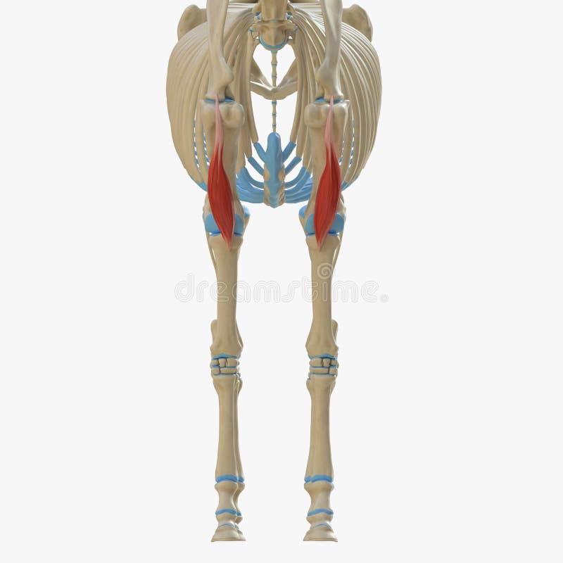
Free with trial 3d rendered medically accurate illustration of the equine muscle anatomy - Semimembranosus. Limb bones 3d illustrations Semimembranosus
Free with trial 3d rendered medically accurate illustration of the equine muscle anatomy - Common Digital Extensor. Limb bones 3d illustrations Common Digital Extensor
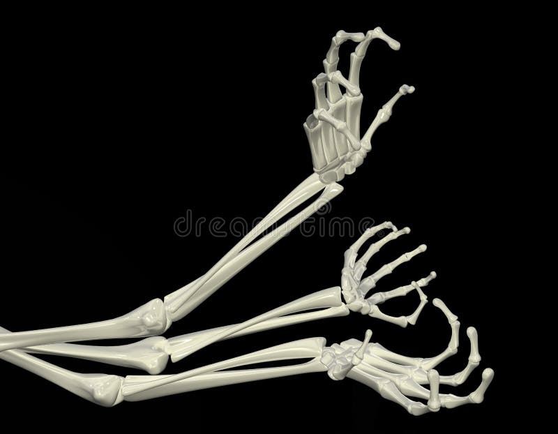
Free with trial 3d skeletal arm, isolated, dark background. Limb bones 3d illustrations Skeletal Arms. 3d skeletal arm, isolated, dark background
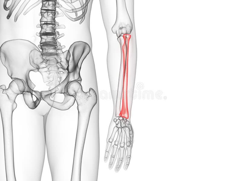
Free with trial 3d rendered medically accurate illustration of the ulna bone. Limb bones 3d illustrations The ulna bone
Free with trial 3d rendered medically accurate illustration of the equine muscle anatomy - Popliteus. Limb bones 3d illustrations Popliteus
Free with trial 3d rendered medically accurate illustration of the equine muscle anatomy - Sartorius. Limb bones 3d illustrations Sartorius
Free with trial 3d rendered medically accurate illustration of the equine muscle anatomy - Brachialis. Limb bones 3d illustrations Brachialis
Free with trial 3d rendered medically accurate illustration of the equine muscle anatomy - Policis Longus. Limb bones 3d illustrations Policis Longus
Free with trial 3d rendered medically accurate illustration of the equine muscle anatomy - Flexor Carpi Ulnaris. Limb bones 3d illustrations Flexor Carpi Ulnaris
Free with trial 3d rendered medically accurate illustration of the equine muscle anatomy - Popliteus. Limb bones 3d illustrations Popliteus
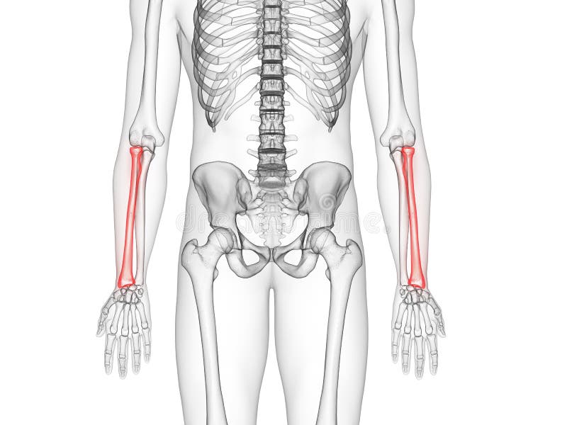
Free with trial 3d rendered medically accurate illustration of the radius bone. Limb bones 3d illustrations The radius bone
Free with trial 3d rendered medically accurate illustration of the radius bone. Limb bones 3d illustrations The radius bone
Free with trial 3d skeletal arm, isolated, dark background. Limb bones 3d illustrations Skeletal Arm, Right Angle. 3d skeletal arm, isolated, dark background
Free with trial The pelvis, also known as the pelvic girdle, is a bony structure located at the base of the spine between the abdomen and the lower limbs. It consists of several bones that are fused together, providing support for the upper body and connecting the spine to the lower limbs. Limb bones 3d illustrations Pelvis or pelvic girdle anatomy 3D illustration. The pelvis, also known as the pelvic girdle, is a bony structure located at the base of the spine between the abdomen and the lower limbs. It consists of several bones that are fused together, providing support for the upper body and connecting the spine to the lower limbs.
Free with trial 12 x-ray renders of male body with skeleton and internal organs - roentgen concept for healthcare - digital high resolution medical 3D illustration isolated. Limb bones 3d illustrations 12 high resolution xray renders in 1 image, man body with skeleton and organs - anatomy colored examination concept - digital. 12 x-ray renders of male body with skeleton and internal organs - roentgen concept for healthcare - digital high resolution medical 3D illustration isolated
Free with trial A translucent 3D anatomical model of a human leg, showcasing bones with red highlights indicating pain in the knee and hip, isolated on a white background. Limb bones 3d illustrations Anatomical 3d rendering of human leg bones with highlighted pain areas isolated on white background. A translucent 3D anatomical model of a human leg, showcasing bones with red highlights indicating pain in the knee and hip, isolated on a white background
Free with trial A detailed 3D render of a human hand skeleton is presented against a clean white background. The image clearly displays the individual bones of the hand, including the metacarpals, phalanges, and carpals, illustrating the intricate structure of the wrist and fingers. The light gray color of the bones provides a stark contrast to the white backdrop, emphasizing the anatomical details. Limb bones 3d illustrations Anatomical 3D Render of a Human Hand Skeleton on a White Background human anatomy bones. A detailed 3D render of a human hand skeleton is presented against a clean white background. The image clearly displays the individual bones of the hand, including the metacarpals, phalanges, and carpals, illustrating the intricate structure of the wrist and fingers. The light gray color of the bones provides a stark contrast to the white backdrop, emphasizing the anatomical details
Free with trial Detailed 3D render of human leg muscles and bones from the knee down to the foot, isolated on a white background. High quality photo AI generated. Limb bones 3d illustrations Detailed 3D render of human leg muscles and bones from the knee down to the foot, isolated on a white background. High quality photo AI generated


![The two hip bones join at the pubic symphysis and together with the sacrum and coccyx the pelvic part of the spine comprise the skeletal component of the pelvis – the pelvic girdle which surrounds the pelvic cavity. They are connected to the sacrum, which is part of the axial skeleton, at the sacroiliac joint. Each hip bone is connected to the corresponding femur thigh bone forming the primary connection between the bones of the lower limb and the axial skeleton through the large ball and socket joint of the hip. [2]. Limb bones 3d illustrations](https://thumbs.dreamstime.com/b/two-hip-bones-join-pubic-symphysis-together-sacrum-coccyx-pelvic-part-spine-comprise-skeletal-component-pelvis-116766762.jpg)