Free with trial Bones of the upper extremity: Clavicle (collar bone), scapula (shoulder blade), humerus, ulna, radius, finger and hand. Detailed medical illustrations. Latin medical terms. Isolated on a white background. Limb bones vectors Bones of the upper extremity
Free with trial The illustration shows the muscles that make up the arm, with its relative movements. Limb bones vectors Arm muscles. The illustration shows the muscles that make up the arm, with its relative movements.
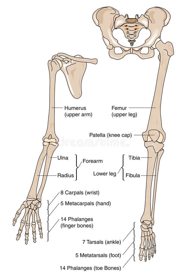
Free with trial Bones of upper and lower human limbs. Limb bones vectors Limb bones. Bones of upper and lower human limbs
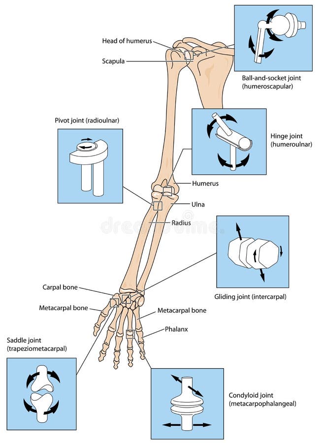
Free with trial Various types of joint, illustrated by the joints of the upper limb from the scapular to the fingers. Created in Adobe Illustrator. Limb bones vectors Joint types in the upper limb. Various types of joint, illustrated by the joints of the upper limb from the scapular to the fingers. Created in Adobe Illustrator.
Free with trial Osteoporosis stage 4 of 4 - upper limb bones. Limb bones illustrations Osteoporosis stage 4 of 4 - upper limb bone - 3d rendering. Osteoporosis stage 4 of 4 - upper limb bones
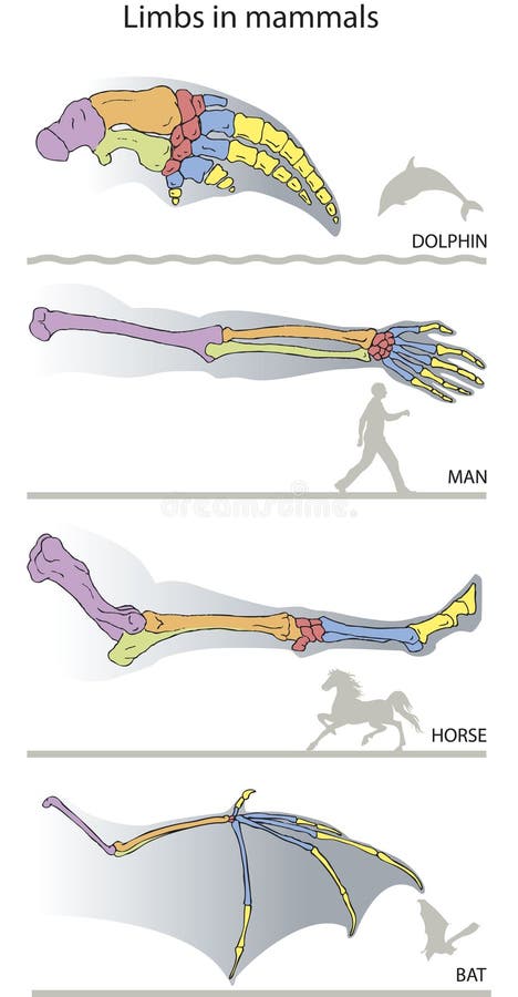
Free with trial Comparative presentation of limb bones in mammals (dolphin, man, horse, bat). Limb bones vectors Limbs. Comparative presentation of limb bones in mammals (dolphin, man, horse, bat).
Free with trial Osteoporosis stage 3 of 4 - upper limb bones - 3d rendering. Limb bones illustrations Osteoporosis stage 3 of 4 - upper limb bones - 3d rendering
Free with trial Osteoporosis upper limb bones - human body. Limb bones illustrations Osteoporosis - upper limb bones - 3d rendering. Osteoporosis upper limb bones - human body
Free with trial Osteoporosis stage 1 of 4 - upper limb bones - 3d rendering. Limb bones illustrations Osteoporosis stage 1 of 4 - upper limb bones - 3d rendering
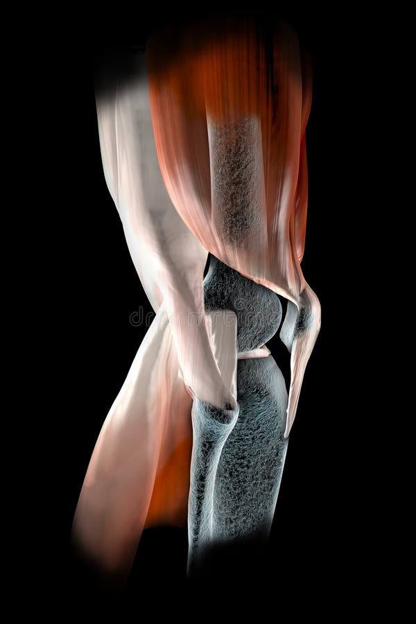
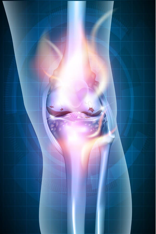
Free with trial Knee seen on x-rays with muscle tendons and bones in evidence. Limb bones illustrations Knee ligaments, tendons, bones, muscles x-ray. Knee seen on x-rays with muscle tendons and bones in evidence
Free with trial Bones of the upper extremity: Clavicle (collar bone), scapula (shoulder blade), humerus, ulna, radius, fingers and hand. Detailed medical illustrations. Isolated on a white background. Limb bones vectors Bones of the upper extremity
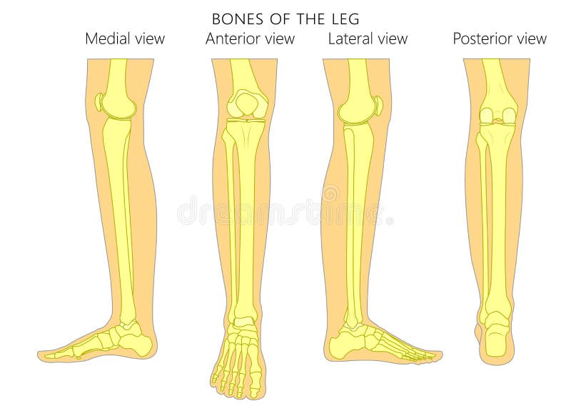
Free with trial Bones of a human leg different views: posterior, frontal, anterior, back, side, lateral, medial with ankle and knee. Vector illustration for advertising, medical health care publications. EPS 10. Limb bones vectors Bone fracture_Leg anatomy bones. Bones of a human leg different views: posterior, frontal, anterior, back, side, lateral, medial with ankle and knee. Vector illustration for advertising, medical health care publications. EPS 10
Free with trial Pain in knee joints. A X-ray of feet of the man. It is isolated on a black background. Limb bones illustrations Pain in knee joints
Free with trial Knee pain abstract background. Healthy joint and unhealthy painful joint with osteoarthritis. Limb bones vectors Knee pain abstract background
Free with trial Burning knee, painful knee and normal knee joint, abstract design. Human legs on a blue checkered background. Limb bones vectors Burning knee
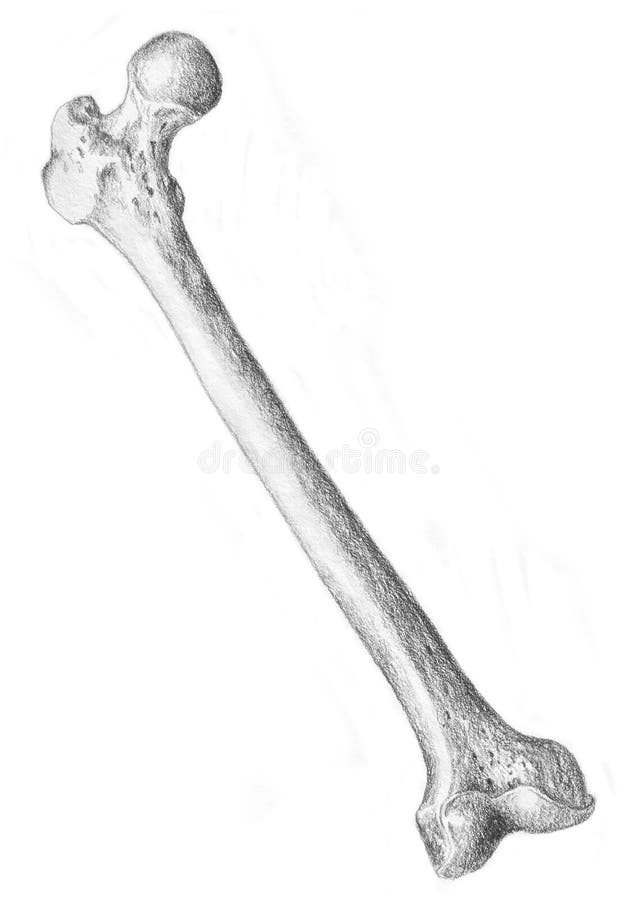
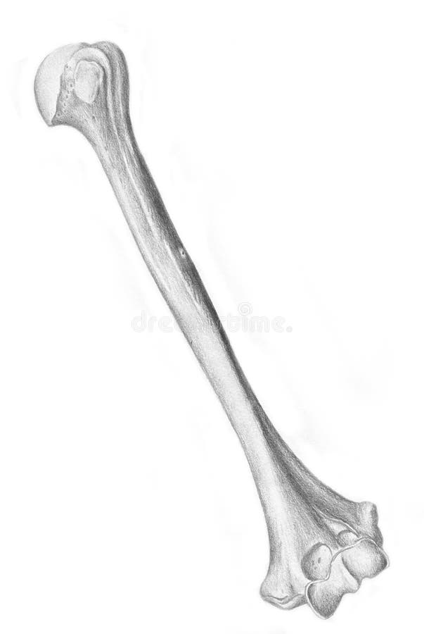
Free with trial A sketch in black and white of the human skeleton. Femur, bone of the thigh. Limb bones illustrations Femur - skeleton. A sketch in black and white of the human skeleton. Femur, bone of the thigh
Free with trial Joint pain, joint anatomy beautiful abstract background. Limb bones vectors Joint pain
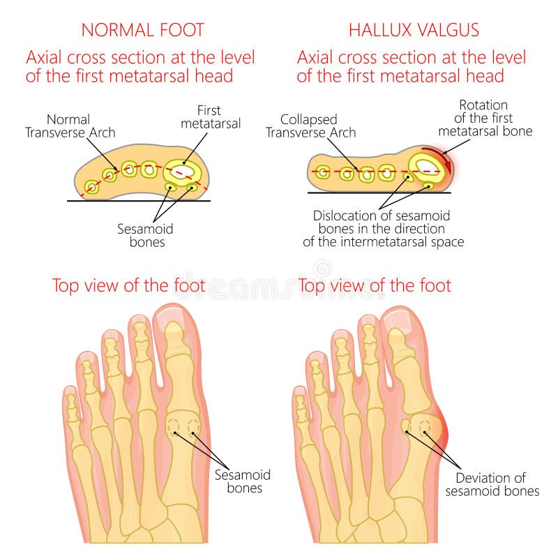
Free with trial Vector illustration of a healthy human forefoot and a foot with hallux valgus, dislocation of sesamoid bones. Top view and cross section of of the foot. For advertising, medical publications. EPS 10. Limb bones vectors Normal foot and Hallux valgus with rotation of the first mematarsal bone. Vector illustration of a healthy human forefoot and a foot with hallux valgus, dislocation of sesamoid bones. Top view and cross section of of the foot. For advertising, medical publications. EPS 10.
Free with trial Skeleton membri superioris. Bones of the upper limb. Anterior view. Human anatomy. Vector illustration isolated on a white background. Limb bones illustrations Skeleton membri superioris. Bones of the upper limb. Anterior view. Human anatomy.
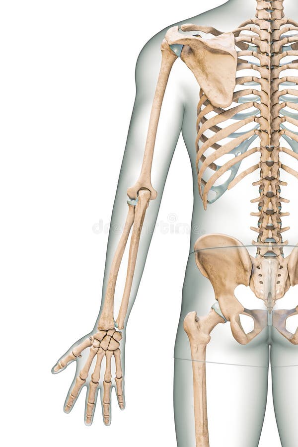
Free with trial Accurate posterior or rear view of the arm or upper limb bones of the human skeletal system with male body contours isolated on white background 3D rendering illustration. Anatomy, osteology concept. Limb bones illustrations Accurate posterior or rear view of the arm or upper limb bones of the human skeletal system with male body contours isolated on. White background 3D rendering. Accurate posterior or rear view of the arm or upper limb bones of the human skeletal system with male body contours isolated on white background 3D rendering illustration. Anatomy, osteology concept
Free with trial The major (long) bones of the human leg are the femur (thigh bone), tibia (shin bone), and fibula (the smaller, rear calf bone). The patella (kneecap) is the bone in front of the knee. Most of the leg skeleton has bony prominences and margins that can be palpated, notable exceptions being the hip joint, and the neck and shaft of femur. Many of these anatomical landmarks are used to define the extent of the leg: most notably the anterior superior iliac spine, the greater trochanter, the superior margin of the medial condyle of tibia, and the medial malleolus. Limb bones illustrations Lower Limb Bones Anterior view. The major (long) bones of the human leg are the femur (thigh bone), tibia (shin bone), and fibula (the smaller, rear calf bone). The patella (kneecap) is the bone in front of the knee. Most of the leg skeleton has bony prominences and margins that can be palpated, notable exceptions being the hip joint, and the neck and shaft of femur. Many of these anatomical landmarks are used to define the extent of the leg: most notably the anterior superior iliac spine, the greater trochanter, the superior margin of the medial condyle of tibia, and the medial malleolus.
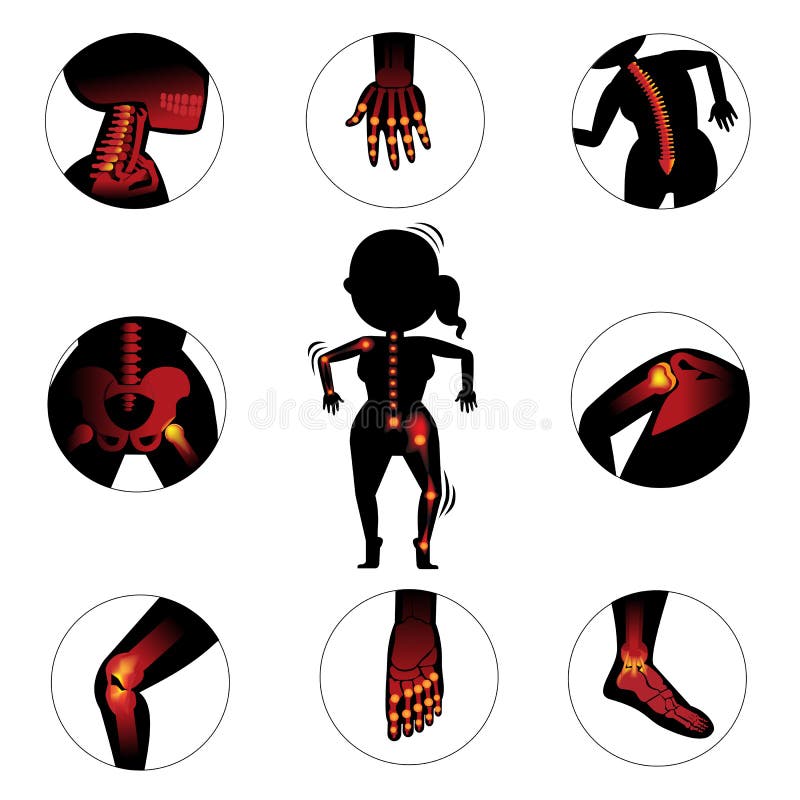
Free with trial Bones and joints in the circle. Vector Illustration. Limb bones vectors Bones and joints
Free with trial Osteoporosis stage 2 of 4 - upper limb bones - 3d rendering. Limb bones illustrations Osteoporosis stage 2 of 4 - upper limb bones - 3d rendering
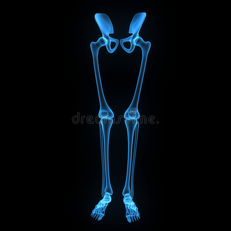
Free with trial The hip joint is one of the most important joints in the human body. It allows us to walk, run, and jump. It bears our body’s weight and the force of the strong muscles of the hip and leg. Yet the hip joint is also one of our most flexible joints and allows a greater range of motion than all other joints in the body except for the shoulder. The hip joint is a ball-and-socket synovial joint formed between the os coxa (hip bone) and the femur. A round, cup-shaped structure on the os coax, known as the acetabulum, forms the socket for the hip joint. The rounded head of the femur forms the ball of the joint. The tibia, sometimes known as the shin bone, is the larger and stronger of the two lower leg bones. It forms the knee joint with the femur and the ankle joint with the fibula and tarsus. The fibula is the long, thin and lateral bone of the lower leg. It runs parallel to the tibia, or shin bone, and plays a significant role in stabilizing the ankle and supporting the muscles of the lower leg. The bones of the ankle and foot form the most distal region of the lower limb in the appendicular skeleton. These bones are responsible for the propulsion, balance, and support of the body’s weight through many diverse activities, such as standing, walking, running, and jumping. Limb bones illustrations Skeleton: Hip, Femur, Tibia, Fibula, Ankle and Foot bones. The hip joint is one of the most important joints in the human body. It allows us to walk, run, and jump. It bears our body’s weight and the force of the strong muscles of the hip and leg. Yet the hip joint is also one of our most flexible joints and allows a greater range of motion than all other joints in the body except for the shoulder. The hip joint is a ball-and-socket synovial joint formed between the os coxa (hip bone) and the femur. A round, cup-shaped structure on the os coax, known as the acetabulum, forms the socket for the hip joint. The rounded head of the femur forms the ball of the joint. The tibia, sometimes known as the shin bone, is the larger and stronger of the two lower leg bones. It forms the knee joint with the femur and the ankle joint with the fibula and tarsus. The fibula is the long, thin and lateral bone of the lower leg. It runs parallel to the tibia, or shin bone, and plays a significant role in stabilizing the ankle and supporting the muscles of the lower leg. The bones of the ankle and foot form the most distal region of the lower limb in the appendicular skeleton. These bones are responsible for the propulsion, balance, and support of the body’s weight through many diverse activities, such as standing, walking, running, and jumping.
Free with trial Osteoporosis upper limb bones - human body. Limb bones illustrations Osteoporosis - upper limb bones - 3d rendering. Osteoporosis upper limb bones - human body
Free with trial A sketch in black and white of the human skeleton. Bones of the foot, side vision. Limb bones illustrations Foot - skeleton. A sketch in black and white of the human skeleton. Bones of the foot, side vision
Free with trial The sketch in white and black realized to hand. The bones of the foot. Limb bones illustrations Foot, man's anatomy. The sketch in white and black realized to hand. The bones of the foot
Free with trial A sketch in black and white of the human skeleton. Bones of the arm, left humerus. Limb bones illustrations Left humerus - skeleton. A sketch in black and white of the human skeleton. Bones of the arm, left humerus
Free with trial The sketch in white and black realized to hand. The bones of the vertebral column. Limb bones illustrations Vertebral column, man's anatomy. The sketch in white and black realized to hand. The bones of the vertebral column
Free with trial Iliacus muscle with hip or groin muscular and skeletal anatomy outline diagram. Labeled educational scheme with human lesser trochanter of femur and iliac fossa bones location vector illustration. Limb bones vectors Iliacus muscle with hip or groin muscular, skeletal anatomy outline diagram. Iliacus muscle with hip or groin muscular and skeletal anatomy outline diagram. Labeled educational scheme with human lesser trochanter of femur and iliac fossa bones location vector illustration.
Free with trial 3d rendered medically accurate illustration of the equine muscle anatomy - Biceps Brachii. Limb bones illustrations Biceps Brachii
Free with trial 3d rendered medically accurate illustration of the equine muscle anatomy - Digitorum Profundus. Limb bones illustrations Digitorum Profundus
Free with trial 3d rendered medically accurate illustration of the equine muscle anatomy - Flexor Carpi Radialis. Limb bones illustrations Flexor Carpi Radialis
Free with trial 3d rendered medically accurate illustration of the equine muscle anatomy - Extensor Carpi Radialis. Limb bones illustrations Extensor Carpi Radialis
Free with trial 3d rendered medically accurate illustration of the equine muscle anatomy - Brachialis. Limb bones illustrations Brachialis
Free with trial 3d rendered medically accurate illustration of the equine muscle anatomy - Superficial Digital Flexor. Limb bones illustrations Superficial Digital Flexor
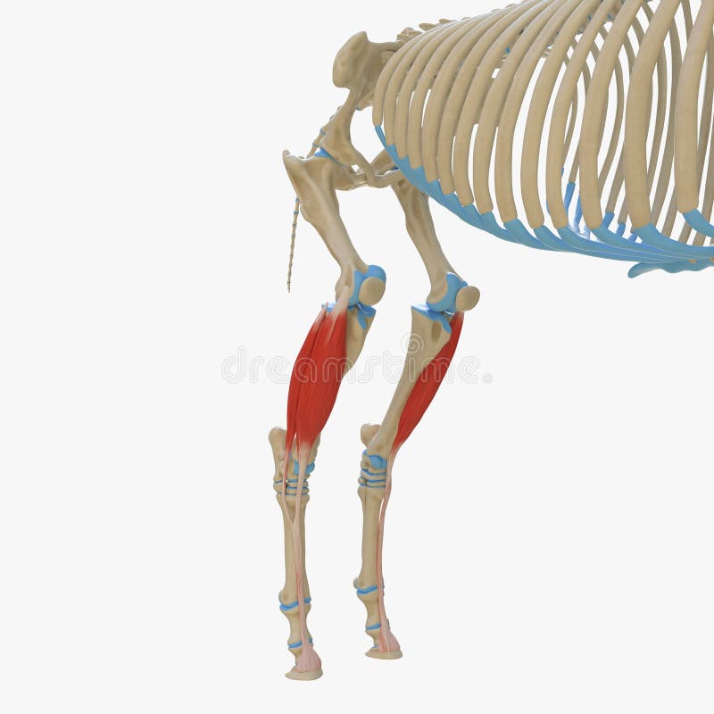
Free with trial 3d rendered medically accurate illustration of the equine muscle anatomy - Gastrocnemius. Limb bones illustrations Gastrocnemius
Free with trial 3d rendered medically accurate illustration of the equine muscle anatomy - Carpi Radialis. Limb bones illustrations Carpi Radialis
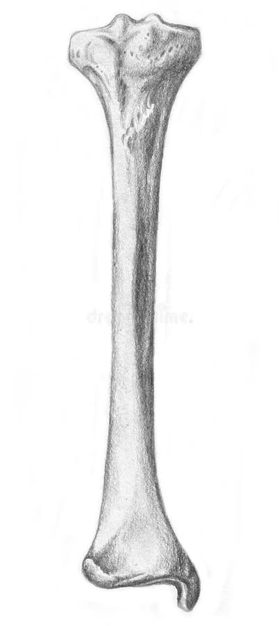
Free with trial A sketch in black and white of the human skeleton. Tibia, bone of the leg. Limb bones illustrations Tibia - skeleton. A sketch in black and white of the human skeleton. Tibia, bone of the leg
Free with trial 3d rendered medically accurate illustration of the equine muscle anatomy - Deep Digital Flexor. Limb bones illustrations Deep Digital Flexor
Free with trial Damaged joint and normal joint design on an abstract grey glowing background. Limb bones vectors Joint
Free with trial A sketch in black and white of the human skeleton. the second lumbar bone. Limb bones illustrations Lumbar bone - skeleton. A sketch in black and white of the human skeleton. the second lumbar bone
Free with trial 3d rendered medically accurate illustration of the equine muscle anatomy - Flexor Carpi Ulnaris. Limb bones illustrations Flexor Carpi Ulnaris
Free with trial Burning leg knee joint on a abstract blue checkered background. Limb bones illustrations Burning leg knee joint
Free with trial 3d rendered medically accurate illustration of the equine muscle anatomy - Common Digital Extensor. Limb bones illustrations Common Digital Extensor
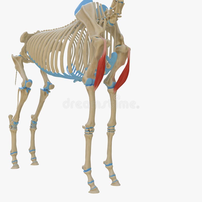
Free with trial 3d rendered medically accurate illustration of the equine muscle anatomy - Gracilis. Limb bones illustrations Gracilis
Free with trial 3d rendered medically accurate illustration of the equine muscle anatomy - Extensor Digitorum Longus. Limb bones illustrations Extensor Digitorum Longus
Free with trial 3d rendered medically accurate illustration of the equine muscle anatomy - Gastrocnemius. Limb bones illustrations Gastrocnemius
Free with trial 3d rendered medically accurate illustration of the equine muscle anatomy - Gastrocnemius. Limb bones illustrations Gastrocnemius
Free with trial 3d rendered medically accurate illustration of the equine muscle anatomy - Lateral Ulnar Muscle. Limb bones illustrations Lateral Ulnar Muscle
Free with trial A sketch in black and white of the human skeleton. Articulation of the shoulder blade and humerus. Limb bones illustrations Articulation of the shoulder blade and humerus - s. A sketch in black and white of the human skeleton. Articulation of the shoulder blade and humerus
Free with trial Normal leg knee joint at the blue background and unhealthy joint at the red background. Limb bones vectors Knee joints healthy and unhealthy. Normal leg knee joint at the blue background and unhealthy joint at the red background
Free with trial 3d rendered medically accurate illustration of the equine muscle anatomy - Tensor Fascia Lata. Limb bones illustrations Tensor Fascia Lata
Free with trial Healthy knee and knee with osteoarthritis on a white background. Limb bones vectors Healthy knee and knee with osteoarthritis
Free with trial Anatomy of the canine dog`s knee joint colorful design, healthy joint info poster illustration. Limb bones vectors Anatomy of the canine knee. Anatomy of the canine dog`s knee joint colorful design, healthy joint info poster illustration
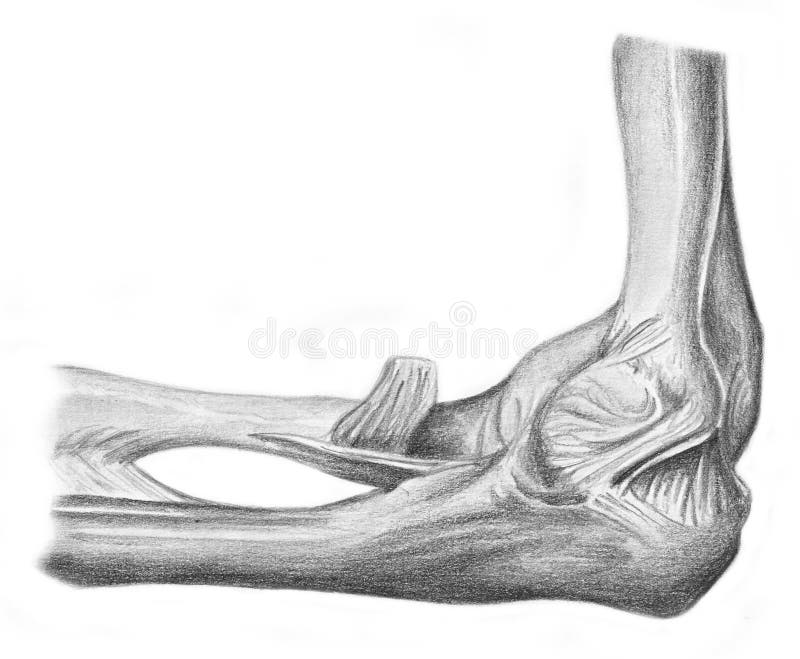
Free with trial A sketch in black and white of the human skeleton. Articulation of the elbow. Limb bones illustrations Articulation of the elbow - skeleton. A sketch in black and white of the human skeleton. Articulation of the elbow
Free with trial Human leg, knee anatomy, abstract grey mesh background. Limb bones vectors Human leg, knee anatomy
Free with trial Human leg knee joint anatomy on a beautiful light blue background. Limb bones vectors Human leg and knee joint. Human leg knee joint anatomy on a beautiful light blue background
Free with trial Knee joint pain abstract blue triangle background. Limb bones vectors Knee joint pain abstract triangle background
Free with trial Knee joint abstract treatment procedure illustration. Doctor with screwdriver and gears in the joint. Limb bones vectors Knee joint abstract treatment
Free with trial Vector illustration of a healthy bones of human leg and a leg with tibial shaft fracture. Twisting, blunt trauma injury. Front view of the foot with knee. For advertising, medical publications. EPS 10. Limb bones vectors Bone fracture_Tibial shaft fracture. Vector illustration of a healthy bones of human leg and a leg with tibial shaft fracture. Twisting, blunt trauma injury. Front view of the foot with knee. For advertising, medical publications. EPS 10
Free with trial Human pelvis hip pain on a dark radial background. Limb bones vectors Human pelvis hip pain
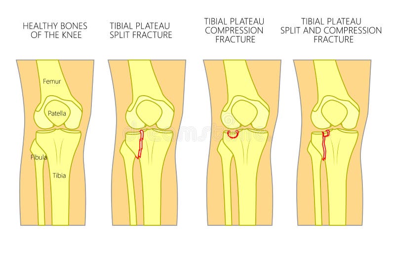
Free with trial Vector illustration of a healthy bones of human knee and a knee with tibial plateau split, compression or depression fractures. Front view of the knee. For advertising, medical publications. EPS 10. Limb bones vectors Bone fracture_Tibial plateau fracture. Vector illustration of a healthy bones of human knee and a knee with tibial plateau split, compression or depression fractures. Front view of the knee. For advertising, medical publications. EPS 10.
Free with trial Vector illustration of a human hand with denominations of palm and wrist bones. Anatomy of dorsal posterior and palmar anterior views of the hand. For advertising or medical publications. EPS 10. Limb bones vectors Anatomy_bones of the human hand. Vector illustration of a human hand with denominations of palm and wrist bones . Anatomy of dorsal posterior and palmar anterior views of the hand. For advertising or medical publications. EPS 10
Free with trial Medical vector illustration of human spine drawing. Limb bones vectors Human Spine 01. Medical vector illustration of human spine drawing
Free with trial Bones of lower limb of human. Anatomy of a human pelvic girdle and legs. Human Skeleton System Lower Limbs Anatomy. Front and back view. For advertising and medical publications. Limb bones vectors Bones of lower limb of human.
Free with trial The major (long) bones of the human leg are the femur (thigh bone), tibia (shin bone), and fibula (the smaller, rear calf bone). The patella (kneecap) is the bone in front of the knee. Most of the leg skeleton has bony prominences and margins that can be palpated, notable exceptions being the hip joint, and the neck and shaft of femur. Many of these anatomical landmarks are used to define the extent of the leg: most notably the anterior superior iliac spine, the greater trochanter, the superior margin of the medial condyle of tibia, and the medial malleolus. Limb bones illustrations Lower Limb Bones Lateral view. The major (long) bones of the human leg are the femur (thigh bone), tibia (shin bone), and fibula (the smaller, rear calf bone). The patella (kneecap) is the bone in front of the knee. Most of the leg skeleton has bony prominences and margins that can be palpated, notable exceptions being the hip joint, and the neck and shaft of femur. Many of these anatomical landmarks are used to define the extent of the leg: most notably the anterior superior iliac spine, the greater trochanter, the superior margin of the medial condyle of tibia, and the medial malleolus.
Free with trial The major bones of the human leg are the femur; thigh bone, tibia; shin bone, and fibula; the smaller, rear calf bone. The patella kneecap is the bone in front of the knee. Most of the leg skeleton has bony prominences and margins that can be palpated, notable exceptions being the hip joint, and the neck and shaft of femur. Many of these anatomical landmarks are used to define the extent of the leg: most notably the anterior superior iliac spine, the greater trochanter, the superior margin of the medial condyle of tibia, and the medial malleolus. Limb bones illustrations Lower Limb Bones Posterior view. The major bones of the human leg are the femur; thigh bone, tibia; shin bone, and fibula; the smaller, rear calf bone. The patella kneecap is the bone in front of the knee. Most of the leg skeleton has bony prominences and margins that can be palpated, notable exceptions being the hip joint, and the neck and shaft of femur. Many of these anatomical landmarks are used to define the extent of the leg: most notably the anterior superior iliac spine, the greater trochanter, the superior margin of the medial condyle of tibia, and the medial malleolus.
Free with trial Bones of the Hand: Carpals, Metacarpals and Phalanges. The Clavicle. The Humerus. The Radius. The Scapula. The Ulna. Limb bones illustrations Bones of the Upper Limb Lateral view. Bones of the Hand: Carpals, Metacarpals and Phalanges. The Clavicle. The Humerus. The Radius. The Scapula. The Ulna.
Free with trial Human upper limb bones infographic diagram name of bones and appendages including clavicle scapula humerus radius ulna carpus metacarpus and phalanges vector for anatomy science education and healthcare. Limb bones vectors Human upper limb bones infographic diagram name of bones
Free with trial Lateral radiograph of human foot or limb. X-ray picture or radiographic image of metatarsus bones and toes, side view. Modern medical radiography. Monochrome vector illustration in flat cartoon style. Limb bones vectors Lateral radiograph of human foot or limb. X-ray picture or radiographic image of metatarsus bones and toes, side view
Free with trial The foot is an anatomical structure found in many vertebrates. It is the terminal portion of a limb which bears weight and allows locomotion. In many animals with feet, the foot is a separate organ at the terminal part of the leg made up of one or more segments or bones, generally including claws or nails. Limb bones illustrations Human Skeleton Foot bones Anatomy. 3d illustration. The foot is an anatomical structure found in many vertebrates. It is the terminal portion of a limb which bears weight and allows locomotion. In many animals with feet, the foot is a separate organ at the terminal part of the leg made up of one or more segments or bones, generally including claws or nails.
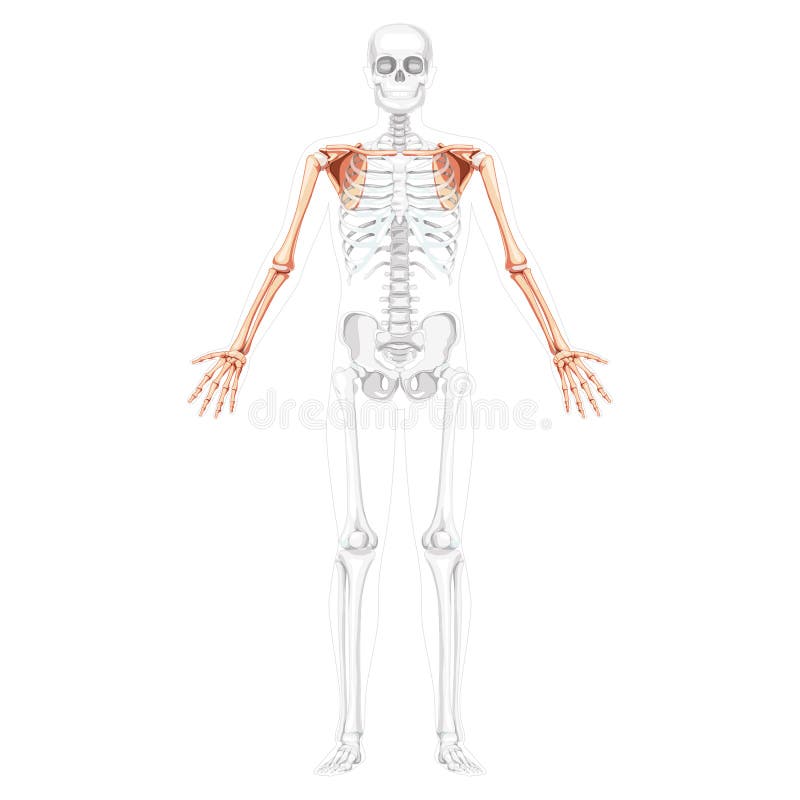
Free with trial Skeleton upper limb Arms with Shoulder girdle Human front view with two arm poses with transparent bones position. Forearms realistic flat Vector illustration of anatomy isolated on white background. Limb bones vectors Skeleton upper limb Arms with Shoulder girdle Human front view with two arm poses with transparent bones position
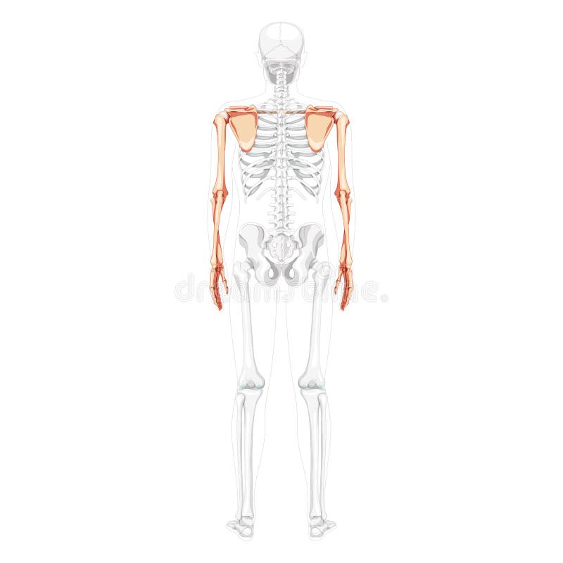
Free with trial Skeleton upper limb Arms with Shoulder girdle Human back view with partly transparent bones position. Hands realistic flat natural color Vector illustration of anatomy isolated on white background. Limb bones vectors Skeleton upper limb Arms with Shoulder girdle Human back view with partly transparent bones position. Hands realistic
Free with trial The bones of the arm and hand have the important jobs of supporting the upper limb and providing attachment points for the muscles that move the upper limb. These bones form joints that provide a wide range of motion and flexibility needed to manipulate objects deftly with the arm and hand. They also provide strength to resist the extreme forces and stresses acting upon the arms and hands during sports, exercise, and heavy labor. Limb bones illustrations Bones of the Arm and Hand
Free with trial 3d rendered medically accurate illustration of the bones of the foot. Limb bones illustrations The bones of the foot