Free with trial A skeletal hand and arm that could be used for medical concepts. Limb bones illustrations Bone Hand And Arm. A skeletal hand and arm that could be used for medical concepts.
Free with trial Shoulder joint anatomy infographic diagram. physiology physiotherapy medical science education. bones muscles ligaments bursa cavity capsule cartilage membrane. human body health vector. Limb bones vectors Shoulder joint anatomy infographic diagram physiology physiotherapy medical science. Shoulder joint anatomy infographic diagram. physiology physiotherapy medical science education. bones muscles ligaments bursa cavity capsule cartilage membrane. human body health vector
Free with trial The sketch in white and black realized to hand. The bones of the hand. Limb bones illustrations Hand, man's anatomy. The sketch in white and black realized to hand. The bones of the hand
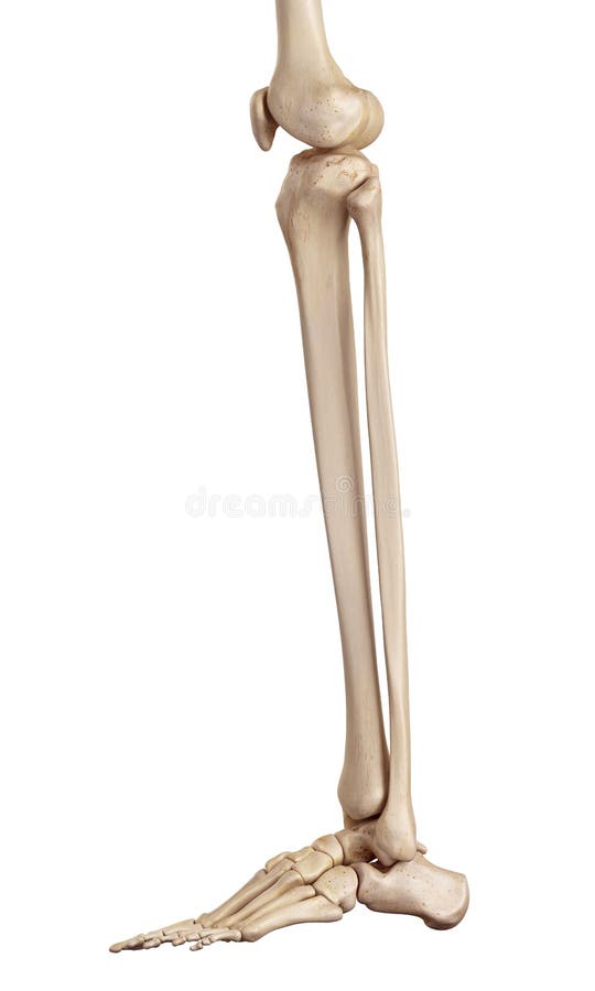
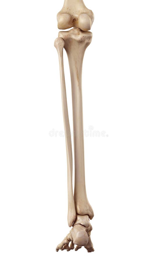
Free with trial Medical accurate illustration of the lower leg bones. Limb bones illustrations The lower leg bones
Free with trial Medical accurate illustration of the lower leg bones. Limb bones illustrations The lower leg bones
Free with trial Medical accurate illustration of the lower leg bones. Limb bones illustrations The lower leg bones
Free with trial Osteoporosis upper limb bones - human body. Limb bones illustrations Osteoporosis - upper limb bones - 3d rendering. Osteoporosis upper limb bones - human body
Free with trial Osteoporosis upper limb bones - human body. Limb bones illustrations Osteoporosis - upper limb bones - 3d rendering. Osteoporosis upper limb bones - human body
Free with trial The human leg, in the general sense, is the entire lower extremity or limb of the human body, including the foot, thigh and even the hip or gluteal region. Limb bones illustrations Lower Limb. The human leg, in the general sense, is the entire lower extremity or limb of the human body, including the foot, thigh and even the hip or gluteal region.
Free with trial The lower limb is subdivided by the hip joint, knee joint, and ankle joint into the regions:buttock (gluteal), thigh, leg (crus), foot. The movements which occur at the hip joint are similar to those that take place at the shoulder joint except their range is more limited. Those at the knee joint occur mainly in one plane. When the knee is bent, it is said to be flexed; when it is straightened out it is extended. There is a small amount of rotation at the joint. The ankle joint is a simpler hinge type joint than the knee. When the foot is bent upwards, it is dorsiflexed or extended; when it is bent downwards, it is plantar flexed or flexed. Through the joints within the foot, the foot can be turned so that the sole of the foot is inwards, inversion or it can be turned so that the sole is turned outwards, eversion. The toes have limited movement when compared to the movements of the fingers, especially the thumb. Limb bones illustrations Right Lower Limb. The lower limb is subdivided by the hip joint, knee joint, and ankle joint into the regions:buttock (gluteal), thigh, leg (crus), foot. The movements which occur at the hip joint are similar to those that take place at the shoulder joint except their range is more limited. Those at the knee joint occur mainly in one plane. When the knee is bent, it is said to be flexed; when it is straightened out it is extended. There is a small amount of rotation at the joint. The ankle joint is a simpler hinge type joint than the knee. When the foot is bent upwards, it is dorsiflexed or extended; when it is bent downwards, it is plantar flexed or flexed. Through the joints within the foot, the foot can be turned so that the sole of the foot is inwards, inversion or it can be turned so that the sole is turned outwards, eversion. The toes have limited movement when compared to the movements of the fingers, especially the thumb.
Free with trial Osteoporosis upper limb bones - human body. Limb bones illustrations Osteoporosis - upper limb bones - 3d rendering. Osteoporosis upper limb bones - human body
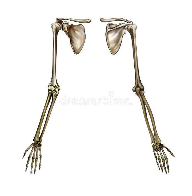
Free with trial Human upper limb anatomy anterior view realistic infographic vector illustration. Limb bones vectors Upper Limb Anatomy
Free with trial Knee seen on x-rays with muscle tendons and bones in evidence. Limb bones illustrations Knee ligaments, tendons, bones, muscles x-ray. Knee seen on x-rays with muscle tendons and bones in evidence
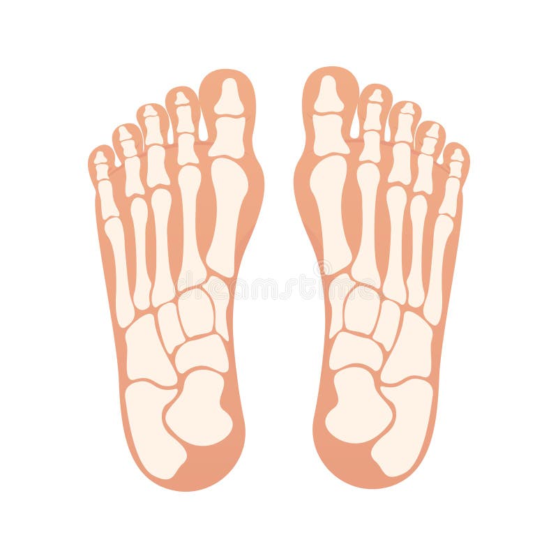
Free with trial The foot plural feet is an anatomical structure found in many vertebrates. It is the terminal portion of a limb which bears weight and allows locomotion. In many animals with feet, the foot is a separate organ at the terminal part of the leg made up of one or more segments or bones, generally including claws or nails. Limb bones illustrations 3d illustration of human body feet bone. The foot plural feet is an anatomical structure found in many vertebrates. It is the terminal portion of a limb which bears weight and allows locomotion. In many animals with feet, the foot is a separate organ at the terminal part of the leg made up of one or more segments or bones, generally including claws or nails.
Free with trial The foot plural feet is an anatomical structure found in many vertebrates. It is the terminal portion of a limb which bears weight and allows locomotion. In many animals with feet, the foot is a separate organ at the terminal part of the leg made up of one or more segments or bones, generally including claws or nails. Limb bones illustrations 3d illustration of skeleton foot bone anatomy. The foot plural feet is an anatomical structure found in many vertebrates. It is the terminal portion of a limb which bears weight and allows locomotion. In many animals with feet, the foot is a separate organ at the terminal part of the leg made up of one or more segments or bones, generally including claws or nails.
Free with trial The foot is an anatomical structure found in many vertebrates. It is the terminal portion of a limb which bears weight and allows locomotion. In many animals with feet, the foot is a separate organ at the terminal part of the leg made up of one or more segments or bones, generally including claws or nails. Limb bones illustrations 3d illustration of skeleton foot bone anatomy. The foot is an anatomical structure found in many vertebrates. It is the terminal portion of a limb which bears weight and allows locomotion. In many animals with feet, the foot is a separate organ at the terminal part of the leg made up of one or more segments or bones, generally including claws or nails.
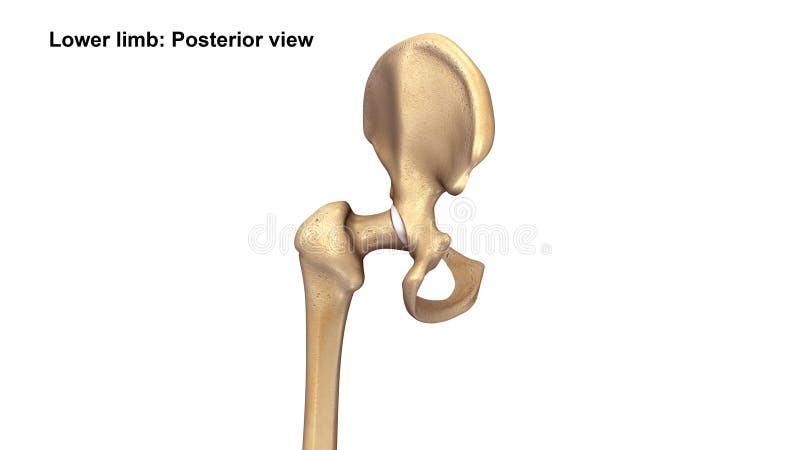
Free with trial The two hip bones join at the pubic symphysis and together with the sacrum and coccyx the pelvic part of the spine comprise the skeletal component of the pelvis – the pelvic girdle which surrounds the pelvic cavity. They are connected to the sacrum, which is part of the axial skeleton, at the sacroiliac joint. Each hip bone is connected to the corresponding femur thigh bone forming the primary connection between the bones of the lower limb and the axial skeleton through the large ball and socket joint of the hip. [2]. Limb bones illustrations 3d illustration of skeleton hip bone anatomy. The two hip bones join at the pubic symphysis and together with the sacrum and coccyx the pelvic part of the spine comprise the skeletal component of the pelvis – the pelvic girdle which surrounds the pelvic cavity. They are connected to the sacrum, which is part of the axial skeleton, at the sacroiliac joint. Each hip bone is connected to the corresponding femur thigh bone forming the primary connection between the bones of the lower limb and the axial skeleton through the large ball and socket joint of the hip.[2]
Free with trial A close up of a leg with bones and muscles in it, AI. Limb bones illustrations A close up of a leg with bones and muscles in it, AI
Free with trial The foot plural feet is an anatomical structure found in many vertebrates. It is the terminal portion of a limb which bears weight and allows locomotion. In many animals with feet, the foot is a separate organ at the terminal part of the leg made up of one or more segments or bones, generally including claws or nails. Limb bones illustrations 3d illustration of skeleton feet bone anatomy. The foot plural feet is an anatomical structure found in many vertebrates. It is the terminal portion of a limb which bears weight and allows locomotion. In many animals with feet, the foot is a separate organ at the terminal part of the leg made up of one or more segments or bones, generally including claws or nails.
Free with trial The foot plural feet is an anatomical structure found in many vertebrates. It is the terminal portion of a limb which bears weight and allows locomotion. In many animals with feet, the foot is a separate organ at the terminal part of the leg made up of one or more segments or bones, generally including claws or nails. Limb bones illustrations 3d illustration of skeleton feet bone anatomy. The foot plural feet is an anatomical structure found in many vertebrates. It is the terminal portion of a limb which bears weight and allows locomotion. In many animals with feet, the foot is a separate organ at the terminal part of the leg made up of one or more segments or bones, generally including claws or nails.
Free with trial Detailed X-ray image of parrot head on black background. Image of bones, head, beak, neck. Concept of medicine, diagnostics, veterinary clinic. Scientific illustration. Limb bones illustrations Detailed X-ray image of parrot head on black background. Image of bones, head, beak, neck. Concept of medicine, diagnostics
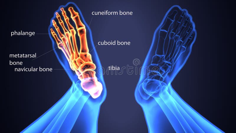
Free with trial X ray human foot. X-ray picture or radiographic monitor image of metatarsus bones and toes. Medical radiology. Vector illustration. Limb bones vectors X ray human foot. X-ray picture or radiographic monitor image of metatarsus bones and toes. Medical radiology. Vector illustration
Free with trial A sketch in black and white of the human skeleton. Bones of the right hip. Limb bones illustrations Right hip - skeleton. A sketch in black and white of the human skeleton. Bones of the right hip.
Free with trial Human hand anatomy, bones of the shoulder girdle, shoulder and forearm, realistic drawing, skeleton structure, front and back view, isolated image on a white background. Limb bones illustrations Human hand anatomy
Free with trial Graphic detailed black and white colorful human skeleton spine bones. Isolated on white background. Vector icon set. Limb bones vectors Graphic human skeleton spine bones vector. Graphic detailed black and white colorful human skeleton spine bones. Isolated on white background. Vector icon set
Free with trial Knee anatomy. Structure of knee joint. Major parts of human leg. Bones, muscles, cartilage, tendon and Ligaments of knee. Side view. Vector poster. Limb bones vectors Knee anatomy. Structure of knee joint
Free with trial Orthopedics Healthcare Concept. Doctor Orthopedist Character Looking on Huge Leg Bones, Knee Joint through Magnifying Glass. Specialist Medical Check Up in Hospital. Linear Vector Illustration. Limb bones vectors Orthopedics Healthcare Concept. Doctor Orthopedist Character Looking on Huge Leg Bones
Free with trial Collection of fractured bones and limbs fixed with metal implantable devices drawn with contour lines on white background. Bundle of osteosynthesis constructions. Monochrome vector illustration. Limb bones vectors Collection of fractured bones and limbs fixed with metal implantable devices drawn with contour lines on white
Free with trial Detailed anatomical illustration of a human hand skeleton, perfect for medical, artistic, or Halloween design projects. The image showcases the intricate structure of bones in the hand and wrist. Limb bones vectors Detailed Hand Skeleton Anatomy Drawing, Graphic Design Element. Detailed anatomical illustration of a human hand skeleton, perfect for medical, artistic, or Halloween design projects. The image showcases the intricate structure of bones in the hand and wrist.
Free with trial Detailed 3D illustration of human knee joint anatomy with labels. Educational medical diagram showing ligaments and bones on white background. Useful for healthcare presentations. Limb bones illustrations Detailed 3D illustration of human knee joint anatomy with labels. Educational medical diagram showing ligaments and bones on white. Background. Useful for. Detailed 3D illustration of human knee joint anatomy with labels. Educational medical diagram showing ligaments and bones on white background. Useful for healthcare presentations.
Free with trial Bones Fracture, Joints Trauma or Injury Treatment Flat Vector Banner or Poster with Broken Human Leg, Hand and Neck in Orthopedic Cast, Gypsum Bandage, Male Doctor Consulting Patient Illustration. Limb bones vectors Broken Limb Bone Treatment Flat Vector Concept. Bones Fracture, Joints Trauma or Injury Treatment Flat Vector Banner or Poster with Broken Human Leg, Hand and Neck in Orthopedic Cast, Gypsum Bandage, Male Doctor Consulting Patient Illustration
Free with trial Medical Health Care Concept. Tiny Orthopedist Doctor Character Install Bandage Brace at Huge Leg with Bones Fracture. Patient Treatment in Orthopedics Hospital or Clinic. Cartoon Vector Illustration. Limb bones vectors Medical Health Care Concept. Tiny Orthopedist Doctor Character Install Bandage Brace at Huge Leg with Bones Fracture
Free with trial This captivating macro photograph offers a detailed side view of a pigeon skeleton, meticulously showcasing the intricate avian anatomy. The image's close-up perspective highlights the delicate structure of the bones, revealing the unique adaptations that allow birds to fly. From the slender ribs to the complex arrangement of the vertebrae, the image provides a fascinating glimpse into the. Limb bones illustrations Unveiling the Avian Architecture A Detailed Macro Study of a Pigeon Skeletons Side View Perfect for Anatomy Research. This captivating macro photograph offers a detailed side view of a pigeon skeleton, meticulously showcasing the intricate avian anatomy. The image's close-up perspective highlights the delicate structure of the bones, revealing the unique adaptations that allow birds to fly. From the slender ribs to the complex arrangement of the vertebrae, the image provides a fascinating glimpse into the
Free with trial The Ilizarov apparatus set. External ring fixation technique in orthopedic surgery to lengthen or reshape limb bones. Flat vector illustration. Limb bones vectors The Ilizarov apparatus set. External ring fixation technique in orthopedic
Free with trial Valgus and varus leg deformities. Diagram showing the deformed bones of the lower extremities. Cosmetic pathology. Vector illustration. Limb bones vectors Valgus and varus leg deformities.
Free with trial The human ankle joint is a complex joint that connects the lower leg bones (tibia and fibula) to the foot. It plays a crucial role in weight-bearing, locomotion, and maintaining balance. The ankle joint allows for up-and-down movements of the foot and also permits limited side-to-side movements. Limb bones illustrations Human ankle joint medical background. The human ankle joint is a complex joint that connects the lower leg bones (tibia and fibula) to the foot. It plays a crucial role in weight-bearing, locomotion, and maintaining balance. The ankle joint allows for up-and-down movements of the foot and also permits limited side-to-side movements.
Free with trial The foot is an anatomical structure found in many vertebrates. It is the terminal portion of a limb which bears weight and allows locomotion. Limb bones vectors Bare Feet with bones isolated vector illustration cartoon graphic. The foot is an anatomical structure found in many vertebrates. It is the terminal portion of a limb which bears weight and allows locomotion
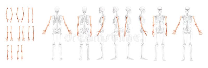
Free with trial Set of Skeleton Arms Human front back side view with partly transparent bones position. Hands, forearms realistic flat natural color concept Vector illustration of anatomy isolated on white background. Limb bones vectors Set of Skeleton Arms Human front back side view with partly transparent bones position. Hands, forearms realistic flat
Free with trial A sketch in black and white of the human skeleton. Left clavicle. Limb bones illustrations Left clavicle - skeleton. A sketch in black and white of the human skeleton. Left clavicle
Free with trial An image of an x-rayed hand that is pushing a word. Limb bones illustrations Bone Hand Pushing Button 22. An image of an x-rayed hand that is pushing a word.
Free with trial Osteoarthritis and healthy knee joint. Healthy part of the joint on a blue background and unhealthy on a red. Limb bones vectors Osteoarthritis
Free with trial A sketch in black and white of the human skeleton. bone of the leg, right fibula. Limb bones illustrations Right fibula - skeleton. A sketch in black and white of the human skeleton. bone of the leg, right fibula
Free with trial 3d rendered medically accurate illustration of the equine muscle anatomy - Common Digital Extensor. Limb bones illustrations Common Digital Extensor
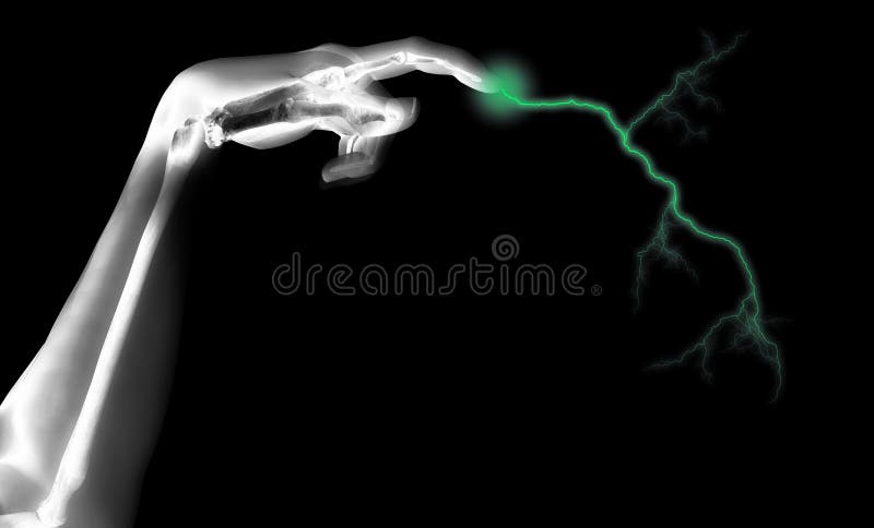
Free with trial A skeleton hand that is shooting out electricity. Limb bones illustrations Electric Skeleton Hand. A skeleton hand that is shooting out electricity.
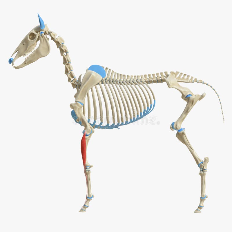
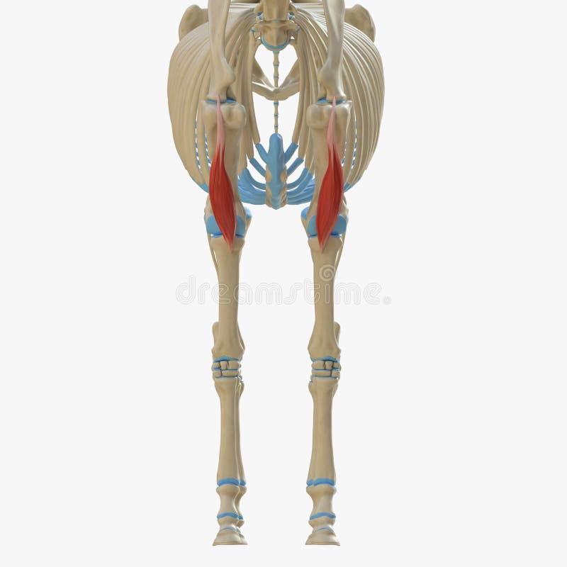
Free with trial 3d rendered medically accurate illustration of the equine muscle anatomy - Gracilis. Limb bones illustrations Gracilis
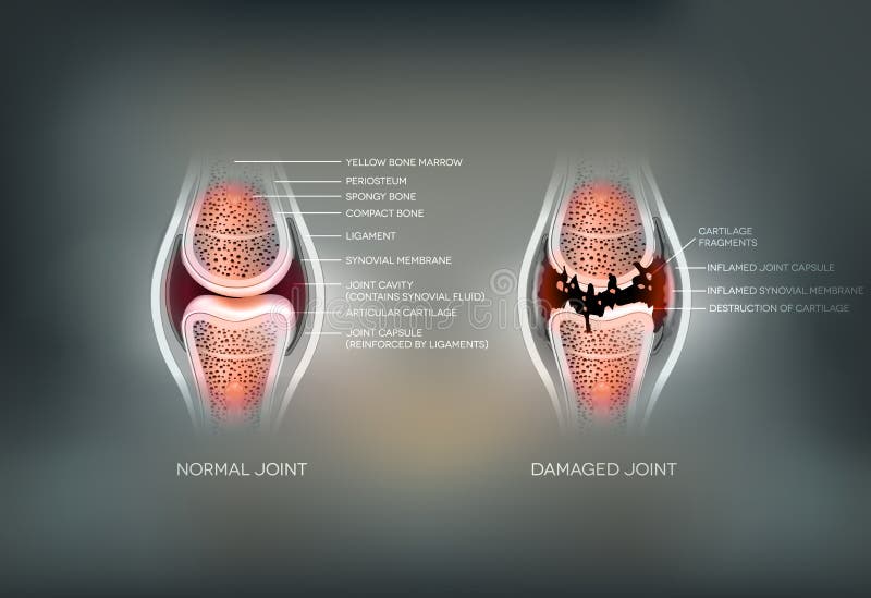
Free with trial Damaged Synovial joint and normal joint colorful design on an abstract grey background. Limb bones vectors Joint
Free with trial 3d rendered medically accurate illustration of the equine muscle anatomy - Adductor. Limb bones illustrations Adductor
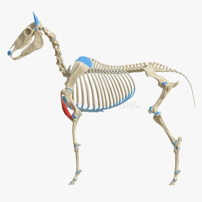
Free with trial 3d rendered medically accurate illustration of the equine muscle anatomy - Popliteus. Limb bones illustrations Popliteus
Free with trial Human legs and knee joint detailed anatomy, painful joint and healthy joint. Beautiful abstract technology background. Limb bones vectors Human legs and knee joint
Free with trial 3d rendered medically accurate illustration of the equine muscle anatomy - Brachialis. Limb bones illustrations Brachialis
Free with trial 3d rendered medically accurate illustration of the equine muscle anatomy - Flexor Digitorum Profundus. Limb bones illustrations Flexor Digitorum Profundus
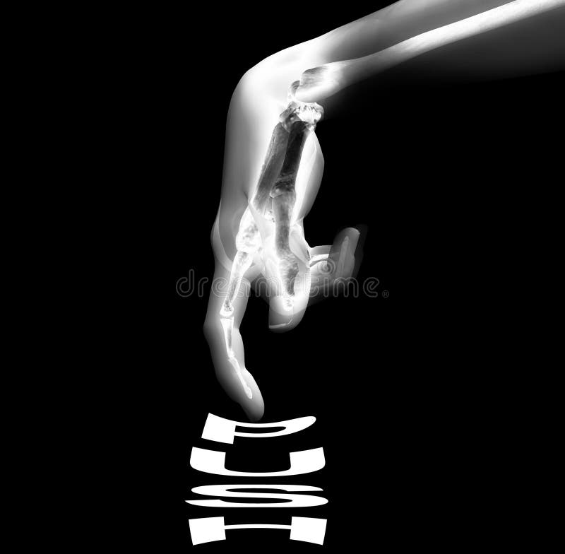
Free with trial An image of an x-rayed hand that is pushing a word. Limb bones illustrations Bone Hand Pushing Button 20. An image of an x-rayed hand that is pushing a word.
Free with trial 3d rendered medically accurate illustration of the equine muscle anatomy - Biceps Brachii. Limb bones illustrations Biceps Brachii
Free with trial 3d rendered medically accurate illustration of the equine muscle anatomy - Policis Longus. Limb bones illustrations Policis Longus
Free with trial 3d rendered medically accurate illustration of the equine muscle anatomy - Biceps Brachii. Limb bones illustrations Biceps Brachii
Free with trial 3d rendered medically accurate illustration of the equine muscle anatomy - Flexor Digitorum Superficialis. Limb bones illustrations Flexor Digitorum Superficialis
Free with trial 3d rendered medically accurate illustration of the equine muscle anatomy - Tibialis Cranialis. Limb bones illustrations Tibialis Cranialis
Free with trial 3d rendered medically accurate illustration of the equine muscle anatomy - Tibialis Cranialis. Limb bones illustrations Tibialis Cranialis
Free with trial 3d rendered medically accurate illustration of the equine muscle anatomy - Digitorum Profundus. Limb bones illustrations Digitorum Profundus
Free with trial 3d rendered medically accurate illustration of the equine muscle anatomy - Deep Digital Flexor. Limb bones illustrations Deep Digital Flexor
Free with trial 3d skeletal arm, over white. Limb bones illustrations Skeleton Arm, Grey Scroll Sign. 3d skeletal arm, over white
Free with trial 3d rendered medically accurate illustration of the equine muscle anatomy - Lateral Ulnar Muscle. Limb bones illustrations Lateral Ulnar Muscle
Free with trial 3d rendered medically accurate illustration of the equine muscle anatomy - Flexor Carpi Radialis. Limb bones illustrations Flexor Carpi Radialis
Free with trial 3d rendered medically accurate illustration of the equine muscle anatomy - Extensor Carpi Radialis. Limb bones illustrations Extensor Carpi Radialis
Free with trial 3d rendered medically accurate illustration of the equine muscle anatomy - Extensor Digitorum Longus. Limb bones illustrations Extensor Digitorum Longus
Free with trial Knee joint blue background, joint treatment technology. Limb bones vectors Knee joint blue background
Free with trial 3d rendered medically accurate illustration of the equine muscle anatomy - Superficial Digital Flexor. Limb bones illustrations Superficial Digital Flexor
Free with trial An computer created image of an x-rayed hand. Limb bones illustrations Bone Hand 12. An computer created image of an x-rayed hand.
Free with trial 3d rendered medically accurate illustration of the equine muscle anatomy - Flexor Digitorum Superficialis. Limb bones illustrations Flexor Digitorum Superficialis
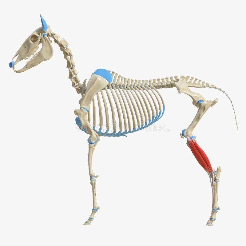
Free with trial 3d rendered medically accurate illustration of the equine muscle anatomy - Semimembranosus. Limb bones illustrations Semimembranosus
Free with trial Osteoarthritis and normal knee joint anatomy on white. Limb bones vectors Osteoarthritis and normal joint. Osteoarthritis and normal knee joint anatomy on white
Free with trial 3d rendered medically accurate illustration of the equine muscle anatomy - Common Digital Extensor. Limb bones illustrations Common Digital Extensor
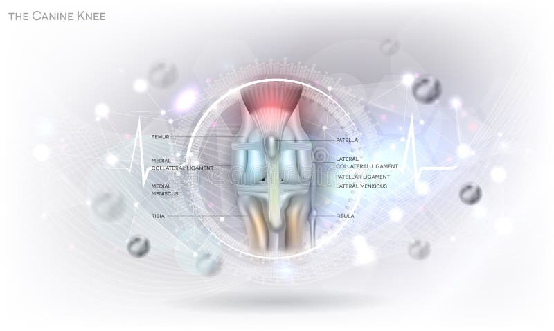
Free with trial Anatomy of the canine dog`s knee joint colorful design on an abstract glowing light grey scientific background. Limb bones vectors Dog knee. Anatomy of the canine dog`s knee joint colorful design on an abstract glowing light grey scientific background
Free with trial The knee joint is one of the strongest and most important joints in the human body. It allows the lower leg to move relative to the thigh while supporting the body’s weight. Movements at the knee joint are essential to many everyday activities, including walking, running, sitting and standing. The knee, also known as the tibiofemoral joint, is a synovial hinge joint formed between three bones: the femur, tibia, and patella. Two rounded, convex processes (known as condyles) on the distal end of the femur meet two rounded, concave condyles at the proximal end of the tibia. The bones of the ankle and foot form the most distal region of the lower limb in the appendicular skeleton. These bones are responsible for the propulsion, balance, and support of the body’s weight through many diverse activities, such as standing, walking, running, and jumping. Limb bones illustrations Knee joint and Ankle& Foot Joint. The knee joint is one of the strongest and most important joints in the human body. It allows the lower leg to move relative to the thigh while supporting the body’s weight. Movements at the knee joint are essential to many everyday activities, including walking, running, sitting and standing. The knee, also known as the tibiofemoral joint, is a synovial hinge joint formed between three bones: the femur, tibia, and patella. Two rounded, convex processes (known as condyles) on the distal end of the femur meet two rounded, concave condyles at the proximal end of the tibia. The bones of the ankle and foot form the most distal region of the lower limb in the appendicular skeleton. These bones are responsible for the propulsion, balance, and support of the body’s weight through many diverse activities, such as standing, walking, running, and jumping.
Free with trial 3d skeletal arm, isolated, dark background. Limb bones illustrations Skeletal Arms. 3d skeletal arm, isolated, dark background


![The two hip bones join at the pubic symphysis and together with the sacrum and coccyx the pelvic part of the spine comprise the skeletal component of the pelvis – the pelvic girdle which surrounds the pelvic cavity. They are connected to the sacrum, which is part of the axial skeleton, at the sacroiliac joint. Each hip bone is connected to the corresponding femur thigh bone forming the primary connection between the bones of the lower limb and the axial skeleton through the large ball and socket joint of the hip. [2]. Limb bones illustrations](https://thumbs.dreamstime.com/b/two-hip-bones-join-pubic-symphysis-together-sacrum-coccyx-pelvic-part-spine-comprise-skeletal-component-pelvis-116766762.jpg)