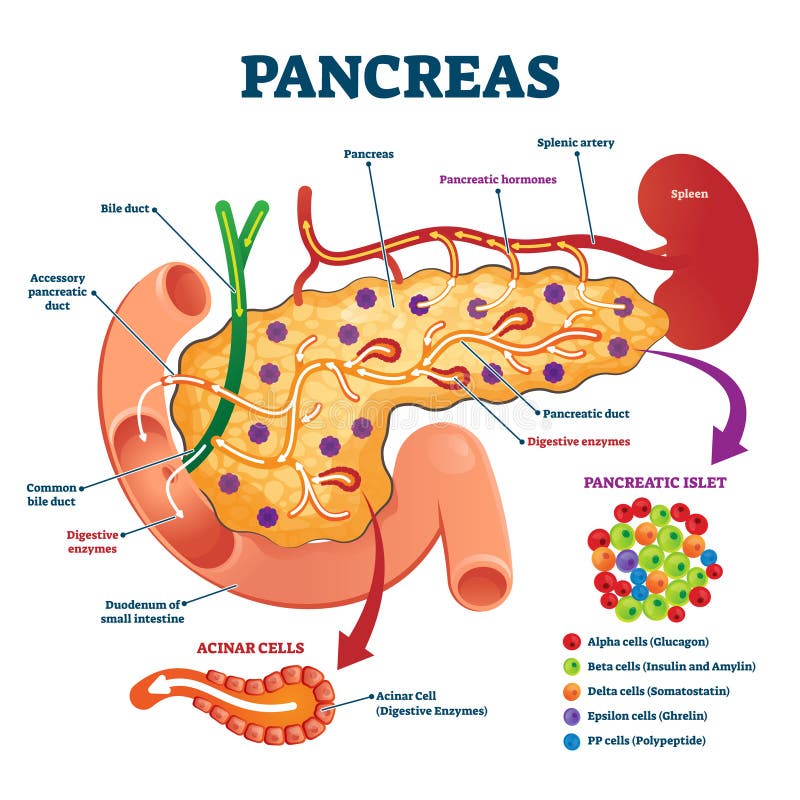
Free with trial Pancreas anatomical cross section model, vector illustration medical example. Blood flow process, cell structure and hormone functions. Digestive enzymes, pancreatic islet and other internal elements. Pancreatic acinar cell vectors Pancreas anatomical cross section model, vector illustration medical example
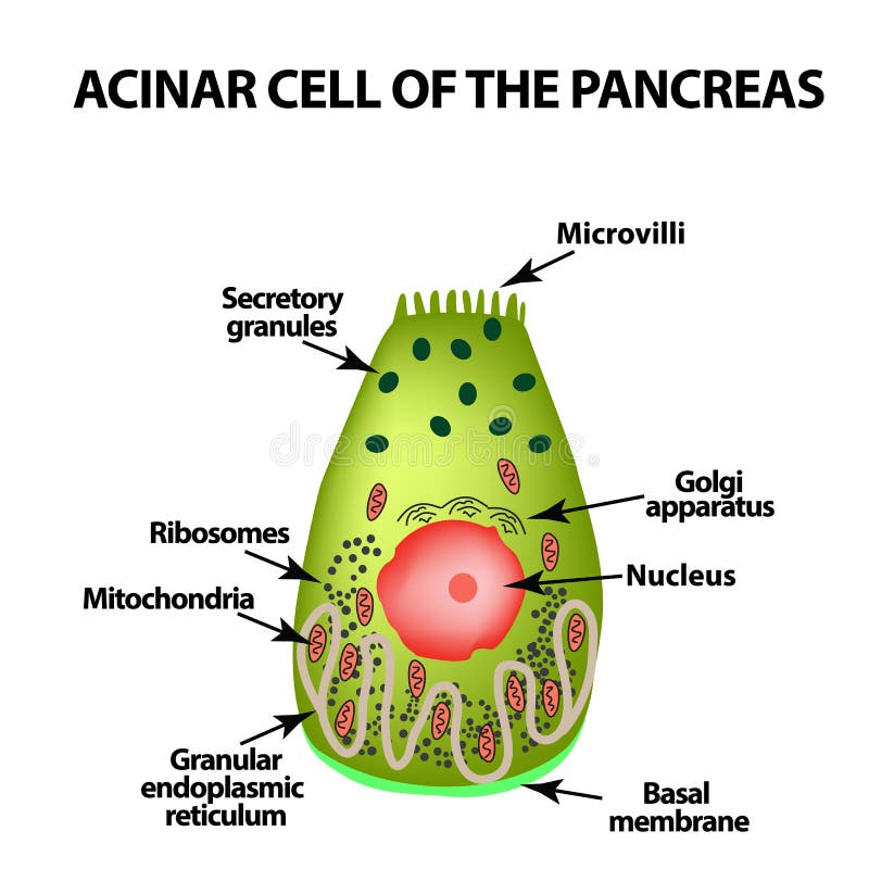
Free with trial Acinar cell of the pancreas. Acinus. Infographics. Vector illustration on isolated background. Pancreatic acinar cell vectors Acinar cell of the pancreas. Acinus. Infographics. Vector illustration on isolated background
Free with trial Pancreatic endocrine system anatomy, alpha, beta and delta cells secreting glucagon, insulin, and somatostatin illustration. Pancreatic acinar cell illustrations Pancreatic endocrine cells anatomy showing endocrine cells involved in secretion of hormones. Pancreatic endocrine system anatomy, alpha, beta and delta cells secreting glucagon, insulin, and somatostatin illustration
Free with trial Acinar cell of the pancreas. Acinus. Infographics. Vector illustration on isolated background. Pancreatic acinar cell vectors Acinar cell of the pancreas. Acinus. Infographics. Vector illustration on isolated background
Free with trial This microscopic image reveals the intricate cellular damage characteristic of pancreatitis. Inflammation is clearly visible within the exocrine pancreas, specifically affecting the acinar cells responsible for enzyme production. The islets of Langerhans, crucial for hormone regulation, are also impacted, highlighting the broad systemic effects of this condition. The image showcases the complex. Pancreatic acinar cell illustrations Microscopic Examination of Inflamed Pancreas Tissue A Deep Dive into Pancreatitis Pathology. This microscopic image reveals the intricate cellular damage characteristic of pancreatitis. Inflammation is clearly visible within the exocrine pancreas, specifically affecting the acinar cells responsible for enzyme production. The islets of Langerhans, crucial for hormone regulation, are also impacted, highlighting the broad systemic effects of this condition. The image showcases the complex
Free with trial Vector illustration graphic diagram presentation slide of the pancreatic islet cells within the Islet of Langerhans inside the pancreas. Pancreatic acinar cell vectors Vector illustration slide of the pancreatic islet cells. vector illustration graphic diagram presentation slide of the pancreatic islet cells within the Islet of Langerhans inside the pancreas
Free with trial Transmission electron microscopy of exocrine pancreatic acinar cell showing zymogen granules at apical pole during regulated secretion. Pancreatic acinar cell illustrations Pancreas Acinar Cell Zymogen Granule Release TEM. Transmission electron microscopy of exocrine pancreatic acinar cell showing zymogen granules at apical pole. Transmission electron microscopy of exocrine pancreatic acinar cell showing zymogen granules at apical pole during regulated secretion
Free with trial This breathtaking microscopic image showcases the intricate cellular architecture of pancreatic islets of Langerhans. Pancreatic islet cells, including beta cells, alpha cells, delta cells, and gamma cells, are vividly depicted, highlighting their critical roles in hormone production. The panoramic view reveals the complex interplay of these endocrine cells within the pancreatic tissue, offering. Pancreatic acinar cell illustrations Pancreatic Islet of Langerhans Cellular Structures Revealed in Stunning Detail via Advanced Microscopy. This breathtaking microscopic image showcases the intricate cellular architecture of pancreatic islets of Langerhans. Pancreatic islet cells, including beta cells, alpha cells, delta cells, and gamma cells, are vividly depicted, highlighting their critical roles in hormone production. The panoramic view reveals the complex interplay of these endocrine cells within the pancreatic tissue, offering
Free with trial This stunning microscopic image reveals the intricate cellular architecture of the pancreas, a vital organ in both the endocrine and digestive systems. The image showcases the pancreas's dual function, highlighting the specific islet cells responsible for insulin production and the acinar cells that synthesize digestive enzymes. Notice the rich detail of the beta cells, which are key in. Pancreatic acinar cell illustrations Detailed Microscopic View of the Pancreas Insulin Production Digestive Enzyme Synthesis and Cellular Processes. This stunning microscopic image reveals the intricate cellular architecture of the pancreas, a vital organ in both the endocrine and digestive systems. The image showcases the pancreas's dual function, highlighting the specific islet cells responsible for insulin production and the acinar cells that synthesize digestive enzymes. Notice the rich detail of the beta cells, which are key in
Free with trial Red and yellow human pancreas and duodenum. Healthcare medical and anatomical concept. Futuristic low poly style. Geometric background. Wireframe light connection structure. Modern 3d graphic. Vector. Pancreatic acinar cell vectors Red and yellow human pancreas and duodenum. Healthcare medical and anatomical concept. Futuristic low poly style.
Free with trial Pancreatic duct concept. Medical vector illustration of the gland in cross section. Pancreatic acinar cell vectors Functions of pancreas concept. Vector illustration. Pancreatic duct concept. Medical vector illustration of the gland in cross section