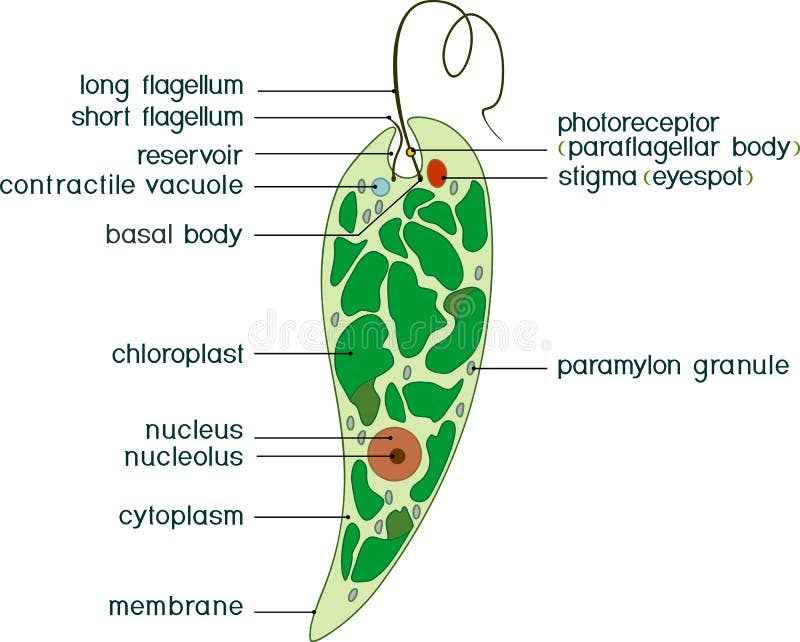
Free with trial Structure of a euglena. Anatomy of a euglena. Euglena freshwater protozoan. It is composed of chlorophyll and has a rudimentary eye. Vector diagram. Flagellate vectors Structure of a euglena. Anatomy of a euglena. Euglena freshwater protozoan. It is composed of chlorophyll and has a rudimentary eye. Vector diagram
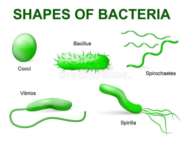
Free with trial Common bacteria infecting human. vector illustration. Bacteria are classified into 5 groups according to their basic shapes: spherical (cocci), rod (bacilli), spiral (spirilla), comma (vibrios) or corkscrew (spirochaetes). Flagellate vectors Types of bacteria. Basic morphological. Common bacteria infecting human. vector illustration. Bacteria are classified into 5 groups according to their basic. Common bacteria infecting human. vector illustration. Bacteria are classified into 5 groups according to their basic shapes: spherical (cocci), rod (bacilli), spiral (spirilla), comma (vibrios) or corkscrew (spirochaetes).
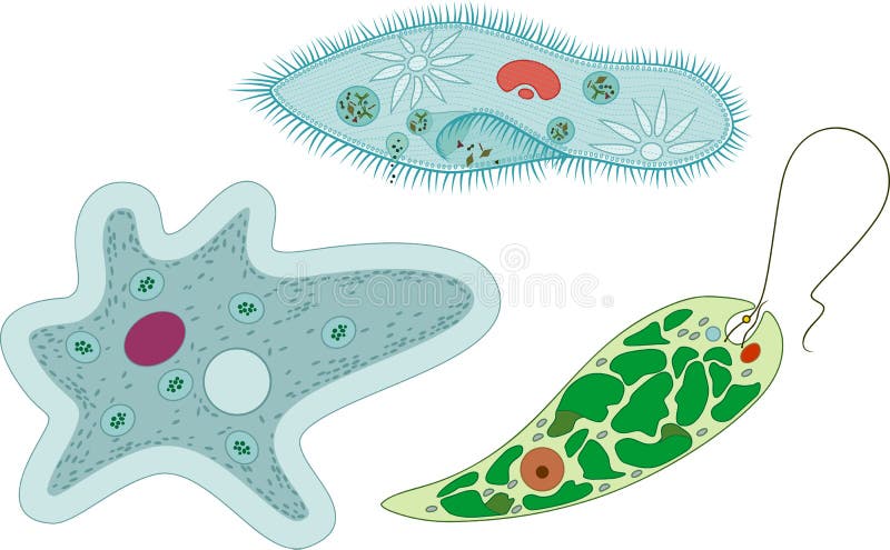
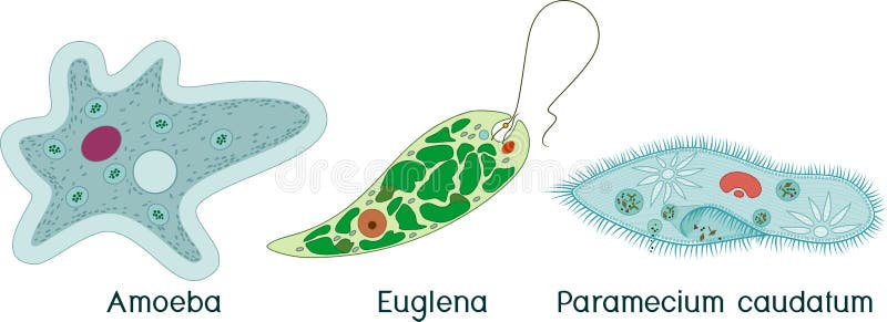
Free with trial Set of unicellular organisms protozoa: Paramecium caudatum, Amoeba proteus and Euglena viridis. Isolated on white background. Flagellate vectors Set of unicellular organisms protozoa: Paramecium caudatum, Amoeba proteus and Euglena viridis. Isolated on white background
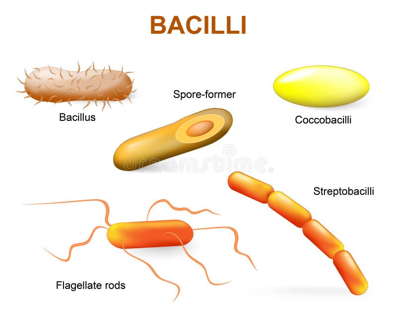
Free with trial Bacillii. Common bacteria infecting human. Flagellate vectors Types of bacteria. bacilli. Bacillii. Common bacteria infecting human. Bacillii. Common bacteria infecting human.
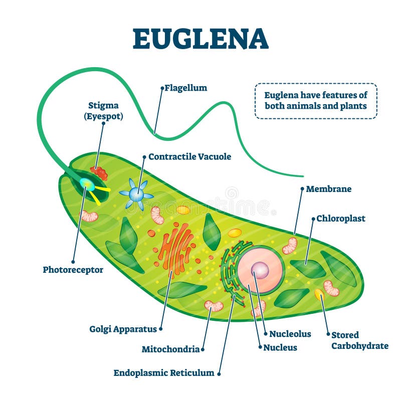
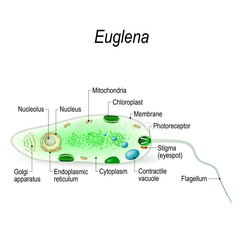
Free with trial Euglena vector illustration. Labeled microorganism structure and description. Biological inner parts scheme for genus of single cell flagellate eukaryotes. Diagram with flagellum and stigma location. Flagellate vectors Euglena vector illustration. Labeled microorganism structure or description. Euglena vector illustration. Labeled microorganism structure and description. Euglena vector illustration. Labeled microorganism structure and description. Biological inner parts scheme for genus of single cell flagellate eukaryotes. Diagram with flagellum and stigma location.
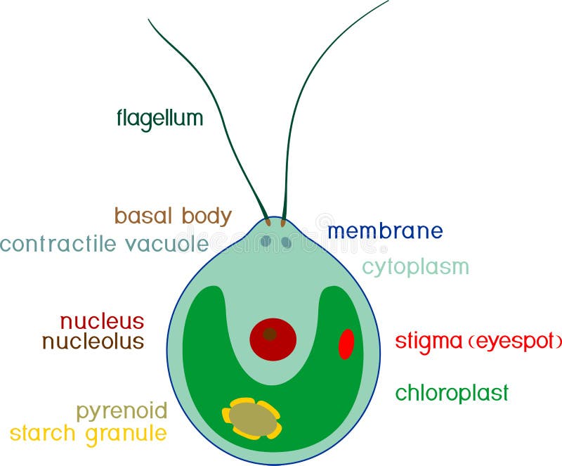
Free with trial Cross section of a Chlamydomonas. Structure of the algae cell. Vector diagram for educational, biological, and science use. Flagellate vectors Cross section of a Chlamydomonas. Structure of the algae cell. Vector diagram for educational, biological, and science use
Free with trial Set of unicellular organisms protozoa: Paramecium caudatum, Amoeba proteus and Euglena viridis. Isolated on white background. Flagellate vectors Set of unicellular organisms protozoa: Paramecium caudatum, Amoeba proteus and Euglena viridis. Isolated on white background
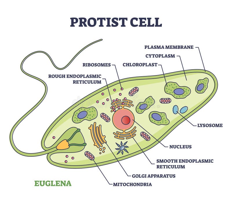
Free with trial Protist cell anatomy with euglena microorganism structure outline diagram. Labeled educational scheme with green organism parts description vector illustration. Eukaryotic biological inner structure. Flagellate vectors Protist cell anatomy with euglena microorganism structure outline diagram. Labeled educational scheme with green organism parts description vector illustration
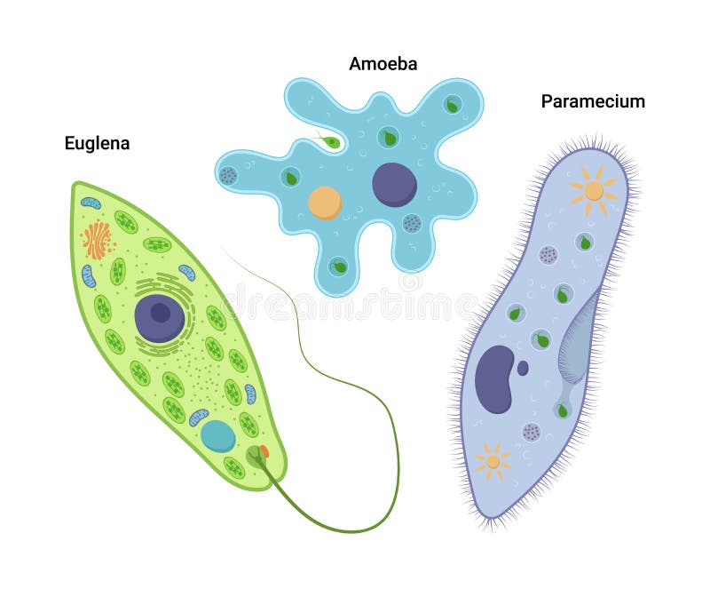
Free with trial Vector illustrationof unicellular organisms. Amoeba proteus Paramecium caudatum and Euglena viridis. Protozoa. Flagellate vectors Vector illustrationof unicellular organisms. Amoeba proteus Paramecium caudatum and Euglena viridis. Protozoa.
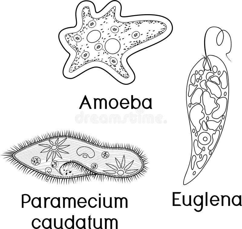
Free with trial Coloring page. Set of unicellular organisms protozoa: Paramecium caudatum, Amoeba proteus and Euglena viridis. Isolated on white background. Flagellate vectors Coloring page. Set of unicellular organisms protozoa: Paramecium caudatum, Amoeba proteus and Euglena viridis. Isolated on white background
Free with trial Diagram of Euglena. Structure of Euglena viridis with different organelles. Structure of Euglena viridis with titles. Flagellate vectors Diagram of Euglena. Structure of Euglena viridis with different organelles. Diagram of Euglena. Structure of Euglena viridis with titles
Free with trial Structure of Chlamydomonas cell. Isolated on white background. Flagellate vectors Structure of Chlamydomonas cell. Isolated on white background
Free with trial Coloring page with structure of Euglena viridis with titles. Isolated on white background. Flagellate vectors Coloring page with structure of Euglena viridis with titles. Isolated on white background
Free with trial Diagram of Euglena. Structure of Euglena viridis with titles. Isolated on white background. Flagellate vectors Diagram of Euglena. Structure of Euglena viridis with titles. Isolated on white background
Free with trial Set of unicellular organisms protozoa: Paramecium caudatum, Amoeba proteus, Chlamydomonas and Euglena viridis. On white background. Flagellate vectors Set of unicellular organisms protozoa: Paramecium caudatum, Amoeba proteus, Chlamydomonas and Euglena viridis. On white background
Free with trial Set of microscopic unicellular organisms: protozoa Paramecium caudatum, Amoeba proteus, Chlamydomonas, Euglena viridis and green algae Chlorella, Spirogyra isolated on white background. Flagellate vectors Set of microscopic unicellular organisms: protozoa Paramecium caudatum, Amoeba proteus, Chlamydomonas,. Euglena viridis and green algae Chlorella, Spirogyra. Set of microscopic unicellular organisms: protozoa Paramecium caudatum, Amoeba proteus, Chlamydomonas, Euglena viridis and green algae Chlorella, Spirogyra isolated on white background
Free with trial Chlamydomonas is a genus of green algae belonging to the division Chlorophyta. These single-celled organisms are known for their flagellated cells and their ability to perform photosynthesis. Chlamydomonas is commonly found in freshwater environments such as ponds, lakes, and rivers, although some species can also thrive in soil or marine habitats. Flagellate vectors Chlamydomonas. Is a genus of green algae belonging to the division Chlorophyta. These single-celled organisms are known for their flagellated cells and their. Chlamydomonas is a genus of green algae belonging to the division Chlorophyta. These single-celled organisms are known for their flagellated cells and their ability to perform photosynthesis. Chlamydomonas is commonly found in freshwater environments such as ponds, lakes, and rivers, although some species can also thrive in soil or marine habitats.
Free with trial Coloring page. Set of unicellular organisms protozoa: Paramecium caudatum, Amoeba proteus, Chlamydomonas and Euglena viridis. On white background. Flagellate vectors Coloring page. Set of unicellular organisms protozoa: Paramecium caudatum, Amoeba proteus, Chlamydomonas and Euglena viridis. On white background
Free with trial Set of microscopic unicellular organisms: protozoa Paramecium caudatum, Amoeba proteus, Chlamydomonas, Euglena viridis, green algae Chlorella, Spirogyra and bacteria isolated on white background. Flagellate vectors Set of microscopic unicellular organisms: protozoa Paramecium caudatum, Amoeba proteus, Chlamydomonas,. Euglena viridis, green algae Chlorella, Spirogyra and. Set of microscopic unicellular organisms: protozoa Paramecium caudatum, Amoeba proteus, Chlamydomonas, Euglena viridis, green algae Chlorella, Spirogyra and bacteria isolated on white background
Free with trial Protozoa Paramecium caudatum, Amoeba proteus, Chlamydomonas, Euglena viridis, green algae Chlorella, Spirogyra under magnifying glass isolated on white background. Flagellate vectors Protozoa Paramecium caudatum, Amoeba proteus, Chlamydomonas, Euglena viridis, green algae Chlorella, Spirogyra. Under magnifying glass isolated on white
Free with trial Parasitic human flagellates protozoa: Trichomonas; Trypanosoma; Leishmania. Black and white hand drawing image. Flagellate vectors Human parasites. Black and white hand drawn illustration. Parasitic human flagellates protozoa: Trichomonas; Trypanosoma; Leishmania. Black and white hand. Parasitic human flagellates protozoa: Trichomonas; Trypanosoma; Leishmania. Black and white hand drawing image
Free with trial Protozoa, protista and amoeba microorganism cells, vector micro organism. Ameba and protist unicellular cells in lab microscope, protozoan eukaryotic organism types. Flagellate vectors Protozoa, protista and amoeba microorganism cells. Vector micro organism. Ameba and protist unicellular cells in lab microscope, protozoan eukaryotic organism
Free with trial Paramecium Caudatum proteus science icon with nucleus, vacuole, contractile. Biology education laboratory cartoon protozoa organism. Bold bright unicellular microorganism. Vector illustration isolated on white. Flagellate vectors Paramecium Caudatum proteus science icon with nucleus, vacuole, contractile. Biology education laboratory cartoon. Protozoa organism. Bold bright unicellular. Paramecium Caudatum proteus science icon with nucleus, vacuole, contractile. Biology education laboratory cartoon protozoa organism. Bold bright unicellular microorganism. Vector illustration isolated on white
Free with trial Green Algae Chlorella proteus science icon with nucleus, vacuole, contractile. Biology education laboratory cartoon protozoa organism. Bold bright unicellular microorganism. Vector illustration isolated on white. Flagellate vectors Green Algae Chlorella proteus science icon with nucleus, vacuole, contractile. Biology education laboratory cartoon. Protozoa organism. Bold bright unicellular. Green Algae Chlorella proteus science icon with nucleus, vacuole, contractile. Biology education laboratory cartoon protozoa organism. Bold bright unicellular microorganism. Vector illustration isolated on white
Free with trial Euglena structure anatomy diagram poster chart, medical illustration vector. Science biology study. Single cell flagellate eukaryotes, chloroplast, nucleolus, mitochondrion, flagellum, photoreceptor. Flagellate vectors Euglena structure anatomy diagram poster chart, medical illustration vector. Science biology study. Single cell flagellate. Eukaryotes, chloroplast, nucleolus. Euglena structure anatomy diagram poster chart, medical illustration vector. Science biology study. Single cell flagellate eukaryotes, chloroplast, nucleolus, mitochondrion, flagellum, photoreceptor
Free with trial Euglena structure anatomy diagram poster chart, medical illustration vector. Science biology study. Single cell flagellate eukaryotes, chloroplast, nucleolus, mitochondrion, flagellum, photoreceptor. Flagellate vectors Euglena structure anatomy diagram poster chart, medical illustration vector. Science biology study. Single cell flagellate. Eukaryotes, chloroplast, nucleolus. Euglena structure anatomy diagram poster chart, medical illustration vector. Science biology study. Single cell flagellate eukaryotes, chloroplast, nucleolus, mitochondrion, flagellum, photoreceptor
Free with trial Set of twelve colorful flat line icons depicting various microorganisms, cells, bacteria, algae, pollen, and botanical elements on white background. Includes stylized virus, bacillus, diatom, water droplets, plant cell, flower, flagellate and ciliate organisms. Bright outline design ideal for educational, scientific, medical, biological concepts, textbooks, presentations, infographics, and laboratory research visuals. Flagellate vectors Colorful Biology Microorganisms Icons Set Illustration. Set of twelve colorful flat line icons depicting various microorganisms, cells, bacteria, algae, pollen. Set of twelve colorful flat line icons depicting various microorganisms, cells, bacteria, algae, pollen, and botanical elements on white background. Includes stylized virus, bacillus, diatom, water droplets, plant cell, flower, flagellate and ciliate organisms. Bright outline design ideal for educational, scientific, medical, biological concepts, textbooks, presentations, infographics, and laboratory research visuals.
Free with trial Chlamydomonas structure anatomy diagram poster chart, medical illustration vector. Science biology study. Green algae unicellular flagellate, basal, chloroplast, pyrenoid, vacuole, eyespot. Flagellate vectors Chlamydomonas structure anatomy diagram poster chart, medical illustration vector. Science biology study. Green algae unicellular. Flagellate, basal. Chlamydomonas structure anatomy diagram poster chart, medical illustration vector. Science biology study. Green algae unicellular flagellate, basal, chloroplast, pyrenoid, vacuole, eyespot
Free with trial Trypanosomes anatomy labeled diagram, educational parasitology poster, Trypanosoma morphology, protozoan forms, epimastigote, trypomastigote, promastigote, amastigote, binary fission. Flagellate vectors Trypanosomes anatomy labeled diagram, educational parasitology poster, Trypanosoma morphology, protozoan forms, epimastigote,. Trypomastigote, promastigote. Trypanosomes anatomy labeled diagram, educational parasitology poster, Trypanosoma morphology, protozoan forms, epimastigote, trypomastigote, promastigote, amastigote, binary fission
Free with trial Tritrichomonas muris Structure labeled diagram, educational parasitology poster, protozoan anatomy, flagella, undulating membrane, costa, axostyle, nucleus, vacuole, intestinal parasite morphology. Flagellate vectors Tritrichomonas muris Structure labeled diagram, educational parasitology poster, protozoan anatomy, flagella, undulating membrane. Costa, axostyle, nucleus. Tritrichomonas muris Structure labeled diagram, educational parasitology poster, protozoan anatomy, flagella, undulating membrane, costa, axostyle, nucleus, vacuole, intestinal parasite morphology
Free with trial Trichomonas vaginalis Structure labeled diagram, educational parasitology poster, protozoan anatomy, flagella, undulating membrane, axostyle, nucleus, vacuole, parasite morphology illustration. Flagellate vectors Trichomonas vaginalis Structure labeled diagram, educational parasitology poster, protozoan anatomy, flagella, undulating membrane. Axostyle, nucleus, vacuole. Trichomonas vaginalis Structure labeled diagram, educational parasitology poster, protozoan anatomy, flagella, undulating membrane, axostyle, nucleus, vacuole, parasite morphology illustration
Free with trial Amoebas are single-celled organisms belonging to the phylum Amoebozoa. They are known for their characteristic shapelessness, often resembling a blob or a drop of water. Amoebas move and feed by extending their pseudopodia, which are temporary projections of their cytoplasm, allowing them to engulf food particles such as bacteria or algae. They primarily inhabit freshwater environments, although some species can also be found in soil and marine habitats. Flagellate vectors Amoeba bio. Amoebas are single-celled organisms belonging to the phylum Amoebozoa. They are known for their characteristic shapelessness, often resembling a blob. Amoebas are single-celled organisms belonging to the phylum Amoebozoa. They are known for their characteristic shapelessness, often resembling a blob or a drop of water. Amoebas move and feed by extending their pseudopodia, which are temporary projections of their cytoplasm, allowing them to engulf food particles such as bacteria or algae. They primarily inhabit freshwater environments, although some species can also be found in soil and marine habitats.
Free with trial Entamoeba histolytica is a microscopic single-celled organism, a protozoan parasite that can cause amoebic dysentery and other intestinal infections in humans. Flagellate vectors E. histolytica. Entamoeba histolytica is a microscopic single-celled organism, a protozoan parasite that can cause amoebic dysentery and other intestinal. Entamoeba histolytica is a microscopic single-celled organism, a protozoan parasite that can cause amoebic dysentery and other intestinal infections in humans.
Free with trial Stentor organisms exhibit a distinctive trumpet or funnel-like shape, with a wide oral end and a narrower posterior end. This shape allows them to efficiently capture food particles and maintain stability in water. Flagellate vectors Stentor anatomy. Stentor organisms exhibit a distinctive trumpet or funnel-like shape, with a wide oral end and a narrower posterior end. This shape allows them. Stentor organisms exhibit a distinctive trumpet or funnel-like shape, with a wide oral end and a narrower posterior end. This shape allows them to efficiently capture food particles and maintain stability in water.
Free with trial Protozoa exhibit a remarkable diversity of forms, lifestyles, and feeding strategies. They can be classified into several groups based on their mode of movement, such as flagellates (which move using whip-like structures called flagella), ciliates (which use hair-like structures called cilia for locomotion), amoebae (which move by extending and retracting pseudopods or "false feet"), and sporozoans (which are typically non-motile and often parasitic). Flagellate vectors Protozoa anatomy. Protozoa exhibit a remarkable diversity of forms, lifestyles, and feeding strategies. They can be classified into several groups based on their. Protozoa exhibit a remarkable diversity of forms, lifestyles, and feeding strategies. They can be classified into several groups based on their mode of movement, such as flagellates (which move using whip-like structures called flagella), ciliates (which use hair-like structures called cilia for locomotion), amoebae (which move by extending and retracting pseudopods or "false feet"), and sporozoans (which are typically non-motile and often parasitic).
Free with trial Stop giardia lamblia infection, crossed out parasite causing giardiasis, diarrhea, and intestinal diseases, healthcare and medical. Flagellate vectors Stop giardia infection, crossed out parasite causing giardiasis. Stop giardia lamblia infection, crossed out parasite causing giardiasis, diarrhea, and. Stop giardia lamblia infection, crossed out parasite causing giardiasis, diarrhea, and intestinal diseases, healthcare and medical
Free with trial Giardia lamblia life cycle showing the trophozoite stage, encystation with the formation of a cyst, and excystation with the release of two trophozoites from the cyst. Flagellate vectors Giardia lamblia life cycle, trophozoite, encystation and excystation stages. Giardia lamblia life cycle showing the trophozoite stage, encystation with the. Giardia lamblia life cycle showing the trophozoite stage, encystation with the formation of a cyst, and excystation with the release of two trophozoites from the cyst
Free with trial Flagellate icon vector from virus collection. Thin line flagellate outline icon vector illustration. Linear symbol for use on web and mobile apps, logo, print media. Flagellate vectors Flagellate icon vector from virus collection. Thin line flagellate outline icon vector illustration. Linear symbol for use on web and mobile apps, logo, print
Free with trial Cartoon Symbol of Flagellate Germ - Science Microorganism Concept - Drawing Sketch Vector Illustration. Flagellate vectors Cartoon Symbol of Flagellate Germ - Science Microorganism Concept Drawing Sketch Vector Illustration. Cartoon Symbol of Flagellate Germ - Science Microorganism. Cartoon Symbol of Flagellate Germ - Science Microorganism Concept - Drawing Sketch Vector Illustration
Free with trial Parasitic human flagellate protozoa Giardia. Black and white hand drawing image. Flagellate vectors Human parasite giardia. Black and white hand drawn illustration. Parasitic human flagellate protozoa Giardia. Black and white hand drawing image. Parasitic human flagellate protozoa Giardia. Black and white hand drawing image.
Free with trial Parasitic human flagellate protozoa Giardia. Black and white hand drawing image. Flagellate vectors Human parasite giardia. Black and white hand drawn illustration. Parasitic human flagellate protozoa Giardia. Black and white hand drawing image. Parasitic human flagellate protozoa Giardia. Black and white hand drawing image.
Free with trial Flagellate Infusoria, vintage illustration. Infusoria is an obsolete collective term for minute aquatic creatures, vintage line drawing or engraving illustration. Flagellate vectors Flagellate Infusoria, vintage illustration. Infusoria is an obsolete collective term for minute aquatic creatures, vintage line drawing or engraving illustration
Free with trial Structure of a euglena. Anatomy of a euglena. Euglena freshwater protozoan. It is composed of chlorophyll and has a rudimentary eye. Vector diagram. Flagellate vectors Structure of a euglena. Anatomy of a euglena. Euglena freshwater protozoan. It is composed of chlorophyll and has a rudimentary eye. Vector diagram
Free with trial Microbiology Protozoa Organisms Vector Set. Flagellate vectors Microbiology Protozoa Organisms Vector Set. Microbiology Protozoa Organisms Vector Collection
Free with trial Vector structure of Paramecium caudatum. Educational illustration. Flagellate vectors Vector structure of Paramecium caudatum. Educational illustration.
Free with trial Vector illustration of Eukaryotic Algal Cell. Educational infographic. Anatomy. Flagellate vectors Vector illustration of Eukaryotic Algal Cell. Educational infographic. Anatomy.
Free with trial Vector illustration of Eukaryotic Algal Cell. Educational infographic. Anatomy. Flagellate vectors Vector illustration of Eukaryotic Algal Cell. Educational infographic. Anatomy.
Free with trial Coloring page with structure of Chlamydomonas cell with titles. Isolated on white background. Flagellate vectors Coloring page with structure of Chlamydomonas cell with titles. Isolated on white background
Free with trial Structure of Chlamydomonas cell with titles. Isolated on white background. Flagellate vectors Structure of Chlamydomonas cell with titles. Isolated on white background
Free with trial Structure of Euglena viridis. Isolated on white background. Flagellate vectors Structure of Euglena viridis. Isolated on white background
Free with trial Structure of Chlamydomonas cell with titles. Isolated on white background. Flagellate vectors Structure of Chlamydomonas cell with titles. Isolated on white background
Free with trial Structure of Chlamydomonas cell. Isolated on white background. Flagellate vectors Structure of Chlamydomonas cell. Isolated on white background
Free with trial Diagram of Euglena. Structure of Euglena viridis with different organelles. Structure of Euglena viridis with titles. Flagellate vectors Diagram of Euglena. Structure of Euglena viridis with different organelles. Diagram of Euglena. Structure of Euglena viridis with titles
Free with trial Structure of Euglena viridis. Isolated on white background. Flagellate vectors Structure of Euglena viridis. Isolated on white background
Free with trial Diagram of Euglena. Structure of Euglena viridis. With different organelles. Flagellate vectors Diagram of Euglena. Structure of Euglena viridis. With different organelles
Free with trial Euglena viridis under magnifying glass. Isolated on white background. Flagellate vectors Euglena viridis under magnifying glass. Isolated on white background
Free with trial A strain of Anthophysa vegetans Stein Chrysophyceae or Chrysomonadida was grown in culture and examined in detail by light and electron microscopy. It was shown to be a typical, though apochlorotic, member of its class; features of interest include a well-developed leucoplast and a starch-like reserve product. Flagellate vectors Image of fine structure of the colourless colonial flagellate Anthophysa vegetans. A strain of Anthophysa vegetans Stein Chrysophyceae or Chrysomonadida was. A strain of Anthophysa vegetans Stein Chrysophyceae or Chrysomonadida was grown in culture and examined in detail by light and electron microscopy. It was shown to be a typical, though apochlorotic, member of its class; features of interest include a well-developed leucoplast and a starch-like reserve product
Free with trial Structure of Chlorella single-celled green algae and Chlamydomonas with titles. Isolated on white background. Flagellate vectors Structure of Chlorella single-celled green algae and Chlamydomonas with titles. Isolated on white background
Free with trial Structure of Chlamydomonas cell. Isolated on white background. Flagellate vectors Structure of Chlamydomonas cell. Isolated on white background
Free with trial Chlamydomonas, euglena viridis and spirogyra charophyte green algae under magnifying glass. Isolated on white background. Flagellate vectors Chlamydomonas, euglena viridis and spirogyra charophyte green algae under magnifying glass. Isolated on white background
Free with trial Educational game: assembling Chlamydomonas from ready-made components in form of stickers. Isolated on white background. Flagellate vectors Educational game: assembling Chlamydomonas from ready-made components in form of stickers. Isolated on white background
Free with trial Structure of Chlamydomonas cell. Coloring page with structure of Chlamydomonas cell. Flagellate vectors Structure of Chlamydomonas cell. Coloring page with structure of Chlamydomonas cell
Free with trial Educational game: assembling Euglena viridis from ready-made components in form of stickers. Isolated on white background. Flagellate vectors Educational game: assembling Euglena viridis from ready-made components in form of stickers. Isolated on white background
Free with trial Black Euglena viridis in flat style. Viridis isolated on white background. Flagellate vectors Black Euglena viridis in flat style. Viridis isolated on white background
Free with trial Black silhouette of Euglena viridis. Isolated on white background. Flagellate vectors Black silhouette of Euglena viridis. Isolated on white background
Free with trial Set of unicellular organisms protozoa: Paramecium caudatum, Amoeba proteus and Euglena viridis. Isolated on white background. Flagellate vectors Set of unicellular organisms protozoa: Paramecium caudatum, Amoeba proteus and Euglena viridis. Isolated on white background
Free with trial Set of unicellular organisms protozoa: Paramecium caudatum, Amoeba proteus and Euglena viridis. Isolated on white background. Flagellate vectors Set of unicellular organisms protozoa: Paramecium caudatum, Amoeba proteus and Euglena viridis. Isolated on white background
Free with trial Black silhouette of Chlamydomonas cell. Isolated on white background. Flagellate vectors Black silhouette of Chlamydomonas cell. Isolated on white background
Free with trial Black silhouette of Chlamydomonas cell. Isolated on white background. Flagellate vectors Black silhouette of Chlamydomonas cell. Isolated on white background
Free with trial Coloring page. Set of unicellular organisms protozoa: Paramecium caudatum, Amoeba proteus, Chlamydomonas and Euglena viridis. Isolated on white background. Flagellate vectors Coloring page. Set of unicellular organisms protozoa: Paramecium caudatum, Amoeba proteus, Chlamydomonas and Euglena viridis. Isolated on white background
Free with trial Black silhouette of Euglena viridis under magnifying glass. Isolated on white background. Flagellate vectors Black silhouette of Euglena viridis under magnifying glass. Isolated on white background
Free with trial Black silhouette of Chlamydomonas cell. Isolated on white background. Flagellate vectors Black silhouette of Chlamydomonas cell. Isolated on white background
Free with trial Set of unicellular organisms protozoa: Paramecium caudatum, Amoeba proteus, Chlamydomonas and Euglena viridis. Isolated on white background. Flagellate vectors Set of unicellular organisms protozoa: Paramecium caudatum, Amoeba proteus, Chlamydomonas and Euglena viridis. Isolated on white background
Free with trial Set of unicellular organisms protozoa: Paramecium caudatum, Amoeba proteus, Chlamydomonas and Euglena viridis. On white background. Flagellate vectors Set of unicellular organisms protozoa: Paramecium caudatum, Amoeba proteus, Chlamydomonas and Euglena viridis. On white background
Free with trial Coloring page with Euglena viridis. Isolated on white background. Flagellate vectors Coloring page with Euglena viridis. Isolated on white background
Free with trial Protista, protozoa or amoeba microorganism, ameba cells and unicellular organism, vector. Bacteria in lab microscope, amoeba protista or protozoan cells structure, protist biology, microbiology. Flagellate vectors Protista, protozoa or amoeba microorganism cells. Protista, protozoa or amoeba microorganism, ameba cells and unicellular organism, vector. Bacteria in lab. Protista, protozoa or amoeba microorganism, ameba cells and unicellular organism, vector. Bacteria in lab microscope, amoeba protista or protozoan cells structure, protist biology, microbiology
Free with trial Euglena viridis under magnifying glass. Isolated on white background. Flagellate vectors Euglena viridis under magnifying glass. Isolated on white background
Free with trial Euglena is a genus of single-celled organisms belonging to the kingdom Protista, specifically the phylum Euglenozoa. These organisms are characterized by their unique ability to photosynthesize like plants in the presence of light but also can behave heterotrophically, consuming organic matter like animals when light is not available. This dual mode of nutrition makes them mixotrophic. Euglena cells are typically elongated and spindle-shaped, with a whip-like tail called a flagellum that they use for movement. They are often green in color due to the presence of chloroplasts, which contain the pigment chlorophyll, allowing them to carry out photosynthesis. However, some species of Euglena lack chloroplasts and are colorless, relying entirely on heterotrophic nutrition. Flagellate vectors Euglena. Is a genus of single-celled organisms belonging to the kingdom Protista, specifically the phylum Euglenozoa. These organisms are characterized by their. Euglena is a genus of single-celled organisms belonging to the kingdom Protista, specifically the phylum Euglenozoa. These organisms are characterized by their unique ability to photosynthesize like plants in the presence of light but also can behave heterotrophically, consuming organic matter like animals when light is not available. This dual mode of nutrition makes them mixotrophic. Euglena cells are typically elongated and spindle-shaped, with a whip-like tail called a flagellum that they use for movement. They are often green in color due to the presence of chloroplasts, which contain the pigment chlorophyll, allowing them to carry out photosynthesis. However, some species of Euglena lack chloroplasts and are colorless, relying entirely on heterotrophic nutrition.
Free with trial Educational game: assembling Euglena viridis from ready-made components in form of stickers. On white background. Flagellate vectors Educational game: assembling Euglena viridis from ready-made components in form of stickers. On white background
Free with trial Protozoa are a diverse group of single-celled eukaryotic organisms that belong to the kingdom Protista. They exhibit a wide range of morphological, physiological, and ecological characteristics, and they are found in various aquatic and terrestrial environments worldwide. Flagellate vectors Purple protozoa. Protozoa are a diverse group of single-celled eukaryotic organisms that belong to the kingdom Protista. They exhibit a wide range of. Protozoa are a diverse group of single-celled eukaryotic organisms that belong to the kingdom Protista. They exhibit a wide range of morphological, physiological, and ecological characteristics, and they are found in various aquatic and terrestrial environments worldwide.
 Sign in